X-rays of the cervical spine are prescribed to obtain clear images that will help identify the cause of the patient’s complaints and subsequently prescribe treatment.
What can diagnostics show?
After receiving the image, the doctor will be able to assess the condition of the cervical vertebrae and the discs between them. Based on the diagnostic results, it will be possible to draw a conclusion about the presence of:
- traumatic injuries (cracks of a vertebral body or several, cracks of processes, displacement of vertebrae and fragments, compression fracture);
- acute torticollis - congenital or acquired pathology, x-ray allows you to identify its type and severity;
- developmental pathologies - underdevelopment of the arches and processes, changes in the shape of the vertebrae, the presence of additional vertebrae and cervical ribs will be visible;
- tumors, but only bone tumors are visible, and destructive foci may indicate metastases;
- spondylolisthesis – displacement of a vertebra relative to neighboring ones;
- osteochondrosis – reduction of the gap between the vertebrae, the appearance of bone growths.
How to quickly understand an x-ray
Every reader who will not spare us precious time will be able to quickly understand X-ray images.
An x-ray is a summary image of the anatomical structures through which x-rays pass. The degree of absorption by tissues varies, so the X-ray image consists of black and white shades of varying intensity (see figure).
Brightness on radiographs of various anatomical structures (according to Matthias Hofer)
Organs and tissues are represented by a cluster of shadows and clearings of varying intensity, to which the eye of a radiologist (radiologist) must “get used to”.
Reading X-rays of the lungs
To read X-rays of the lungs, you should study the structural elements of the chest: pulmonary fields, mediastinum and bony skeleton. When training radiologists at the Leningrad School, professors used a practical approach and recommended that the doctor look at the maximum number of normal images of the lungs at the initial stages. Then they had to determine by touch which anatomical element of the skeleton was in their hands. Only after a few months it was possible to begin studying radiological syndromes. In an accelerated training course, we propose to study the structural components of a chest x-ray according to the diagram (see figure).
Scheme for reading an x-ray of the lungs and schematic x-ray anatomy (according to Hofer), where 23 are the collarbones, 27 are the shoulder blades, 26 are the spine, 22b are the ribs. Dark areas on the right and left of the chest are pulmonary fields (white areas appear in them during pathology)
How to Read X-Rays of the Spine
In the above figure, number 26 shows the spine. On an x-ray, it is represented by intense shadows of the vertebrae, between which there are light areas - intervertebral discs, which have a cartilaginous structure, and therefore do not absorb x-rays. With pathological changes, the vertical axis can:
- deviate to the side (scoliosis);
- “overgrow” with bone spines (spondylosis);
- have a reduced height of intervertebral spaces (osteochondrosis).
In diseases, a decrease in the intensity of the vertebrae is also observed (osteoporosis, hemangioma).
X-ray of the lungs: preparation, technique, meaning, how to read, problems in the images
Chest X-ray (X-ray) is a simple, effective test that has remained relevant for over 100 years in helping to see any abnormalities in the chest cavity.
The value of the method is that it can show almost all serious pathologies at an early stage, suitable for emergency diagnosis and treatment monitoring. Therefore, it is a frequently performed method and can be performed on an outpatient basis or in a department using a stationary or portable X-ray machine.
Snapshot of healthy lungs
In a healthy person, the R-image clearly shows the fields of the lungs located on the sides of the spine. They do not block X-rays, so they look uniform and have no spots.
There is a heart in the center; on the right side its shadow should protrude no more than one centimeter. The collarbone is visible at the top. The dome-shaped diaphragm is clearly visible in the lower part. Normally, one half of it is raised relative to the other. Horizontal shadows are edges.
The bronchi are usually not visible. Their appearance indicates “smoker’s lungs” or chronic diseases (bronchiectasis or cavitary formations in them).
Also, the vascular pattern should not be visible; its strengthening indicates a violation of blood circulation, an increase in pressure in them, which is typical for cardiac pathology. Normally, the vessels are visible only near the roots in the central part. The roots should be clearly visible, have a standard size, and not be expanded. The trachea should be centrally located.
Important
If the radiologist sees a similar picture (without disturbances in blood supply, cavitary and cystic formations, signs of stagnation), then in the description protocol he will write a conclusion: the pulmonary pattern is not changed, has a clear shape, no pathological formations have been identified.
When required
Radiography, unlike fluorography, is a more informative method. Allows you to resolve doubts that arose after FLG. Therefore, doctors often refer for x-rays if strange “findings” are detected on fluorography. In these cases, you cannot do without an overview photo.
It is also preferable if a person works in hazardous work or has contact with a patient with tuberculosis.
X-rays are required in the following conditions:
- difficulty breathing, shortness of breath;
- prolonged coughing;
- chest injury and pain;
- elevated temperature of unknown origin;
- severe sweating;
- monitoring the effectiveness of treatment of various pulmonary diseases, such as pneumonia, cancer;
- the presence of blood or pus in the sputum;
- chest pain for no apparent reason;
- after an accident with multiple bruises.
What diseases does it detect?
Pathologies
- Chronic obstructive diseases (COPD) - emphysema and chronic bronchitis.
- Air outside the lungs (in the pleural cavity). The pathology is called pneumothorax.
- Infection (pneumonia).
- An abscess is a ring-shaped shadow that indicates a cavity.
- Fluid in an organ, which is a sign of heart failure
- Fluid around (pleural effusion).
- Blackouts (cancer, tuberculosis infection).
- Edema.
Chest bone problems:
- Chest contusion.
- Rib fractures.
- Clavicle fracture.
Heart problems:
- Dilated and enlarged (cardiomegaly) indicates heart failure, problems with the valves.
- Abnormal position of the heart (dextrocardia), when it is located in the right half of the chest
- To monitor the operation of pacemakers and the position of catheters
- Fluid around the heart (pericardial effusion) is pericarditis.
- Salt deposits - calcifications of the heart or blood vessels.
- Dilation or narrowing of the arteries, which suggests an increase in blood pressure in the pulmonary arteries, indicates pathology of the right side of the heart.
Aperture problems:
- Lowering or raising its dome.
- Protrusion (hernia).
- Wall sealing.
- The presence of gas or liquid under the diaphragm dome.
Normally, it has the shape of a slightly concave dome. Its right half should be higher than the left because of the liver. The difference is about 3 cm. Its outline should be smooth. A flattened (drooped) diaphragm is often seen in patients with chronic obstructive pulmonary disease (COPD) or pneumothorax.
Important
Chest X-ray allows you to practically make the correct diagnosis, start treatment in a timely manner and save a person’s life. It reveals the pathology of all organs of the chest, starting with the lungs and ending with the diaphragm. On an x-ray, shadows of 2 mm in size can be distinguished, and with fluorographic examination - at least 5 mm.
What kind of preparation
The study does not require special preparation. Smoking does not affect the diagnosis. It is advisable to reduce food intake, since with a full stomach the diaphragm moves slightly upward, therefore, the result may be distorted.
Pregnant women and children are of concern. Usually the attending physician decides on the need for x-rays in this category of patients.
Before the procedure, you should inform about the presence of a pacemaker or other implants. Leave jewelry at home, remove glasses, remove dentures.
Technique
X-rays are similar to photography. It is performed by a radiologist in a special room. The entire stage of implementation includes several mandatory steps:
- The patient is registered in the journal.
- Frees the upper half of the body from jewelry and clothing.
- If necessary, covers areas of the body that are not subject to examination with a lead apron.
- Placed between the film screen and the X-ray tube. Presses your chest or back tightly against the device.
- Takes a deep breath and holds it for a few seconds while the photo is taken.
At this time, the specialist is in another room and turns on the emitting device.
The procedure takes no more than five minutes and does not cause discomfort.
The examination can be performed in a standing, lying or sitting position. The radiologist begins the description immediately and gives the result to the patient. The image can then be shown to your doctor or any other specialists if necessary.
General radiography is usually done, but the direction may indicate the need for targeted R-graphy. It provides more information on a specific area, allowing a better study of the pathological area.
Why do you need x-rays in two projections?
There are two types of R-images - frontal and lateral projections. In a straight position, it is not always possible to see infiltrates of the upper lobe of the lung. A side shot allows you to study them in more detail. It also shows the paths to the roots better, indicating, for example, tuberculosis.
The direct position does not always “see” pneumonia, since the lung consists of segments, and in the direct projection they are layered on top of each other. Diagnostics from both sides allows you to better determine the size of the heart. To more accurately identify central and peripheral cancer.
Also, on lateral photographs, small foci of infiltration, abscesses, and cysts are more clearly visible; in this case, they are not covered by the sternum.
Are X-rays dangerous?
X-rays are a form of radiation such as light or radio waves. They pass well through most objects, including the body. Some patients are concerned that they are contributing to cancer.
The amount of radiation a person receives from a chest x-ray is much less than the natural radiation from the Earth that we are exposed to every day.
Radiation is dangerous if its total dose exceeds a threshold level (more than 1 millisievert in one session).
During FLG, the patient receives a dose of up to 0.8 mSv, and for one x-ray – 0.3 mSv. A 16-hour flight is equivalent to one x-ray.
The highest dose is received from a CT scan (computed tomography) - about 4 mSv, which is 30-40 times higher than the dose from an X-ray. So, CT scans must be done according to strict indications.
For life-threatening diseases, harm from radiation will be the least evil for the patient. The diagnosis has no contraindications, but there are age restrictions. It is not advisable to perform the procedure on children under 14 years of age and pregnant women.
How to “read” an R-image
To correctly describe an image, you need knowledge of various forms of diseases and extensive practical experience. It is difficult for the average person to “read” an x-ray. Even doctors sometimes disagree on the images they receive.
Still, some concepts are worth getting acquainted with. Various types of compactions, white spots (accumulation of air or liquid), shadows in the area of the lung fields are considered dangerous. They talk about pathological processes.
Different parts of the body transmit rays differently. Dense bone (ribs and spine) absorbs most of the radiation and appears white in the image, while soft tissue such as muscle, fat, and organs allows x-rays to pass through and absorbs less, so they appear darker.
As a result, on an x-ray we see bones as white, soft tissue as gray, and air as black.
Do not panic if a shadow is detected in the lung. It can indicate not only a dangerous disease, but also a defective film. The occurrence of shadows can be caused by many reasons and are not always life-threatening.
Radiologists have a whole classification of shadows according to size, degree of compaction, shape, and location. For example, a spot that is ominous for us may not cause alarm to a radiologist, and may be interpreted as residual effects of a killed infection. Distinguishing signs that seem insignificant at first glance will tell him about this.
The table below presents a list of diagnoses with the corresponding x-ray pattern for each of them:
| Disease | Characteristic changes |
| Tuberculosis | A large number of small blackouts. A large, round shadow may indicate advanced tuberculosis. |
| Edema | Unevenly scattered shadows resembling flakes. |
| Exudative pleurisy | A thin dark line, usually along the lower edge of the costal arch. The trachea is displaced or stretched |
| Venous stagnation | The basal zones resemble the wings of a moth, which indicates stagnation of blood in the pulmonary circulation (or basal pneumonia). |
| Cancer | Round formations, different in size, having a strictly defined line along the contour. |
| Emphysema | Consolidation of the diaphragm, high airiness of the lung fields. |
| Peritonitis | Due to the concentration of gases in the peritoneal area, there is no clearing under the dome of the diaphragm. |
| Abscess | Ring shadow. |
| Atelectasis (collapsed lung) | Intense (black) uniform darkening of the entire lung, lobe or segment. On the affected side, the dome of the diaphragm is raised high. |
| Heart failure | The heart's shadow takes on a rounded shape on the right or left. With an enlarged right ventricle, darkening is visualized on the left |
| Silicosis (dust) | Finely scattered diffuse nodular opacities with relative sparing of the lower lung zones |
What diseases look like on R-images
| Problems: | |
| 1. Chronic obstructive pulmonary diseases (COPD) - emphysema and chronic bronchitis. The image shows flattening of the diaphragm, excessive clearing of the lung fields, and expansion of the roots. An x-ray will not show acute bronchitis. | |
| 2. Air behind the lungs (in the pleural cavity). As a result of air entering the pleural cavity, we see partial or complete collapse. The pathology is called pneumothorax. | |
| 3. Infection (pneumonia). An area of clearing is visible. | |
| 4. Abscess. A ring-shaped shadow indicates a cavity. | |
| 5. Fluid in the organ (heart failure). Bilateral pleural effusions are present in 70% of patients with congestive heart failure. | |
| 6. A lesion in the lungs (cancer, tuberculosis infection). We see a spot. | |
Chest bone problems: | |
| 1. Chest contusion. There are small inflammatory infiltrates in both lungs. | |
| 2. Rib fractures. | |
| 3. Fracture of the collarbone. | |
Heart problems: | |
| 1. Expansion. Enlarged (cardiomegaly). Occupies 50% of the size of the chest, an increase suggests its expansion (cardiomegaly) or pericardial effusion. | |
| 2. Calcium deposits. Calcium deposits in the organ are most often associated with an old, cured infection. This nodule appears firm and white and has the same density as bone. | |
Aperture problems: | |
| 1. Flattening, indicating dilated lungs, characteristic of COPD and emphysema. | |
| 2. Right-sided pneumothorax without mediastinal displacement and diaphragmatic hernia (indicated by an arrow). | |
| 3. Free air under the diaphragm (indicated in yellow) | |
Types of R-visualization, their advantages
Most often, conventional film X-rays are used in medical institutions. For this, it is necessary to purchase film on which X-rays passing through the human body form an image of the lung.
Such a film requires development using harmful chemicals and exists in a single copy. The main disadvantage of analog X-rays is the higher radiation dose and the contact of medical personnel with harmful substances during the development process. Artifacts of the film itself due to pulsation of large vessels and movements during the act of breathing cannot be excluded.
Digital X-ray is a completely different matter. Instead of film, a digital receiver is used, which converts the rays into an image on a computer. It can be copied, processed, sent electronically, and recorded on other digital media. The radiation dose on a digital device is 8-10 times less.
Recording an image on a digital medium provides a unique opportunity to view images over time (important for chronic diseases), make the required number of copies, highlight individual areas, enlarge and print the resulting image.
The method will be of great help to specialists in the therapeutic department, accurately detecting pneumonia, bronchitis, pleurisy, and tuberculosis. Cardiologists will show heart defects.
A gastroenterologist will help diagnose a diaphragmatic hernia, and a pediatrician will help diagnose spinal curvature. Endocrinologists using a digital device will be able to identify thyroid diseases at an early stage.
Important
Digital radiography increases the efficiency of research and has a low radiation dose. Due to the high resolution, the images are clearer and more contrasty, which allows you to better assess the nature of the compaction and identify the shadow. Diagnose a tumor in the initial stage of development.
In severe cases, when the patient is not transportable, it is possible to take an x-ray at home or in the ward. The procedure is performed on a portable mobile or portable X-ray device. The device runs on a battery. It is suitable for all radiographic procedures in emergency and intensive care wards, ideal for pediatric wards.
Another radiological method is fluoroscopy. It allows visualization of the organ in real time. You can examine the pathology in the breathing process and evaluate the mobility of the diaphragm. R-copy is used in case of loss of elasticity of the lungs and suspected bronchial obstruction.
In controversial and unclear situations, this additional examination method is used. The decision to conduct it remains with the attending physician.
What are the benefits and risks of R-graphy
Chest radiography greatly aids in diagnosis by providing general guidance as an initial diagnostic test and is particularly useful in diagnosing pneumonia, cancer, and chronic obstructive disease.
Advantages of the method:
- accessible and relatively safe;
- inexpensive, highly informative method;
- acts as a document important in resolving issues of disability;
- especially useful in emergency diagnosis;
- free of side effects in the typical diagnostic range;
- The equipment used is inexpensive and therefore available for emergency departments, doctors' offices, ambulatory care centers, and nursing homes.
At the same time, the risks are minimal, and the benefits significantly outweigh the harm. Don't be afraid of R-examination. It should be done in case of sudden complaints of shortness of breath, persistent cough with hemoptysis, chest pain associated with breathing, despite the fact that FLG or radiography was recently done.
Be attentive to yourself and your health, use this informative and safe diagnostic method in a timely manner.
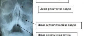
Learning to describe x-rays of the sinuses
Anyone can describe X-ray images of the sinuses with sinusitis (inflammatory accumulation of fluid) independently if they study the X-ray syndromes of the disease. It is enough to remember the normal intensity of the clearings formed by the frontal and maxillary sinuses in order to learn how to identify sinusitis or cysts (cavity formations filled with fluid) on an x-ray.
X-ray of the paranasal sinuses. The arrows indicate the maxillary (maxillary) and frontal sinuses
It will be easy for the reader to read the radiograph if he remembers that the sinuses on it are black. With pathological accumulations of fluid, white shadows appear (see picture).
A fragment of a radiograph shows how to detect bilateral sinusitis
If you compare Figure 2 with the previous one, you can detect intense darkening (white) in the projection of both maxillary sinuses against the background of bilateral sinusitis. They are formed by the accumulation of fluid.
To summarize: you can easily read X-ray images of the lungs, nose and even teeth only after studying the x-ray anatomy of the normal areas of study. This is what representatives of the Leningrad School of Radiologists advise, and we agree with them. Practical experience is required to identify fractures on photographs.
X-ray and general information about it
Many health problems cannot be identified without an accurate diagnosis or x-ray. For example, when we suspect pneumonia, we often hear from the doctor: “we need to do a fluorography.” What is an X-ray examination? And why do doctors often recommend it?
Fluorography is the most popular type of x-ray.
What is an x-ray? In professional medical terms, an x-ray is a detailed study of the internal structure of the body by shining through it with x-rays and recording the image on a special film or digital detector, i.e. x-rays penetrate through the tissues of the body without damaging them and form a picture of the condition of human organs.
What does an x-ray show? In the pictures you can see (depending on the purpose of the device) various pathologies: inflammation, fractures, neoplasms (tumors), degenerative-dystrophic changes, destructive changes, developmental anomalies, etc. X-ray methods are used to examine the lungs, bones, soft tissues, and internal organs (stomach, kidneys, etc.).
After an X-ray examination, the doctor can make an accurate diagnosis in a number of complex diseases.
The color of the image of organs depends on their density. Different tissue captures X-rays differently. Bones, muscles, lungs - will appear differently; the denser the tissue, the lighter it will be on the x-ray image.
How often can an x-ray be taken?
X-rays can be preventive or diagnostic. For preventive purposes, fluorography or radiography of the chest organs is done (at least once a year), mammography (at least once every two years). Diagnostic x-rays (including fluorography) are done if the presence of any diseases is suspected; it is prescribed by the attending physician. Radiation dose limits for patients (and, accordingly, the number of x-ray procedures) for diagnostic purposes are not established (SanPiN 2.6.1.1192-03).
What norm is acceptable?
The standard for preventive radiation during preventive medical X-ray examinations is 1 mSv per year. Radiation dose limits for patients for diagnostic purposes are not established (if the doctor considers it necessary, then it is necessary). When the accumulated dose of medical diagnostic radiation to a patient reaches 500 mSv (the average dose for one x-ray examination is approximately 0.001-0.5 mSv), measures should be taken to further limit his radiation exposure if radiation procedures are not dictated by vital indications.
Is it necessary to remove radiation from the body after an X-ray examination?
After X-ray examinations, there is no need to remove radiation, since the radiation dose is negligible. Even after scintigraphy, in which a radioactive drug is injected into a vein, it is only recommended to drink more fluids.
An important role is played by high-quality modern equipment and competent work with a specialist’s apparatus.
At Sanas MC, X-rays are taken using the best new generation Japanese equipment, Shimadzu SONIALVISION G4. This is the best in its class and the only multi-complex in the Far East, which, in addition to standard radiographic functions, has unique functions - tomosynthesis (layer-by-layer examination) and SLOT radiography (panoramic image of the spine or lower extremities). Provides the highest quality images and detailed transmission of information with a minimum radiation dose.
7 indisputable advantages of Shimadzu SONIALVISION G4 over other devices:
- SONIALVISION G4 is a premium universal remote-controlled X-ray diagnostic complex . The all-in-one multi-purpose system sets new standards for flexible imaging systems, increasing radiology room productivity compared to conventional systems.
- SONIALVISION G4 recognized as the best in its class universal X-ray unit. Independent analysis company KLAS awarded Shimaszu Medical Systems the “2015 Best in KLAS award” in the X-ray equipment segment.
- The world's first remote-controlled device with the function tomosynthesis is a radiographic research method that produces a layer-by-layer image of the area under study with a slice thickness of 0.5 mm, which allows you to see the smallest pathological changes up to 1 mm. The diagnostic capabilities of this method are much wider than with conventional digital radiography.
Tomosynthesis significantly expands the detection limits of smaller pathological changes than traditional radiography. 74% of focal-like shadows (focal-like shadows can occur with tumors, metastases, tuberculosis and other pathological processes) are detected with tomosynthesis compared to 25% with standard radiography, which indicates a threefold increase in detection sensitivity with tomosynthesis. Digital radiography failed to detect metastatic changes in the lungs in 21.3%, which were determined by tomosynthesis. The information value of tomosynthesis in detecting peripheral lung cancer has been proven by scientists at the Research Center for Cancer Prevention and Screening (Tokyo, Chiba).
- The low radiation dose allows tomosynthesis to be used as a screening method, in contrast to computed tomography. In the low-dose mode (20 sections), the dose does not exceed 0.001 mSv, which corresponds to radiation safety standards.
- Another advantage of tomosynthesis over computed tomography is the ability to examine patients with metal implants without artifacts.
- SLOT radiography – (also known as panoramic radiography, slit radiography, axial radiography, teleradiography). This method allows you to produce a panoramic image of the entire spine including the pelvis or lower extremities including the pelvis in one image with one pass of the X-ray tube. The image is obtained with true anatomical dimensions, in contrast to the image stitching method. Slot radiography is effectively used to diagnose: scoliosis, shortening and deformation of the lower extremities, distortion and rotation of the pelvic bones. This method is necessary for the work of orthopedic doctors and chiropractors.
- The well-thought-out design of the device ensures that all studies are carried out without moving the patient; the head-to-foot coverage is 202 cm.
The Sanas MC employs experienced doctors - radiologists and x-ray technicians who will take and describe high-quality X-ray images of the desired organ and give truthful information about the state of your health.