Quite often in their practice, ENT doctors are faced with such a problem as paranasal sinus cyst. Despite the fact that the disease occurs frequently, in almost every tenth person, it is mainly discovered by chance, for example, during an X-ray examination for a completely different reason. This is explained by the fact that a cyst in the nose may not bother the patient for a long time and may not manifest any symptoms. Therefore, it is important to understand that timely treatment of cysts is necessary to avoid possible complications and to improve the patient’s quality of life.
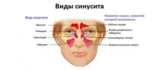
What is a paranasal sinus cyst?
A paranasal sinus cyst is a formation with an elastic, elastic wall filled with fluid. Its size and location can be very different, which is reflected in clinical manifestations (patient complaints).
According to the classification, there are several types of cysts:
- Retention (true): occur due to blockage of the ducts of the glands of the mucous membrane;
- Pseudocysts (false): appear under the influence of an allergen, due to an infectious disease or due to an inflammatory process in the roots of the teeth of the upper jaw.
In addition, cysts are classified depending on what contents they are filled with, i.e. mucous, purulent and serous.
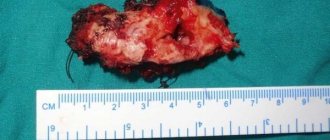
Foreign body in the maxillary sinuses
A foreign body in the maxillary sinuses , like a maxillary sinus cyst, is often detected by chance, but turns out to be the cause of severe forms of sinusitis.
A foreign body can get into the maxillary sinus either through an open wound or after filling teeth (as a rule, these are the so-called “sixes” of the upper jaw). The dental canals communicating with the maxillary sinuses bring pieces of filling material into these accessory cavities, where they “get stuck” and become factors of severe purulent inflammation.
How does a sinus cyst appear?
The mechanism of development of paranasal sinus cysts is quite simple.
Let's start with the fact that in the mucous membrane of the paranasal sinuses there are glands that produce mucus. Each such gland has its own excretory duct for secreting secretions, which opens on the surface of the mucous membrane of the sinus.
Under the influence of various unfavorable factors, the mucous membrane thickens and the excretory ducts become clogged. At the same time, the gland continues its work with the same force, mucus is formed, but the exit for it is blocked. Due to wall pressure, the glands begin to fill with mucus, stretch and form a cystic neoplasm. Approximately the same effect is obtained if you put an inflatable balloon in a water tap and open the tap - water will flow and inflate the balloon.
There are several UNFAVORABLE FACTORS that will contribute to the development of this process, namely changes in the thickness of the mucosa and blockage of the ducts:
- exposure to an allergen and the occurrence of allergic reactions,
- polyps,
- infectious diseases,
- chronic diseases developing in the nasal cavity (rhinitis) or in its paranasal sinuses (sinusitis: sinusitis, sinusitis, etc.),
- diseases of the teeth of the upper jaw, anatomical features of the structure of the nose.
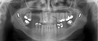
What medical errors lead to perforation?
The presence of the problems described above does not guarantee that the tooth will independently enter the maxillary sinus. This usually occurs when there is a perforation - a break in the thin wall between the mouth and the nasal sinus. In the vast majority of cases, this occurs during dental procedures. And if monitoring the condition of teeth and preventing osteoporosis is the responsibility of the patient, then mechanical perforation is always the responsibility of the doctor. Of course, it is much more difficult for the latter to work if at least one of the factors described above is present. That is why good health (including teeth) is the result of cooperation between the doctor and the patient.
Patients often complain that the pain from sinusitis radiates to the teeth. A detailed examination reveals a radically opposite picture. Perforation of the bottom of the maxillary sinus is detected, which causes pain. The fact is that periodontitis may not make itself felt for a long time, but sinusitis debuts quickly. Therefore, it seems that the problem is in the nose, and not in the mouth.
Perforation in most cases appears as a result of a medical error made during the treatment or extraction of teeth.
Perforation of the maxillary sinus during tooth extraction
Removing a tooth if its root is located in the maxillary sinus or is separated from it by a thin layer of bone is always difficult. Especially when it comes to 2-3 molars with massive and intertwined roots. Since the dental surgeon applies physical force during the operation, there is a high probability of damaging the already thin layer of bone.
In this case, it is recommended to take a photo at the end of the procedure. Treatment of perforation of the maxillary sinus immediately after tooth extraction is simple. The doctor will be able to quickly restore the tightness of the sinus, avoiding complications. To exclude the possibility of a fistula forming in the nasal sinus after some more time, it is recommended to take a repeat photo after a month.
Perforation during implantation
A perforation of a sinus near a tooth during prosthetics is always a very serious complication that can cast doubt on the success of the entire operation. To prevent this, additional bone augmentation using various methods is performed before implantation.
Before implanting the implant, the doctor must take a panoramic photo. If the roots of a wisdom tooth or other molars and premolars are located in the maxillary sinus or in close proximity to it, then a sinus lift is prescribed, during which the bottom of the sinus is raised and strengthened.
If perforation does occur, then implantation is postponed until complete healing.
Perforation during endodontic treatment
Acute sinusitis can also develop after routine dental treatment. During canal treatment, it is easy to go beyond the apex with a metal instrument or filling material. To eliminate the possibility of complications and promptly eliminate perforation after each stage of endodontic treatment, it is important to take an image. Modern radiovisiographs have a very low radiation dose, so they cannot harm health in any way.
Perforation during removal of the apex or root of a tooth completely
From the point of view of perforation, tooth-saving operations can also pose a danger, for example, removing the root of a tooth with a cyst on it that has grown into the maxillary sinus. Such operations are relatively safe for teeth 4-5, while for teeth 6-7 they are no longer advisable. The risk of perforation is too great. It is worth clarifying that we are talking about cases with a pathologically thin bone septum between the nasal sinus and the oral cavity. If the bone thickness is within normal limits, then apectomy is performed for any tooth.
Symptoms of a maxillary sinus cyst
So, what symptoms should you pay attention to? In what cases is it necessary to consult a doctor?
Of course, first of all, it should be said that a maxillary sinus cyst (both right and left) can develop asymptomatically (that is, its appearance and constant presence in the maxillary sinus does not cause discomfort for a person) with a diameter of less than 1 cm.
In this case, usually, the detection of a cyst occurs either accidentally (for example, an x-ray examination to confirm the diagnosis of sinusitis), or with further significant growth of the tumor (more than 1 cm in girth).
If a cyst in one of the sinuses causes discomfort (it is larger than 1 cm), then the following symptoms are observed:
- difficulty breathing (especially after exercise),
- constant nasal congestion (a common symptom for children),
- local pain over the affected sinus (usually the maxillary, right or left),
- false symptoms of increased intraocular pressure (caused by a cyst with a volume of more than 1 cm, hence the unpleasant sensations),
- purulent discharge (if concomitant diseases are headed by sinusitis),
- symptoms of innervation damage (hypo- or anosmia - loss of smell),
- excruciating headaches that quickly recover even after strong analgesics (compression of the trigeminal nerve branch).
All of these listed symptoms are a reason to contact an ENT doctor in order to avoid complications that such a process can cause!
Removal of a maxillary sinus cyst at the ENT clinic of Dr. Korenchenko
See also Treatment of ENT diseases Cyst in the maxillary sinus Treatment of a cyst in the maxillary sinus Surgery to remove a cyst in the maxillary sinus
Endoscopic ENT surgeries are not performed in all clinics. After all, they require modern equipment, the doctor having the appropriate skills and certificates. Dr. Korenchenko’s ENT clinic is a modern, specialized and well-equipped medical center. Our specialists are highly qualified and have rich clinical experience, all the necessary certificates and skills. When treating patients, we use only modern, clinically proven and highly effective techniques.
Endoscopy at Dr. Korenchenko’s Clinic is an important and widely used therapeutic and diagnostic procedure. It is included in the basic examination of all patients who apply and are observed, which allows doctors to receive reliable and accurate information about the current condition of the ENT organs. Our specialists also perform removal of maxillary cysts and most other operations endoscopically, with high results and without long-term rehabilitation of patients.
Treatment at Dr. Korenchenko’s ENT clinic is a modern and competent approach, using effective technologies and effective therapeutic regimens.
Diagnosis of sinus cysts
In addition to the standard questioning and examination of the patient, all doctors at the Lor-Plus clinic are proficient in endoscopic examination techniques. Using an endoscope - a long thin tube with a miniature video camera, the doctor can conduct the most complete examination of the ENT organs, determine the presence of additional structural features of the nasal cavity, changes in the nasal mucosa in the deep sections that may interfere with normal breathing.
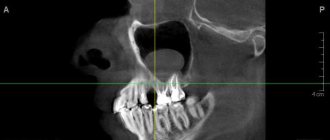
Currently, the gold standard for diagnosing sinus cysts is computed tomography . This method allows you to determine the size of the cyst and its location in the sinus with millimeter accuracy, which is very important for choosing a method for removing the cyst.
Treatment
The choice of treatment for maxillary sinus perforation depends on the changes that occur as a result of the rupture. Non-surgical treatment seems possible in case of violation of the integrity of the tissues during tooth extraction, when the pathology was detected immediately, and according to the results of the radiographic examination it is clear that there is no infection of the sinus cavity and there are no foreign bodies in it. With such a clinical picture, the doctor, as a rule, tries to preserve the blood clot formed in the hole as much as possible. In addition, preventative measures are taken to prevent wound infection.
For this purpose, a small gauze swab soaked in an iodine solution is inserted into the hole. As a rule, the tampon is self-fixed in the wound cavity. However, sometimes it may be necessary to place a stitch in the gum. Treatment with an iodide solution should be carried out for 6-7 days until the defect disappears and full-fledged granulations are formed. It is not recommended to remove the tampon from the socket during treatment, as this can damage the clot and lead to infection of the wound.
When a tooth root is found in the maxillary sinus, everyone should know what to do. In some cases, the doctor decides to close the defect with a special plastic plate, which is secured to adjacent teeth with clasps. Thus, it is possible to achieve separation of the sinus and oral cavity.
Diagnostics and treatment in our clinic
In our specialized medical center you can receive a wide range of services at the level of world standards. We offer examination and treatment by specialists with extensive experience who undergo regular training, advanced training, certification and licensing in Russia and abroad. Highly professional doctors at the AMC-Medionika clinic in Moscow will understand your problems and provide high-quality and effective care.
Call the clinic's contact numbers or make an appointment online. We will restore and maintain your health!