Modern dentistry cannot be imagined without accurate diagnostics. A set of measures to establish a diagnosis is indispensable in the treatment of teeth and gums; it plays a key role in implantation and prosthetics, and is also important during orthodontic treatment.
Diagnostics is carried out at all stages of preventive and therapeutic tasks. The dentist-therapist resorts to full diagnostic measures when the patient comes to the clinic. The specialist also uses operational diagnostic measures as an intermediate stage of treatment after installing a filling. Upon completion of prosthetic procedures, full diagnostics allows you to evaluate the results and correct them in a timely manner.
Dental diagnostics is, without exaggeration, 50% of the success of treatment and prevention.
Diagnostics involves the following activities:
- History - information about the problem from the patient himself. Mandatory primary stage of diagnosis. The patient indicates the area of concern - which tooth hurts, where there is increased sensitivity, etc.
- Introduction to medical history . Modern technologies make it possible to provide a specialist with images and examination results taken earlier. The information in the medical history card can greatly facilitate the doctor’s work when developing the optimal treatment strategy, so it is important to have it with you whenever possible.
- A thorough visual examination and examination of the condition of the teeth and gums. The specialist determines the problem by the appearance of the tissue.
- Instrumental (X-ray) examination is a study using special equipment. A key element of modern dental diagnostics.
X-rays of teeth
Main stages
Today, dentists use different methods, the choice of which depends on the condition of the oral cavity, the patient’s complaints and other factors. Basic measures include:
- Anamnesis is collected, including an oral interview and examination;
- fillings, crowns and other structures are checked;
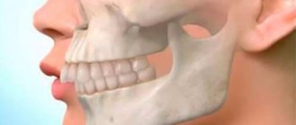
- analysis of occlusion, identification of defects in the dentofacial apparatus;
- undergoing an X-ray examination;
- computed tomography (performed if necessary, before implantation);
- making a diagnosis, drawing up a treatment plan.
Diagnostics is an important condition for a full assessment of the clinical picture, identifying problems, and selecting effective therapeutic methods.
X-ray diagnostics
The use of X-rays and other high-precision equipment in diagnostics allows us to obtain, in real time, accurate images of the desired areas of the oral cavity in optimal resolution.
What equipment is used for instrumental diagnostics:
- Radiovisiograph (visiograph, videograph, dental imaging apparatus) is a device that converts X-ray data into a digital image on a computer in real time. The dentist, using a visiograph, controls the treatment procedure without leaving the chair with the patient. The device allows you to quickly take a photo and obtain an image of the problem tooth.
- Digital 3D orthopantomograph - allows you to take a panoramic image of the desired area of the skull or an individual tooth. The device is indispensable for prosthetics, treatment, implantation, bite correction (orthodontics), etc.
- Rheodentograph is a device for accurately assessing the functional state of the pulp (tissue containing nerve endings) of a tooth according to hemodynamic indications. Allows you to quickly, painlessly and accurately determine the extent of tissue damage. The device is important in the gentle treatment of pulpitis.
- A tool for diagnosing early and latent caries. Pathological processes of hard tissues in the early stages are often difficult to determine. At the same time, treating caries in the early stages allows you to solve the problem with minimal impact on the tissue. The device allows you to effectively identify crisis areas on the tooth.
The capabilities of the equipment allow you to print the results or transfer them to electronic media.
Diagnostic measures include control modeling methods, as well as taking impressions. These methods can significantly increase the effectiveness of orthodontic treatment and prosthetics.
The main task of the dentist is to maintain the health of teeth and gums, maximize tissue preservation, and ensure functionality and aesthetics. All these problems cannot be solved without competent dental diagnostics.
Goals of diagnostics in dentistry
Research in the field of dentistry is needed to assess the oral cavity, identify diseases and pathologies, and analyze the condition of the dental system. An examination is also required to monitor the intermediate or final result of therapy.
Basic goals:
- the ability to quickly assess the muscles, ligaments and joints of the jaw apparatus;
- determine pathologies or dystrophic processes in the bone, structural features, identify inflammation, the course of pathological processes;
- assessment of mucous and gingival tissues;
- carrying out analysis of such individual areas as crown, roots, pulp, dentin and others;
- control of treatment, the effect of actions taken;
- preparation before prosthetics, installation of implants or surgery.
EXAMINATION OF A SURGICAL DENTAL PATIENT
The purpose of examining the patient is to establish a diagnosis. It consists of a thorough history taking and an objective examination of the patient.
During the interview, complaints and medical history are ascertained. During the interview, it is necessary to establish confidential contact with the patient, determine his neuropsychic status, intelligence, and on this basis analyze complaints and the course of disease development. The doctor should use leading questions to help the patient outline the medical history.
The examination begins with clarification of complaints. The most characteristic of them are complaints of pain, which can be constant or temporary, sharp or dull, localized or diffuse, spontaneous or associated with touching a tooth, an area of facial tissue, jaws and other irritations. Pathological processes developing in the maxillofacial area, in most cases, are signs of inflammation, often of an odontogenic nature. They differ in a certain nature of pain, which can serve as a basis for differential diagnosis of certain diseases. Thus, with pulpitis, acute diffuse pains are observed, often night pains radiating along the nerve branches and trunks. Acute periodontitis is characterized by acute pain localized in the tooth and pain when biting. Then they intensify, become permanent and radiate along the branches of the sensory nerves. Acute purulent periostitis of the jaw is manifested by the spread of pain from the causative tooth to the area of the jaw, i.e. the pain is diffuse in nature. Pain in acute osteomyelitis of the jaw, depending on the localization of the process and the extent of bone damage, is varied: acute, radiating along the nerves, drilling, diffuse. Abscesses, acute lymphadenitis, specific inflammatory processes of the head, neck, jaws are characterized by aching pain localized in the area of the affected tissue. They may intensify upon palpation. With phlegmon, adenophlegmon, boils, carbuncles, the pain is diffuse and constant. Subsequently, the intensity of the pain increases, it becomes twitching and throbbing. In addition to local pain, inflammatory processes cause headaches, malaise, loss of appetite, sleep, chills and other manifestations that reflect the degree of intoxication.
Pain may occur when moving the lower jaw, tongue, swallowing, breathing, talking. This is observed in inflammatory and oncological diseases, injuries of soft and bone tissues of the face, and oral organs. There may be disturbances in chewing, swallowing, mouth opening, taste, and breathing. Complaints of difficulty swallowing and breathing are a serious symptom and in these cases, immediate further examination of the patient is required.
Patients may complain of soreness and swelling of the salivary glands, dry mouth, and an unpleasant salty taste associated with food intake, which is typical for diseases of the salivary glands.
Patients often complain of impaired facial symmetry. This can occur due to swelling, neoplasm of facial tissue, jaws, and oral organs. By comparing complaints of pain with the nature of the swelling, in some cases we can talk about diseases of an inflammatory nature, in others - about a tumor or tumor-like formation.
Patients may complain about a defect or deformation of the face, causing functional and aesthetic disturbances. In such cases, the nature of the defect or deformation (congenital or acquired) should be clarified. In case of an acquired defect, it is important to establish its cause (trauma, inflammatory, inflammatory processes, previous operations, etc.).
During the questioning, the dynamics of the disease are clarified:
when the first symptoms appeared, what they were, who noticed them (the patient, others, the doctor), where the patient went for help, what treatment was carried out and with what result. You should familiarize yourself with the documentation available to the patient regarding the examination (extract from the medical history, data from laboratory and other studies, radiographs, consultants’ opinions).
If you complain of pain and swelling in the maxillofacial area, you should clarify how the process developed and establish the source of infection. With an increase in general and local symptoms of the inflammatory process, hospitalization and, possibly, emergency operations are necessary.
In the presence of specific inflammatory foci, ulcers, defects of the maxillofacial area and oral mucosa, information about heredity, lifestyle, contacts with patients, animals, and others should be collected to exclude tuberculosis, syphilis, anthrax, HIV infection, and also clarify the results of examinations carried out for these diseases.
When the process is localized in the area of the salivary glands, it is necessary to find out from the anamnesis whether there was swelling of the gland and whether it is associated with food intake. It is necessary to clarify the development of the disease after operations on internal organs, especially the abdominal cavity, pelvis, after a viral or other infection, as well as diseases of internal organs.
If there is an injury, it is necessary to clarify under what circumstances it occurred, whether the patient lost consciousness and for how long, whether there was nausea, dizziness, vomiting, bleeding from the nose or ears, and what assistance was provided . It is necessary to find out whether the patient was administered antitetanus serum or tetanus toxoid, how, when and in what doses. The fact of injury while intoxicated requires clarification .
When a patient contacts you about bleeding associated with trauma, surgery (including tooth extraction), you must ask about the duration of bleeding due to previous operations, cuts, and bruises.
For pain that characterizes diseases and damage to the nerves of the face and jaws, you need to know the data on your neurological status. When patients contact us for pain and dysfunction of the temporomandibular joint, it is necessary to find out the connection of the process with diseases of the cardiovascular system, musculoskeletal system, etc.
In case of tumors and tumor-like lesions of the face, jaws, oral cavity organs, it is necessary to clarify the connection of the process with other diseases - internal organs, ENT organs, skin, etc., to clarify the characteristics of the growth of the tumor (widespread or limited), accompanying symptoms (pain and their nature, dysfunction, etc.).
In the case of birth defects, it is necessary to find out family history (heredity), features of the course of the first half of pregnancy and childbirth, development at an early age and later. If a defect is acquired, it is important to find out its cause (trauma, burn, inflammatory, specific or oncological processes, previous operations, etc.). In case of acquired defects and deformations, it is necessary to exclude diseases such as syphilis, chronic osteomyelitis of the jaw, actinomycosis, and noma.
Anamnesis of life
consists of information about the characteristics of childbirth, the health of parents, working conditions, living conditions, nutrition, recreation, physical education, identification of bad habits (smoking, drinking alcohol, medications, drugs). This allows you to get a correct idea of physical and mental health. It is necessary to find out what diseases the patient suffered, how they progressed, what treatment was carried out and its results. It is necessary to clarify whether the patient has immunopathological diseases and conditions. Identification of primary or secondary immunodeficiency states and accompanying diseases in connection with the dental pathological process will allow us to correctly assess the functional state of the body and clarify the diagnosis.
The examination of the patient begins with a general examination and determination of his condition (satisfactory, moderate, severe and extremely severe).
In a hospital setting, the examination is carried out taking into account all the rules accepted in clinical medicine. In the clinic, you should evaluate the patient’s physique, determine the presence of defects and
hell
body deformations, determine pulse, blood pressure, moral and mental state.
If an acute infection, syphilis, erysipelas, tumor and other diseases are suspected, the skin of the whole body is examined (the presence of rashes on it), the occipital, lateral cervical, subclavian, axillary lymph nodes are palpated, the pupillary reflex, Kernig's sign, etc. are examined.
Examination of the maxillofacial area
includes external examination, palpation, examination of the oral cavity, instrumental examination (probes, blunt and sharp needles, etc.). If necessary, a clinical examination can be supplemented by taking a scraping, performing a puncture or biopsy, biochemical, microbiological, immunological studies, radiography, tomography, etc.
Inspection
The patient is placed in a dental chair. His head should be well fixed on the headrest, you can raise and lower the chair, change the position of its back (straight, at an obtuse angle) and the headrest (the patient’s head is thrown back or the chin is close to the chest). In cases of moderate or severe condition, the patient is examined in bed, on a table in a dressing room, or in a dental chair placed in a horizontal position.
For examination, use a tray with sterile instruments: a spatula (for retracting iy6, cheeks and examining the vestibule and the oral cavity itself, retracting the tongue and examining the sublingual area, body of the tongue, tonsils, pharynx) and dental or anatomical tweezers (to determine the mobility of teeth and their percussion ). During the examination, a dental mirror is used (to examine the teeth, sublingual area, palate), a dental probe, often at an angle (to probe defects in the crown of the teeth, gingival papillae, gingival margin; the probe handle can also be used to percussion the teeth), a thin Bauman probe, special salivary probes (for probing ducts, fistula tracts), a button probe (for probing wounds, fistulas, perforation communications with the maxillary sinus, palate defects, etc.). It is better to examine the nasal cavity, pharynx, and external ear using a frontal reflector, nasal and ear mirrors.
The examination consists of determining the symmetry of the face - its relief, caused by the connection of the bones of the facial skeleton, the level of development of the subcutaneous fat layer, the condition of the cartilaginous part of the nose, eye and mouth slits, ears and skin covering. The normal face is often asymmetrical. It is important to determine the violation of its symmetry due to inflammatory, traumatic, tumor and other changes. In case of diseases and injuries of the maxillofacial area, attention should be paid to the nature of the violation of the symmetry of the face and neck (swelling, infiltration, tumor formation, deformation, etc.).
It is necessary to tilt, turn, and throw back your heads to determine its movements.
Palpation examination
allows you to clarify the boundaries of pathological changes, tissue consistency, the ability of the skin to fold, the presence of scars, fistula tracts. If there is swelling of the perimaxillary soft tissues, its consistency, the adhesion of the skin to the underlying tissues, and its color are determined. If the blunt end of the instruments leaves a mark under pressure, this indicates swelling of an inflammatory nature. It can occur with various inflammatory diseases and trauma to the face and jaws.
If, upon palpation, the perimaxillary soft tissues are compacted, painful, the skin is fused with the underlying tissues, is difficult to form a fold or does not form it, the color changes from intense pink to bright red or purple-blue, the temperature of the tissues is increased, then this indicates the presence of infiltrate and can be observed with abscess, phlegmon, lymphadenitis and other inflammatory diseases in the perimandibular soft tissues. In this case, it is necessary to mark the boundaries of pathological changes, determine the areas of greatest pain and fluctuation, the adhesion of the affected tissues to the underlying bones of the facial skeleton, and the presence of fistulas.
The configuration of the face can be changed due to a posterior displacement of the lower jaw or retraction in the zygomatic region, lengthening of the middle part of the face, retraction of the nasal bridge and other disorders caused by trauma. Pay attention also to bruises, abrasions, wounds, hematomas.
A comparative palpation study of the bones of the facial skeleton is carried out along the bony contours of the face and mainly at the junctions of bones, paying attention to atypical bone irregularities and pain on palpation.
When the jaws or zygomatic bone are fractured, the function of mouth opening is impaired in the form of restriction, displacement of the lower jaw to the side, etc. The temporomandibular joint is examined by palpation - the head of the condylar process, its articulation with the glenoid cavity, and the range of movements of the lower jaw during opening and closing is determined mouth, to the sides.
Palpation determines the sensitivity of the exit of the peripheral branches of the trigeminal nerve at Balle's points (supraorbital, infraorbital and mental nerves). Various diseases and damage to the nerves of the face and jaws are accompanied by pain and sensory disturbances. During palpation examination, the pain may intensify or an attack may develop. During the examination, a violation of the sensitivity of the facial skin (anesthesia, paresthesia, hypoesthesia, hyperesthesia) is also noted.
If cancer is suspected, deep palpation is used. Tumors and tumor-like diseases can have different consistencies: doughy, densely elastic, cartilaginous, etc., smooth or bumpy surface, clear or poorly defined boundaries.
Palpation of regional lymphatics is important
nodes - submandibular, mental and other cervical, mental, facial, etc. To palpate the submandibular nodes, the doctor tilts the patient’s head down with his right hand, and with his left hand he consistently feels them with three fingers, tilting the patient’s head in the appropriate direction; he palpates the mental nodes in the same position with his index finger , and mastoid - with the second finger, moving it forward to the posterior edge of the branch of the lower jaw and posteriorly - to the anterior edge of the sternocleidomastoid muscle. Facial lymph nodes (buccal, nasolabial, molar, mandibular) are palpated bimacularly - with the fingers of the right hand from the side of the mouth and the left - from the outside. The parotid lymph nodes are palpated in the projection of the surface of the branch of the lower jaw, in the retromandibular region - in the thickness of the salivary gland and bimanually - along the anterior edge of the parotid salivary gland. The lateral cervical lymph nodes are palpated with 2-3 fingers anterior and posterior to the sternocleidomastoid muscle, from the mastoid process down to the clavicle. Next, standing behind the patient, three fingers (II, III, IV), placed on the collarbone, palpate the supraclavicular lymph nodes.
Oral examination
consists in determining the opening of the mouth, examining the vestibule and the oral cavity itself, the pharynx.
The opening of the mouth is noted (normally it should be 5 cm or three diameters of the II, III, IV fingers inserted between the central incisors); determine whether it is free and painless, whether there is a crunch in the joint, what is the displacement of the lower jaw to the side. Inflammatory processes involving the masticatory muscles make opening the mouth difficult and painful. In such cases, the reduction of the jaws should be noted (inflammatory contracture of the masticatory muscles of I, II and III degrees).
Inspection of the vestibule of the oral cavity begins with the lips, noting the nature of the color of the border, examining the mucous membrane, its color, degree of moisture, palpating the cheek, the area of the fatty body of the cheek.
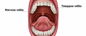
Examination of the oral cavity itself is carried out in good lighting. It is better to examine it using a forehead reflector or a dental mirror with a light bulb built into it. Examine the mucous membrane of the gums (gingival margin, gingival papillae, gingival sulcus), labial frenulum, sublingual folds and papillae, incisive papilla, transverse palatal folds. When examining the oral mucosa, attention is paid to the small salivary glands - labial, buccal, molar, palatine, lingual, etc. The alveolar processes of the jaws (alveolar arches, alveolar elevations), the palatine process of the upper jaw (nasal ridge, incisive suture, palatine spines) are examined by palpation and grooves), tissue behind the tubercle of the upper jaw.
The teeth are examined, they are percussed, the presence of supernumerary or primary teeth in the permanent dentition, the eruption of lower wisdom teeth are noted, the nature of teeth closure is determined, and the dental formula is recorded. The gingival tubercles, pockets and the nature of the discharge from them are examined. Pulling the cheek with a spatula, mark
condition of the parotid gland papillae; lifting the tongue to the palate, examine the ducts of the submandibular and sublingual (large and small ducts) salivary glands • their secretion of saliva.
When doing a massage of the salivary glands, you should pay attention to possible characteristic changes: thick consistency of saliva, cloudy color, the presence of flakes, clots, salivary blood clots in it.
In case of diseases of the salivary glands, probing of the ducts is carried out, which makes it possible to determine their direction, the presence of stenosis, stricture or its complete obliteration, or a stone in the duct.
When examining the tongue, pay attention to its shape, size, condition of the mucous membrane, its color and degree of moisture, and the severity of the papillae. The tongue is palpated by moving it forward, grasping the tip with a gauze napkin. Examining the pharynx, examine the soft palate (uvula, palatoglossus, velopharyngeal arches), tubal-palatal fold, palatine tonsil, etc., and determine the pharyngeal reflex.
When examining and examining the sublingual areas, bimanual palpation is used: the deep tissues of the floor of the mouth are examined from the sublingual fold and submandibular region.
In the case of perforation of the bottom of the maxillary sinus during tooth extraction, the hole is examined and fluid enters the nasal cavity through the mouth. The depth of the sinus is determined by inserting a probe into it.
When examining a patient with a trauma to the facial bones, pathological mobility, painful teeth, and rupture of the mucous membrane are determined. When palpating the fragments, their mobility, crepitus and pain are noted. Pay attention to the closure of the teeth and the displacement of the lower jaw when opening the mouth.
If a tumor or tumor-like disease is suspected, it is necessary to clarify the localization of the formation, its size, consistency, mobility, connection with the teeth, etc. In case of an ulcer, the density of its edges and the condition of the bottom are examined.
Inspection of the oral cavity for defects and deformations of the face and jaws begins with the oral fissure (the shape of the lips), pay attention to the opening of the mouth, examine the alveolar and palatine processes of the upper jaw, the palatine bone and the soft palate. The location and size of the defect and the condition of the surrounding mucous membrane are determined.
The clinical examination of the patient should include a number of diagnostic methods and studies. Their type and volume depend on the nature of the disease or injury in the maxillofacial area and on the conditions of the examination (in a clinic or hospital), as well as on the level of equipment of the medical institution.
X-ray examinations of the teeth, jaws and other bones of the face and skull, maxillary and frontal sinuses, temporomandibular joints, and glands of the oral cavity are important for diagnosis. For this purpose, contact intraoral radiography of teeth, alveolar and palatal processes, and the floor of the oral cavity is used, which makes it possible to clarify the localization and nature of changes in the periodontium and bone, and to note the presence of a calculus. Vaerrot radiography is used for research
upper and lower jaws, zygomatic, frontal, nasal, temporal and other bones of the skull, maxillary and frontal sinuses, maxillary and mandibular joints. The following radiography projections are used: direct, lateral, semi-axial, axial, as well as oblique contact and tangential projections.
A promising method of x-ray examination is orthopantomography, which allows you to obtain an overview layer-by-layer image of the teeth and jaws. X-ray examination of the teeth, jaws and other bones of the facial skeleton is of fundamental importance for judging the presence of carious cavities in the teeth, the shape of the roots, the degree of filling them with filling mass, the condition of the periodontium, bones, etc.
X-ray examination plays a decisive role in diagnosing trauma to the teeth, facial bones and skull. In combination with other methods, it helps to recognize tumors and tumor-like diseases. Diagnosis of defects and deformations of facial bones, planning of reconstructive operations are also based on radiography data. In this case, teleradiography - radiography with a large focal length - is of great help. It is imperative to obtain identical images, from which changes in the bone image are noted and compared with the accepted norm for the correct structure of the facial bones.
In some cases, to clarify inflammatory lesions of soft and bone tissues, localization of foreign bodies, tumor boundaries, in diseases of the temporomandibular joint, layered radiography is indicated - tomography, which is performed on a tomograph or on a tomographic attachment to a universal X-ray machine, as well as on a computed tomography machine . In recent years, magnetic resonance and computed tomography have become of great importance for diagnosis.
For diseases of the salivary glands, in some cases with chronic sinusitis of the maxillary sinus, radicular cyst, chronic osteomyelitis of the jaw, braonchiogenic fistula, contrast sialo-sinus and fistuloradiography are used.
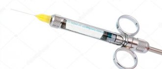
In various pathological processes: inflammation, trauma, tumors, there is a need to determine the viability of the dental pulp using the method of electroodontodiagnosis. Indicators up to 8-10 mA indicate the normal state of the pulp, from 10 to 60 mA and more than 100 mA - about its change and even death. Irritation thresholds from 100 to 200 mA indicate periodontal irritation by electric current.
Laboratory testing for diagnostic purposes includes a large number of different methods, carried out both in a clinic and in a hospital. In a clinic setting, their use is limited, and they are limited to conducting general blood and urine tests, determining their sugar content, cytological and morphological studies. In basic dental and general clinics they can be supplemented by bacteriological, immunological, biochemical
| 2—1184 |
mic and other research. In the hospital, in addition to the methods listed above, laboratory tests are required to test for HIV infection, syphilis, and determine the blood type and Rh factor. In addition, a variety of functional (rheography, capillarography, electromyography) and other studies (biochemical, immunological, hormonal) are used to diagnose the disease and monitor treatment.
For long-term non-healing ulcers, painless infiltrates, palate defects, dental anomalies and other disorders, an examination is carried out for tuberculosis, syphilis, deep mycosis, and HIV infection.
Cytological studies—taking smears, fingerprints, scrapings, punctate, and washings—are important to confirm the nature of the disease.
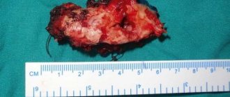
A more reliable answer is obtained by taking material by biopsy - excision of a piece of tissue, which is fixed in a 10% solution of neutral formalin and sent to the pathology laboratory with a special accompanying form. Often, in order to clarify the diagnosis during surgery, an emergency biopsy (express biopsy) is used.
Purulent secretions obtained from patients must be examined natively, which makes it possible to detect radiata fungus drusen, cholesterol crystals, etc.
In both hospital and clinic settings, there is often a need to conduct microbiological studies. Sowing purulent exudate under aerobic and anaerobic conditions, isolating the main pathogen, determining its properties, and obtaining antibiograms are important for the diagnosis and treatment of inflammatory diseases.
In case of diseases of the salivary glands, a study of the secretory-excretory function, qualitative and cytological analysis of saliva is carried out. In the presence of special equipment, radiosialography, scanning of the salivary glands, scintigraphy, echosialography, and thermal imaging acquire important diagnostic significance.
Immunological tests are mandatory in hospitals and, if possible, in basic dental clinics.
In some cases, when examining a surgical dental patient, as well as when preparing the patient for surgery, orthopedic measures are carried out: taking impressions and making protective plates, orthopedic devices (Vankevich splints, Weber splints, with an inclined plane, wire splints with Schroeder hinges, Pomerantseva-Urbanskoy etc. .), jaw models, face masks.
All of the above examination methods are included in the medical history, which is an important legal document, including for forensic medical examination.
It should be noted that compliance with the rules of deontology and ethics is the first condition for successful diagnosis and treatment of a surgical dental patient.
X-ray
To make a correct diagnosis in dentistry, classical and modern diagnostic methods are used. The classic ones include visual inspection and targeted X-rays. X-ray is a painless procedure with a radiation dose that is safe for the patient. It is suitable for the need to diagnose one specific tooth.
A small plate is placed near the tooth of interest and held in position for several seconds. A targeted image is immediately displayed on the computer monitor, showing the condition of the tissues hidden under the mucous membrane and in the jaw bone.
Comprehensive diagnostics at the Estetika clinic
Our clinic in Novosibirsk has modern German dental equipment installed. High-precision equipment allows for high-quality radiation diagnostics, which is one of the informative research methods in dentistry. We also have a modern digital device “Gendex” for three-dimensional diagnostics.
The articles published on the site are for informational purposes only, and the services described may not correspond to the list of services provided in the dental clinic. Please check with the administrator for the availability and cost of procedures.
Diagnostic options
Today, various methods are used for examination, including the following:
- initial examination, collection of complaints and information about symptoms;
- assessment of teeth, mucous membranes, closure of rows;
- examination of the coronal part using a mirror, microscope or scanner, probe (for enamel analysis), tapping and percussion are also performed;
- hardware examination methods using special equipment;
- laboratory research.
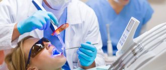
Visual inspection
The examination of the Patient begins with an initial appointment and visual examination. In this case, the doctor takes into account the following parameters:
- pain syndrome, characteristics, frequency, intensity of pain;
- presence of acute symptoms;
- reaction to touch, temperature changes, sour or salty;
- injuries and others.
To perform this stage, dental instruments, palpation methods, and tapping are used. If the problem is identified immediately and does not require further investigation, the doctor can begin treatment immediately. This usually happens when caries is detected, with large accumulations of stone or plaque.
Microscopy
For diagnosis, special equipment can be used in the form of a microscope or intraoral scanners. This allows you to examine the internal tissues in detail, identify hidden problems, and caries at the initial stage. Special optical equipment with high resolution quality allows you to identify problems that are not visible during a normal external examination. Most often, this is required to detect areas of erosion, microtrauma, and hidden cavities due to caries.
Radiology
This diagnostic method allows you to determine the characteristics of bone, tooth, and soft tissue. Indications for using the method are:
- detection of caries, inflammatory processes, pulpitis;
- analysis of bone volume, structure of the dentition;
- collecting information to monitor achieved results before prosthetics and implant installation;
- checking for voids and pores in the canals after filling.
The following equipment and research options are used:
- dental intraoral photograph of a small area or individual teeth;
- panoramic image to obtain data on the structure of the jaw, the presence of hidden pathologies, periodontitis;
- volumetric image before bone grafting, sinus lifting, and other surgical interventions.
CT scan
Using CT, you can obtain a three-dimensional image of the dentofacial apparatus, examine individual areas in detail, identify hidden problems and prescribe the most effective therapy. Today the following types of examination installations are used:
- equipment for cone beam examination;
- devices for sequential processing of the resulting layers;
- spiral examination.
CT is indicated for the following cases:
- detection of inflammation, caries, injuries;
- study of tissue structure;
- search for formations, cysts, tumors;
- checking for impacted units;
- assessment of the condition of the oral cavity before prosthetics and installation of implants.
Electroodontometry and other methods
This method is used to assess the condition of the neurovascular bundle of the pulp. To determine the viability of tissues, an electric current is applied, after which the presence or absence of a pain reaction in the Patient is assessed.
In addition to these diagnostic methods, others can be used if necessary to analyze the condition of tissues. These include temperature tests, color markers, and fluorescent methods with UV radiation. All of them help to conduct a full examination, identify pathologies and begin correct, effective treatment, eliminating complications.
Stages of dental examination
The patient undergoes examination in three stages.
- clarification of complaints and medical history;
- examination using physical methods (inspection, palpation, percussion, auscultation);
- research using special methods (laboratory, x-ray).
The final clinical diagnosis is made after clarifying complaints and other aspects of the disease and collecting additional information about the patient. All this will allow appropriate treatment to be carried out at the next stage.
Basic examination methods
Patient interview. This is done at every appointment. The doctor collects information about complaints, existing symptoms, past illnesses, medications taken, etc.
Inspection. The dentist examines the mucous membranes, teeth, evaluates the structure of the face, the closure of the dentition, the presence of swelling, edema, and other signs of inflammation. After a general examination, the condition of each of the teeth in the upper and lower jaw is assessed.
Crowns are examined using a dental mirror (allows you to see hard-to-reach areas and direct light to them). A sharp probe is used to assess the condition of the enamel. If there is a possibility of inflammation, percussion is performed, tapping the tooth (along the cutting edge or chewing surface). Normally it should be painless.
To assess the condition of the periodontium, the gums are examined, the presence of swelling, reddened, swollen, and injured areas is checked. Shallow probing may be performed to detect bleeding. If there is swelling or swelling, palpation (feeling) is performed. It is also used to assess tooth mobility.
Pain symptom
The pain symptom is an important indicator for making a diagnosis. By clarifying complaints, the patient is questioned. The dentist determines the causes of pain, its nature (pulsating, twitching, aching pain), time of occurrence (at night or during the day), duration (constant or attacks), and where pain occurs.
All this information forms the basis for making a diagnosis. The duration of existence of symptoms is established, the dynamics of the pathological process is formed. After this, it is necessary to collect information about the treatment being carried out. Was it carried out and, if so, how effective was it? The patient is questioned about previous diseases, allergy and epidemiological history, and working conditions.
An objective dental ( and not only ) examination consists of examination, palpation (basic methods), percussion and a number of additional methods.
Surgical dentistry
So, surgical dentistry is one of the most significant and complex sections of modern medical science. Many people mistakenly believe that the competence of specialists in this field includes only performing simple tooth extraction operations. In fact, doctors of this profile perform a huge variety of different manipulations, ranging from tooth-preserving procedures to plastic surgery.
The competence of specialists in the field of dental surgery includes:
- treatment of inflammatory processes in the oral cavity (periostitis, periodontitis, osteomyelitis, abscesses, phlegmon, etc.);
- carrying out implantation;
- treatment of tumor diseases of the oral cavity;
- carrying out tooth-preserving procedures;
- treatment of pathologies of the temporomandibular joint;
- tooth extraction;
- combating diseases of the trigeminal nerve;
- performing operations on periodontal tissues;
- elimination of malfunctions of the salivary glands;
- primary treatment of wounds in the neck, face and oral cavity;
- carrying out reconstructive and plastic surgeries.
Dental surgery is directly related to such branches of medical science as implantology, orthodontics, orthopedics and aesthetic dentistry. This is why dental surgeons work closely with specialists in these fields.
Therapeutic dentistry
Therapeutic dental treatment is reduced to eliminating the manifestations of dental diseases using conservative methods.
The following areas of development of this industry are identified:
- treatment of non-carious dental diseases (hypoplasia, fluorosis and enamel erosion, wedge-shaped defects, hyperesthesia and pathological abrasion of teeth, necrosis of hard dental tissues, injuries);
- fight against caries and its complications (pulpitis, periodontitis, periostitis);
- treatment of diseases of the oral mucosa (traumatic, infectious, symptomatic and specific stomatitis, glossitis, cheilitis, etc.);
- aesthetic restoration of teeth;
- dental hygiene.
It is worth noting that therapeutic sanitation of the oral cavity is the starting point for any dental procedures, both orthodontic or orthopedic, and surgical.
Periodontitis
Caries
Pulpitis
Wedge-shaped defect
Content
- X-ray
- Panoramic shot
- Computer radiovisiography
- Tomography
PROMOTION
Dental restoration, installation of fillings
from 2200 rub.
The importance of correct diagnosis in dental treatment is difficult to overestimate. The effectiveness of treatment, its duration, and long-term results directly depend on how correct the doctor’s assumption regarding your clinical picture is. Diagnostic methods in dentistry depend on the level of equipment of the clinic you choose.
Panoramic shot
An orthopantomogram is a panoramic image that gives the doctor accurate information about the condition of the patient’s teeth, bone tissue, jaw joints and maxillary sinuses.
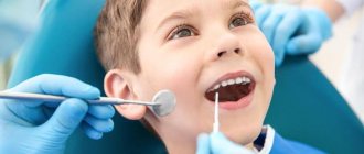
Often, a panoramic photograph is taken for children to detect supernumerary teeth or, on the contrary, the absence of the rudiments of permanent teeth. An orthopantomogram shows all restorations, fillings, any pathological formations in tissues, and at a very early stage
Indications for a panoramic photo:
- Therapeutic treatment - the image will qualitatively visualize each tooth, carious lesions, destroyed fillings, granulomas and cysts, if any.
- Periodontal treatment - in the image, the periodontist will be able to see the depth of the periodontal pockets, the thickness of the bone tissue and other details necessary for the treatment of periodontal disease
- Implantation - an orthopantomogram will allow you to develop an effective implantation plan and determine the most suitable points for implantation.
- Correction of the bite - before choosing a corrective system and placing braces on the patient, the orthodontist needs to understand the position and direction of growth of the roots of each tooth.