If your teeth have lost their shine, then you may be experiencing enamel demineralization. With demineralization, teeth lose mineral components. This is a reversible process, but if it proceeds too quickly and the enamel does not have time to recover naturally, the teeth become fragile and hypersensitive, stains and cracks appear on them, and caries develops.
This problem occurs especially often in children. Those with a sweet tooth and lovers of carbonated drinks are also at risk. In this article we will talk about the causes of enamel demineralization, the consequences of this pathology, and also consider ways to prevent and treat it.
Signs of demineralization
The lack of minerals appears gradually. At first, the crowns simply become matte. The beautiful pearly shine that characterizes a healthy smile disappears.
Then more noticeable defects are added:
- the enamel becomes rough;
- white chalky spots appear;
- light spots darken and turn brown;
- the enamel takes on a “porous” appearance.
All this “beauty” most often appears between the teeth - on the side surfaces of the crowns, at the edge of the gums, on the fissures - the chewing grooves of the tooth. To see the first signs of demineralization, just look at your smile in the mirror. And it would be better to notice the problem right away, because at the stage of brown spots, others will notice it.
Demineralized tooth in section
When is remineralization prescribed?
A set of restorative procedures is necessary if caries often appears, if the enamel reacts to cold or hot, sour and sweet, with pathological abrasion of teeth, metabolic disorders in the body, and also after orthodontic treatment. In addition, teeth become sensitive in adolescents during puberty and in expectant mothers during pregnancy.
Etiology
The main reason at the local level is the presence in the mouth of:
- Cariogenic bacteria - these can be streptococci, lactobacilli and some other species.
- Residues of carbohydrate foods, mainly sugars, where these microorganisms multiply well and comfortably.
As a result of their “violent activity,” the acid-base balance of the oral cavity decreases to a critical level of pH 5.5 and below. Normally, the indicator should be at a pH level of 6.8–7.4. That is, the environment becomes too acidic.
Next, the acids dissolve crystals of hydroxyapatite, a substance that makes up 75% of all enamel minerals. The destruction occurs so quickly that the teeth do not have time to “absorb” all the necessary elements supplied with food.
There is also a second reason - drinking water with a low fluoride content (less than 0.5 mg/l). If the body systematically lacks fluoride, thinning of the enamel will occur regardless of the acidity of the oral fluid.
Demineralized enamel also lacks phosphorus, magnesium, sodium, chlorine, and potassium.
Factors that provoke enamel demineralization
- failure to comply with hygiene rules;
- insufficient secretion of the salivary glands;
- eating soft foods high in simple carbohydrates;
- inflammation of the gums;
- wearing dentures or orthodontic appliances, which prevents the thorough removal of hard deposits;
- crowded teeth - if they overlap each other, it becomes difficult to clean the interdental spaces;
- diseases such as rickets, diabetes, pathology of the adrenal glands;
- hormonal changes.
Detection methods
Visual inspection
After cleaning the plaque, the teeth are isolated from saliva and dried. The dentist notes demineralization in the presence of white matte spots with a rough surface. Most often, pigmentation is localized in the cervical region (at the gingival margin).
Vital staining of enamel (Borovsky-Aksamit test)
A swab soaked in a two percent solution of methylene blue is applied to the cleaned and dried teeth for three minutes. As a result, the demineralized areas become stained. The brighter the shade of blue, the deeper the damage to the enamel.
Ultraviolet research
Under ultraviolet rays, the affected areas glow darkly. They contrast with healthy enamel, illuminated blue. Such diagnostics can be carried out using the Pluraflex device.
Laser examination
The DiangoDENT device is used. Mineral-deficient enamel reflects laser waves above 680 nm, and the device emits a characteristic signal.
Focal demineralization of tooth enamel is a widespread disease that occurs in more than 97% of the population of our country [14]. The development of new methods for diagnosing focal demineralization of tooth enamel is determined by the requirements of clinical practice related to the early and timely diagnosis of this pathology in order to use the least invasive treatment methods. The question of assessing the relationship between objective diagnosis of demineralization and adequate treatment in each specific case, taking into account the characteristics of the course of the disease, is also relevant. Most existing methods for diagnosing focal demineralization of tooth enamel are relatively imperfect, since they are based on subjective perception. In this regard, it is relevant not only to introduce objective diagnostics of enamel demineralization into practice, but also to create a clear scale for assessing its degree, which would reflect the depth and area of the lesion.
Focal demineralization of enamel is a systemic lesion of the cervical areas of teeth in the form of spots as a result of underdevelopment of tooth tissue and a decrease in the amount of calcium in it. This condition is formed during the period of maturation of tooth tissue after its eruption and corresponds to the ICD-10 code K02.0 - “Enamel caries. Stage of “white (matte) spot” [initial caries].” Caries in the spot stage is characterized by changes in color (matte surface) and then texture (roughness) of the enamel resulting from demineralization, which have not spread beyond the enamel-dentin border, in the absence of a carious cavity [2].
A feature of focal demineralization is that almost all teeth are affected. This process is the only reversible one. Enamel is a mesoporous substance; it does not contain cells and is not capable of regeneration when damaged, but there is a constant exchange of mineral ions in it, which come from saliva through the pores and are adsorbed on its surface, which makes it possible to carry out remineralization therapy [2]. The fundamental local factor in the development of this pathology is a local change in pH on the tooth surface due to the accumulation of dental plaque on it containing acid-forming streptococci ( Streptococcus mutans
,
S. sanguis
,
St.
mitis ,
St.
salivarius ), which are characterized by anaerobic fermentation, and some lactobacilli [21]. Caries develops cyclically, the rate of its development depends, firstly, on the frequency of action of lactic acid produced by microorganisms on the tooth, and, secondly, on the degree of demineralization, i.e., on the penetration of calcium and phosphorus ions into the hard tissues of the tooth (the process catalyzed by fluorine). In addition to fluoride deficiency, for demineralization of tooth tissue to occur, the pH of the oral environment must be below a critical point. On average, the critical pH point is estimated at 5.5 (Stefan curve) [1, 3].
Pathomorphological changes are characterized by varying degrees of enamel demineralization. On thin sections of the enamel, the lesion looks like a triangle with the base facing its surface. When studying enamel in polarized light, G. Gustafsonson (1975) identified 5 zones, depending on the intensity of demineralization. The zone of hypermineralization with the disappearance of structural details of the enamel is located deepest in the thickness of the enamel. In the 2nd zone, a decrease in its hardness was noted due to the partial dissolution of minerals, in the 3rd - an increase in mineralization. In the subsurface, 4th demineralization zone, minerals are washed out almost completely. In the superficial, 5th zone, complete disintegration is possible, but the enamel remains fairly mineralized and intact for a long time, even if the carious process extends to more than half the thickness of the enamel. A. Darling (1958) indicates that if in normal enamel microspaces constitute 0.5% of its volume, then in the body of the lesion they reach 25% of the volume, in dark enamel - 4%, and in transparent enamel - 2% [1].
With a white spot area of up to 3 mm2, changes reach 1⁄2 the thickness of the enamel layer. With more extensive lesions, changes in the enamel are observed down to the enamel-dentin junction. The formation of brown pigment in the spot is associated with the accumulation of the amino acid tyrosine with its further conversion into the pigment melanin. Under a white carious spot, changes in the enamel-dentin junction have not yet been detected; under a brown one, its damage may already be observed [1, 3, 21].
Focal demineralization of enamel is a borderline pathology for which both conservative and invasive treatment is possible. At the same time, there are no clear algorithms for determining a particular treatment plan, despite the wide variety of definitions of the degree of demineralization in enamel caries.
Existing methods for determining demineralization are divided into qualitative and quantitative. Qualitative assessments include clinical assessment. The main quantitative methods for determining enamel demineralization include the following:
1. TER-test (enamel resistance test; V.R. Okushko, L.I. Kosareva, I.K. Lutskaya, 1983). This test reflects the predisposition to caries (based on the functional resistance of enamel to acid). The TER test method boils down to isolating the tooth from saliva, cleaning it from plaque, and drying it. Then 1 drop with a diameter of 1-2 mm of 1% HCl is applied to the prepared tooth. After 5 seconds, the drop is washed off with distilled water, dried, and 1 drop of a 1% solution of methylene blue is applied. The dye is removed with a dry cotton swab in one erasing motion. The etched area is painted blue, the intensity of staining is assessed on a 10-point scale (1-10 points; the maximum score corresponds to the least caries resistance of the enamel)[6].
2. KOSRE-test (clinical assessment of the rate of enamel remineralization according to the method of T.L. Redinova et al.). This test is based on assessing both the condition of tooth enamel and the remineralizing properties of saliva. The enamel surface of the tooth being examined is thoroughly cleaned of plaque with a dental spatula and a 3% hydrogen peroxide solution and dried with compressed air. Then a drop of hydrochloric acid buffer pH 0.3-0.6 is applied to it, always in a constant volume. After 1 minute, the demineralizing solution is removed with a cotton swab. The etched area of tooth enamel is also exposed for 1 minute to a cotton ball soaked in a 2% solution of methylene blue. The compliance of enamel to the action of acid is assessed by the intensity of staining of the etched area of tooth enamel. The degree of staining is judged by the blue topographic tint scale (10-point scale: the least stained part is 10%, the most saturated is 100%). After 1 day, the etched area of tooth enamel is re-stained without repeated exposure to the demineralizing solution. If the etched area of tooth enamel becomes stained, this procedure is repeated again after 1 day. The loss of the etched area's ability to stain is regarded as a complete restoration of its mineral composition. The degree of resistance of tooth enamel to the action of acid is taken into account as a percentage, and the remineralizing ability of saliva is calculated in days. Resistance to caries is characterized by low compliance of tooth enamel to acid action (<40%) and high remineralizing ability of saliva (from 24 hours to 3 days), and caries susceptibility is characterized by high resistance of tooth enamel to acid action (≥40%) and low remineralizing ability. ability of saliva (>3 days) [5].
3. Determination of the initial level of mineralization of erupting teeth using the electrometric method (L.P. Kiselnikova, 1990). The degree of mineralization was assessed using the Dentest electrometry apparatus (ZAO Geosoft). The principle of the electrometric method is that in the hard tissues of fully mineralized teeth and without signs of demineralization, the electrical conductivity is 0. In teeth with reduced mineralization processes, the electrical conductivity values increase. The lower the degree of mineralization, the higher the electrical conductivity. An electric current of 1.8 to 4 μA passes through areas of focal demineralization, localized on the visible surfaces of permanent teeth with enamel mineralization ending. Permeability impairment from 1 to 8 points on the methylene blue staining intensity scale corresponds to an increase in the current value from 1.68 to 5.17 μA. With an increase in the size of the spots, the magnitude of the current passing through the tooth tissue significantly increases from 2.55 to 3.31 μA [7].
The electrometric method makes it possible to determine the general mineralization of the tooth crown, but can only be used to a limited extent for local changes. The absolute values of electrical conductivity vary significantly depending on the tooth, the local thickness of the enamel layer, the ability to carry out insulation and other parameters. The electrometric method is applicable for the relative assessment of changes in the mineralization of a spot on a specific tooth in a specific patient with complete repetition of measurement conditions, which during a clinical appointment can lead to quite serious errors in the results obtained.
4. Determination of focal demineralization of tooth enamel using infrared fluorescence using the DIAGNOdent apparatus.
The DIAGNOdent device (KaVo, Biberach, Germany) was introduced in 1998 by Hebst and Gall as an aid to the detection of caries and focal demineralization of tooth enamel on occlusal surfaces in addition to inspection and radiographic examination. This device works using light emanating from a diode laser; wavelength 655 nm, peak power 1 mW. Light travels through the fiber assembly to the tip of the handpiece. The tip of the handpiece is placed on the surface of the tooth and the laser beam passes through the tooth. Both organic and inorganic molecules in the tooth absorb light. Infrared fluorescence is used. Fluorescence, as well as return light, passes through the tip and enters through the ascending tissues to the photodiode sensor. The fluorescent light is measured and its intensity indicates the size and depth of the tooth's decay or focal demineralization. Fluorescence intensity is presented in the range from 0 to 99. According to prof. Reich (Univ. Hamburg), digital values from 5 to 25 correspond to carious damage to the enamel, 25-35 to damage to half the thickness of dentin, ≥35 to deeper damage to dentin. According to O.A. Krasnoslobodtseva and L.Yu. Orekhova (2000), the indicators for caries in the spot stage corresponded to 9.0±2.0, for superficial caries - 15.0±3.0; average caries - 50.0±30.0. Differences in digital indicators characterizing the condition of dental tissues are explained by different degrees of initial mineral maturity of the diagnosed teeth (EA Kidd, D. Beighton, L. Zoitopoulos, 2001). The reason for the fluorescent glow in the tooth is the presence of protoporphyrins and mesoporphyrins, waste products of bacteria (Hibst and Paulus, 1999, 2000). DIAGNOdent is widely used to detect caries on occlusal surfaces and focal demineralization on smooth tooth surfaces (Aljehani et al., 2006, 2007; Antonnen et al., 2003; Bamzahim et al., 2004, 2005; Lussi et al., 2003, 2006 ). Studies have shown that the sensitivity of the DIAGNOdent device for detecting caries and focal demineralization of enamel ranges from 0.17 to 0.78, and in some cases from 0.72 to 0.98 (Lussi et al., 1999; Shi et al., 2000) [8, 16].
Most studies highlight that DIAGNOdent is much more sensitive to pathology detection than traditional methods. But the increasing number of false-positive diagnoses makes it impossible to use this device as the only reliable one (Bader et al., 2004). Therefore, a definitive diagnosis cannot be made based on DIAGNOdent data alone.
5. Transillumination of focal demineralization of enamel - bright illumination of the coronal part of the tooth to study its structural components; carious lesions stand out as a dark shadow against the background of tooth enamel. The transillumination method also makes it possible to identify enamel cracks and assess the condition of tooth tissue around previously applied fillings [4, 12—14].
This method can only identify pathology, but does not evaluate it qualitatively and quantitatively, which deprives it of objectivity.
Treatment of focal demineralization of tooth enamel has been studied for decades and has a serious scientific basis. Theoretically, remineralization therapy for focal demineralization is justified by the preservation of the protein matrix in the tooth enamel in the early stages of caries (white carious spot), as well as the possibility of remineralization. Many studies have noted that after a course of restorative therapy, the enamel surface becomes more uniform and microporosity decreases. Thanks to the remineralization of enamel, it is possible to stabilize the initial process of its demineralization and even eliminate the chalk stain. The pigment spot remains preserved when the process stabilizes [9].
There are a huge number of remineralizing products - remineralizing pastes, gels, mousses. Let's look at some of them.
Profilak JSC "StomaDent" is created on the basis of domestically produced cedar balsam using a special technology that allows you to maintain the active component - fluorine in a dispersed state, preventing its precipitation.
Significant factors that unite fluoride-containing varnishes are the transformation of hydroxyapatite into fluorapatite and the formation of calcium fluoride in the outer layer of the tooth. After applying fluoride-containing varnishes, the tooth surface is protected by an acid-resistant layer of calcium fluoride.
Clinical studies of the use of fluoride-containing varnishes have proven their high effectiveness in the progression of proximal caries. They should be used immediately after the eruption of permanent teeth, since there are no contraindications for their use.
The use of Profilak is also indicated:
— for the prevention of tooth root caries (2-3 times a year);
— for the treatment of hypersensitivity of teeth (course of 1-3 procedures over 7-10 days);
— on low-calcified teeth (4–8 times a year) [15–17].
Remineralizing gel ROCS Medical Minerals. It is a source of bioavailable calcium, phosphorus and magnesium compounds that strengthen enamel. Thanks to special additives, it forms a stable invisible film on the teeth, ensures gradual penetration of minerals into the tooth tissue, and allows prolongation of the exposure of active components. After brushing your teeth, apply it to your teeth with a brush and refrain from eating or drinking for 30 minutes. Recommended course duration is 2 weeks. From 1 to 3 courses are conducted per year. It is possible to use the gel on an ongoing basis, as it is harmless and does not cause side effects [20].
GC Tooth Mousse is an application cream containing calcium and phosphorus, used by dentists in accordance with the recommended indications. GC Tooth Mousse is a water-soluble cream containing Recaldent™* CPP-ACP (casein phosphopeptide - amorphous calcium phosphate). In the oral environment, CPP-ACP binds tightly to biofilm, plaque, bacteria, hydroxyapatite and soft tissue, delivering calcium and phosphorus. Saliva increases the activity of CPP-ACP, and the pleasant taste of the mousse increases salivation. The longer CPP-ACP and saliva are in the oral cavity, the more effective the result. Recaident™ CPP-ACP is derived from cow's milk casein and should not be used in patients with milk protein allergies. There are known cases of recovery from enamel hypersensitivity after taking GC Tooth Mousse [18, 19].
One of the innovative areas for the treatment of focal demineralization is the method of caries infiltration of an area of pathological enamel with a viscous-flowing composite material Icon, developed by the German company DMG in 2009. The method is based on layer-by-layer “impregnation” (infiltration) of the focus of enamel demineralization with a light-curing resin, which is carried out after preliminary etching of the surface, relatively highly mineralized “pseudo-intact” layer of enamel. According to foreign authors [22], the infiltrate penetrates into the intercrystalline pores of the enamel throughout the entire volume of the affected area and, after hardening, strengthens (“reinforces”) the demineralized area of the enamel, generally preventing further progression of the disease [10]. Currently, laboratory and clinical studies are being actively conducted all over the world, which confirm the prospects of using the infiltration method for the treatment of caries in the spot stage [11].
Analysis of publications devoted to methods of diagnosis and treatment of focal demineralization of tooth enamel allows us to conclude that, despite numerous studies in this direction, many unclear questions remain. There are many recommendations and subjective methods for diagnosing this pathology of tooth enamel, but there are no objective standards and a method that gives an accurate quantitative assessment of the lesion. The question of optimizing the indications for the use of certain methods and means of remineralizing tooth enamel, depending on the degree of demineralization of its structure, remains open.
The authors declare no conflict of interest.
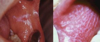
Focal demineralization of enamel in children
Focal demineralization of teeth is the initial degree of caries, in which spots appear not only in the cervical area, but also on the chewing surface - fissures. Usually, 1 month of excessive consumption of sucrose in conditions of poor hygiene is enough for focal demineralization to make itself felt.
The pathology most often affects children aged 9–11 years – from 15 to 40% of the child population, depending on the region of residence. Sweets, crackers and chips contribute to this - carbohydrates that provide food for bacteria. In addition, not every child follows all the rules of oral hygiene.
Focal demineralization in children
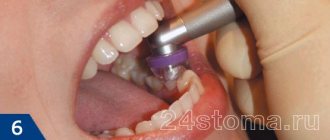
What is remineralization therapy
The remineralization therapy technique involves applying specialized compounds to the surface of tooth enamel that restore the structure. Usually we are talking about drugs based on fluorine, calcium, phosphates and other useful components.
In dentistry, rem therapy is performed in several ways:
- An express method, the essence of which is to put on disposable mouth guards filled with gel with a fluoride-containing base;
- Application of specialized varnishes that eliminate minor damage and restore the enamel structure;
- Reusable trays for remineralization at home.
The duration of such therapy depends on the degree of damage to the enamel, but usually several sessions are enough to achieve the expected result. Remineralizing dental therapy is a unique and affordable technique, but such treatment is carried out only in combination.
Development forecast
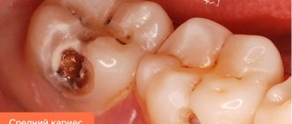
Under conditions of proper nutrition and corrected hygiene, in 20-30% of patients, the destruction of enamel stops spontaneously, and the enamel is restored naturally.
In other cases, the loss of minerals leads to the development of caries: the lesion begins to progress from the stage of focal demineralization. On average, 12-18 months pass from the very beginning of the process until the appearance of a carious cavity.
With timely treatment, caries may not develop, teeth can be kept intact and healthy.
Focal loss of minerals is the only reversible stage of caries.
Secondary medical education
- General midwife
- Dental assistant
- Dentist
- Dentist
- Dental Technician
- General expert research laboratory assistant
- Blood service laboratory technician
- AIDS service laboratory technician
- Laboratory assistant of forensic biological departments
- Laboratory assistant of forensic histology departments
- Laboratory assistant of physical and technical departments
- Laboratory assistant at chemical and toxicological departments
- Gynecological nurse
- Neonatology nurse
- General practice nurse
- Nurse of the intensive care and anesthesiology departments
- Ophthalmic nurse
- Pediatric nurse
- Physical therapy nurse
- Nutritional Nurse
- Massage nurse
- Care nurse
- Healthy lifestyle nurse
- Tuberculosis service nurse
- Psychoneurological nurse
- Blood service nurse
- AIDS service nurse
- Dental nurse
- Therapeutic nurse
- Physiotherapeutic nurse
- Surgical nurse
- Medical statistician
- Laboratory assistant doctor
- Nurse's assistant
- X-ray technician
- Paramedic laboratory assistant
- Narcology paramedic
- General practitioner
- Paramedic of the public education system
How to reduce demineralization
Diet
Preference should be given to products containing calcium, fluoride, vitamin D, since without it calcium is not absorbed:
- fermented milk cheeses with a fat content of at least 5% and hard cheeses;
- milk with a fat content of at least 2.5%;
- fatty fish (salmon, catfish, etc.);
- meat, liver;
- leguminous crops (peas, beans, lentils, beans, beans, chickpeas, etc.);
- nuts;
- green vegetables;
- eggs (chicken, quail).
Fluoridated water will be useful: but only as prescribed by a doctor, in order to avoid fluoride poisoning.
Rules for safe consumption of carbohydrates
- Avoid sweet snacks between main meals;
- do not end your meal with dessert (after sweets you should eat solid vegetables or fruits - they clean your teeth);
- do not eat sweets before bedtime;
- give up sticky sweets (toffees and sucking sweets stay in the mouth for a long time and have the most pronounced cariogenic effect).
According to the recommendations of the World Health Organization, preschool children should limit their daily intake of sugar to twenty grams.
Hygiene
You should brush your teeth every time after eating. It is recommended to alternate pastes containing calcium (President Unique, Splat “Biocalcium”, ROCS, etc.) with fluoride pastes (President Classic, Silca Herbal Complete and Natural Extracte, El-ce med Total Care, Splat “Arktikum”).
It is also useful to use rinses with sodium fluoride (fluoride concentration - 250 pm).