Oral lichen planus is a disease that is allergic in nature. That is, in essence, it is an allergy with a chronic course. Lichen planus is a fairly common disease, affecting not only the oral mucosa, but also the skin and nails. The study of lichen planus began at the end of the 19th century, therefore, over two centuries of studying this dermatosis, several hypotheses and theories of the occurrence of lichen planus have appeared.
Etiology of lichen planus
As I said earlier, the etiology of lichen planus is based on several theories.
Of these, the following etiological theories are important:
- Neurogenic theory of the occurrence of lichen planus;
- Toxic-allergic theory of the occurrence of lichen planus;
- Viral theory of the occurrence of lichen planus.
Despite a number of theories put forward about the occurrence of lichen planus, this type of allergy is considered semi-etiological, that is, one specific factor that provokes the occurrence of pathology is not identified.
Attempts to isolate a specific type of virus that is present in patients diagnosed with lichen planus have also been unsuccessful.
Doctors consider the presence of a history of previous neurological diseases or conditions to be important in the occurrence of lichen planus. That is, the possibility of a person developing lichen planus is influenced by stress, lack of sleep, depression, neuroses, panic conditions, epilepsy, etc.
Interesting fact: it was noticed that in people with diseases of the gastrointestinal tract (erosions, gastritis and stomach ulcers), that is, those diseases where there are changes in the mucous membrane, the risk of developing an erosive form of lichen planus is higher than in those who suffer other gastrointestinal disorders. However, the mechanism of dependence has not been established.
In the etiology of lichen planus, it should also be noted that the patient has:
- Diabetes mellitus;
- Cardiovascular failure;
- Hypertension;
- Pathologies of the liver and kidneys;
- Genetic predisposition;
- Immune suppression;
- Trauma to the oral mucosa (poor quality fillings, dentures, chronic trauma, chemical, mechanical trauma);
- Long-term use of medications;
Lichen planus most often occurs in middle-aged women, or in young people, children and the elderly.
Elements of defeat
Elements of the lesion in lichen planus may appear first on the oral mucosa, then on the red border of the lips, and only then on the skin. But it should be remembered that there is NO specific order and dynamics of occurrence. I have given only the more common options.
In general, lichen planus is a disease related to papular diseases in which metabolic processes in the oral mucosa and skin are disrupted. That is, it is a complex of dystrophic and inflammatory processes at the same time.
Skin papules in lichen planus
Skin papules with lichen planus are violet-bluish in color, up to 2 mm in diameter, dense in the center, with the phenomenon of keratinization.
Papules of the red border of the lips in lichen planus
Papules of the red border in lichen planus first appear in separate forms, which over time are connected to each other by keratinization bridges. After which they imagine a single huge papule rising above the intact surface of the red border of the lips.
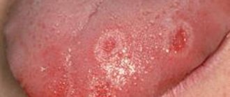
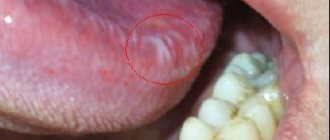
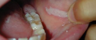
Papules on the oral mucosa with lichen planus
Papules on the oral mucosa with lichen planus are most often grouped, forming circles, lines, and lace. Individual papules are connected to each other by keratotic bridges. The tops of the papules become keratinized and acquire a milky bluish color (this is the most accurate sign for the differentiated diagnosis of lichen planus and leukoplakia, since in leukoplakia the plaques have a yellow tint).
In some cases, the process of keratinization does not occur, but rather necrosis. Then there will be erosion in place of the papule, followed by an ulcer.
If we talk about the more frequent localization of papules in the oral cavity, the order is as follows:
- Buccal mucosa in the retromolar region;
- Tongue (lateral and dorsal surfaces);
- Lips;
- Gum;
- Mucosa of the alveolar processes;
- Solid sky.
Material and methods
The study was carried out with the participation of 147 patients with various forms of LP of the oral mucosa, aged from 24 to 70 years, who sought advice from dental clinics at the Ural State Medical University and the Bashkir State Medical University.
The examination of patients with LP of the oral cavity included a thorough collection of anamnesis of this disease, clarification of hereditary predisposition taking into account previous diseases, identification of comorbid conditions and their relationship with the main one, the stages of manifestation of clinical symptoms of LP (periods of exacerbation and remission), allergic factor, and, if necessary, cytological and histological research.
When examining the mucous membranes, tongue and lips, attention was paid to the presence of signs of inflammation, elements of damage, and their location. An important diagnostic value is the presence of an isomorphic reaction in LP (Koebner's symptom), manifested by the appearance of fresh primary elements characteristic of this disease at the site of irritation of the skin or mucous membrane by any exogenous factor.
Forms of lichen planus
Six main clinical forms of lichen planus should be distinguished:
- Typical form of lichen planus;
- Keratotic form of lichen planus;
- Exudative - hyperemic form of lichen planus;
- Erosive - ulcerative form of lichen planus;
- Bullous form of lichen planus;
- Atypical lichen planus.
Although lichen planus is a chronic disease, there are moments of exacerbation and then the clinical picture may worsen, depending on the form of lichen planus, immunity and the presence of other somatic diseases in the patient.
Typical form of lichen planus
The typical form of lichen planus, also known as the classic form, is recognized by clinicians. The patient does not report any complaints in the typical form of lichen planus. There may be complaints of pain when eating spicy or hot food. More often, such complaints arise a couple of days before the appearance of visible papules on the oral mucosa.
The typical form of lichen planus is characterized by the appearance of papules on the unchanged oral mucosa, and, as you remember, these papules are grouped, resembling lace or fern leaves. Papules in the typical form of lichen planus are small, up to 2 mm in diameter. In the center there is an area of keratosis, so the papules are dense on palpation.
Hyperkeratotic form of lichen planus
The hyperkeratotic form of lichen planus consists of huge foci of keratinization at typical sites of localization for lichen planus. Patients with the hyperkeratotic form do not make any complaints. They may complain about aesthetic defects, dryness and flaking.
Exudative-hyperemic form of lichen planus
The exudative-hyperemic form of lichen planus occurs in 25% of all patients diagnosed with lichen planus. Complaints, as with other forms of lichen planus, and with the exudative-hyperemic form, are vague: burning and pain when eating spicy food, dry mucous membranes. Papules have a typical structure, shape and location. Only in the exudative-hyperemic form is the mucous membrane swollen and hyperemic.
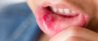
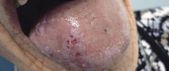
Erosive-ulcerative form of lichen planus
The erosive-ulcerative form of lichen planus is the most severe clinically and difficult to treat form of lichen planus. More often it occurs as complications of typical or exudative-hyperemic forms of lichen planus. With this form of lichen planus, patients report complaints of pain when eating, talking, and a burning sensation.
On the hyperemic and edematous mucosa, in typical places of localization of lichen planus papules - erosions of irregular and unclear shape, often covered with fibrinous plaque, there may be granulations. If these are single ulcers, then the pain will not be so intense. But in most cases, the ulcers merge with each other, affecting most of the oral mucosa. If erosions are left untreated for a long time, they turn into ulcers with keratinization lines.
Bullous form of lichen planus
The bullous form of lichen planus is the rarest form of lichen planus, recorded in 3% of patients with lichen planus.
The bullous form of lichen planus is described not only by the presence of papules, but also by the presence of blisters, the size of which can range from the head of a pin to a kidney bean. The blisters can remain on the mucous membrane for up to several days, then they open, and erosion occurs in the place of the opened blisters. Erosion in bullous form of lichen planus is irregularly shaped, with a bleeding bottom. Often covered with fibrinous plaque.
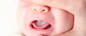
Atypical form of lichen planus
An atypical form of lichen planus is most often observed on the mucous membrane of the lips and gums. On the lip, two symmetrical areas with congestive hyperemia are often identified; these areas are slightly higher than the normal mucous membrane of the lips and gums. Often areas of the epithelium are covered with a whitish coating, which is difficult to remove during diagnosis.
You need to understand that the described forms of lichen planus can transform one into another depending on the influence of both local and general factors. It can occur simultaneously on the mucous membranes of the mouth, lips, and skin. Or vice versa. Forms of lichen planus can become malignant, especially when localized on the mucous membrane of the cheeks and the red border of the lips (erosive-ulcerative form of lichen planus).
Treatment
For different forms of the disease, a different course of medications is prescribed. If the disease is asymptomatic, drug treatment may not be necessary. If erosions and ulcers have already formed, it is necessary to begin treatment as quickly as possible to avoid worsening the condition.
Inflammatory processes are controlled by the use of corticosteroids. Local treatment is carried out to relieve pain and accelerate tissue regeneration. To avoid the development of candidiasis, antifungal drugs are prescribed.
Electrophoresis and phonophoresis are also effective. In rare cases, when erosions do not heal for a long time, surgical intervention is indicated.
In all cases, without exception, the oral cavity is sanitized and the source of inflammation is removed (for example, periodontitis, caries or pulpitis).
What does lichen look like?
As the disease progresses, rashes appear - most often they take the form of small red or brown-purple lumps (on the skin) - a characteristic sign is the shine of the affected areas.
- On the scalp, lichen planus may take the form of small itchy bumps.
- In the mouth - on the surface of the mucous membrane - it can appear as a reticular change in white color.
- On the nail plate, lichen can form cavities and yellowish grooves, and in extreme cases, lead to atrophy of the nail plate.
Lichen planus is an inflammatory disease. It is worth emphasizing that this is not an infectious disease.
Lichen planus
Lichen planus of the oral mucosa
Lichen planus - medicinal
Lichen planus hypertrophic form