Oral syphilis
The diagnosis of “oral syphilis” is based on the patient’s complaints, medical history, clinical examination, and the results of additional research methods. With primary syphilis of the oral cavity, the dentist usually identifies one hard chancre. On palpation, the resulting ulcerative surface is painless, regular rounded, red in color with smooth, raised edges and an infiltrated sebaceous bottom. Lymph nodes are compacted, enlarged, painless, and not fused to the skin and surrounding tissues. With secondary syphilis of the oral cavity, residual syphilomas are found, as well as a roseolous-papular rash on the palate, arches, and tonsils. Scraping the papules leads to exposure of erosive surfaces. In case of relapse of secondary syphilis of the oral cavity, fewer rash elements are formed, papules and roseolas are pale in color, grouped, forming figures resembling garlands and lace.
With secondary syphilis of the oral cavity, polyadenitis is detected. Unlike catarrhal tonsillitis, pain when swallowing and high temperature reaction are not observed with syphilitic lesions. In tertiary syphilis of the oral cavity, a gummous infiltrate is detected, after the disintegration of which a deep crater-shaped ulcerative surface is formed. The integrity of the jaws and nasal bones is compromised. The affected areas become scarred, leading to permanent deformities. There is no enlargement of regional lymph nodes. The detection of treponema pallidum in scrapings or in the contents of lymph nodes confirms the diagnosis of oral syphilis. To identify syphilitic lesions, serological reactions are also used, which in patients become persistently positive, starting from 4 weeks from the moment of formation of chancre. The first 3 weeks of primary oral syphilis are a seronegative period, since at this time it is not possible to confirm the diagnosis using serological reactions.
Radiographically, in patients with tertiary syphilis of the oral cavity, zones of rarefaction of bone tissue in areas corresponding to gummous lesions, as well as sclerotic changes along the periphery, are diagnosed. There is destruction of the cortical bone layer and signs of periostitis ossificans. Oral syphilis is differentiated from a decubital ulcer, a malignant tumor, tuberculous and actinomycotic lesions, tonsillitis, chancriform pyoderma, Setton's aphthae, lichen planus, and leukoplakia. The patient is examined by a dentist or dental surgeon. If a specific syphilitic infection is suspected, the patient is referred for consultation to the dermatovenerological department.
Causes of syphilis in the mouth
Treponema pallidum - the causative agent of syphilis
There is congenital and acquired syphilis. The first occurs when a pathogen passes through the placenta from an infected mother to the fetus. The second develops when Treponema pallidum infects a person, penetrating through injured mucous membranes and skin during sexual intercourse.
Experts pay attention to factors that contribute to causing syphilis in the mouth:
- The use of non-sterile medical instruments in dentistry and ENT practice.
- Entry of the pathogen into the bloodstream during blood transfusion, during injections, during surgery.
- Using the same household items (cutlery, toothbrush, bath accessories).
- Oral sex.
There are circumstances that increase the risk of infection. These include:
- Injuries to the mucous wall of the oral cavity.
- Unhealthy lifestyle.
- Weak general and local immunity.
Treatment
Do not self-medicate under any circumstances!
The main objective of the therapeutic course is the elimination of Treponema pallidum from the body, relief of symptoms and prevention of pathology. Treatment is carried out exclusively in the venereology department of specialized medical institutions.
The treatment course consists of:
- Local therapy , which consists of washing syphilitic lesions with antiseptic drugs. Most often, the main active component of such antiseptics is chloramine. If the ulcers are bleeding, they are treated with white mercury ointment. Calomel or xeroform powders provide effective symptomatic relief. A solution of chromic acid or 10% lapis will help stop the growth of hypertrophic ulcers.
- Antibacterial therapy . Long-term use of antibiotics is prescribed. Most often, a long course of penicillin injections is given. If you are intolerant to penicillin, take tableted antibiotics of various groups.
- Taking immunomodulators helps strengthen the body's protective functions.
- Symptomatic treatment is selected taking into account the stage of development of syphilis and accompanying symptoms. For this purpose, antipyretics, painkillers, antihistamines, and agents to stimulate tissue regeneration are prescribed.
- Sequestrectomy is performed if the disease clinically subsides completely.
Important! Secondary and tertiary forms of syphilis are practically untreatable. Regular examination for this pathology will allow timely identification of the disease and its effective treatment.
The prognosis for recovery after diagnosis of syphilis in the mouth can be positive, subject to timely detection and immediate treatment of the pathology. When treating this disease, it is important to complete the full course and not mistake a temporary reduction in symptoms for recovery.
Syphilis of the tongue in the secondary period of the disease
During the secondary period of syphilis, erosive papules most often appear on the mucous membrane of the tongue - papular syphilide.
Rice. 7. Papules on the tongue are oval in shape, bright red in color, painless and highly contagious.
Rice. 8. The photo shows syphilis of the tongue in the secondary period of the disease. The papules are round, dark pink, single or multiple, devoid of papillae (“mown meadow symptom”).
Rice. 9. Secondary period of syphilis. Papules on the tongue.
Possible complications
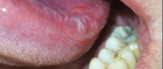
Complications of syphilis in the mouth often develop due to untimely treatment and diagnostic examination. In this connection, the pathogenic effect of spirochetes extends beyond the tissues in the oral cavity and to systemic organs inside the body.
A clear example of a complication of syphilis in the mouth.
As a result, various complications arise, manifested by:
- Damage to the skeletal system and organs.
- Necrosis of the muscular skeleton at the site of the lesion.
- Local bleeding.
- Dysfunctions of the circulatory system.
- Asymmetry of the face with damage to the facial and cervical muscles.
- Destruction of the cellular structure of the brain.
It is important not to delay visiting a venereology clinic in order to prevent the development of severe consequences of this insidious disease. And it must be remembered that immunity to this venous disease is not formed and therefore, if any rash appears on the mucous membrane of the cheeks, gums, or tongue, you should seek medical advice from specialists.
Syphilis of the tongue in the tertiary period of the disease
In the tertiary period of syphilis, single or multiple gummas (nodular glossitis) more often appear on the tongue, diffuse (spread) sclerosing glossitis develops less often. Sometimes, isolated gummas appear against the background of sclerosing glossitis.
The gummous infiltrate is large in size (about the size of a walnut), quickly disintegrates with the formation of a deep ulcer and an uneven bottom, surrounded by a shaft of dense infiltrate. The developed scar tissue significantly deforms the tongue.
Sclerosing glossitis is characterized by the development of diffuse infiltration in the thickness of the tongue. The tongue becomes dense, acquires a dark red color, and the mucous membrane thickens. As a result of rapidly developing sclerosis, when muscle fibers are replaced by dense connective tissue, the tongue contracts and becomes smaller in size, its surface is smoothed (loses papillae), becomes bumpy, and becomes significantly denser (“wooden” tongue). There is increased salivation (salivation). Appearing cracks often become infected, which leads to the appearance of erosions and ulcers that are prone to malignancy. The disease occurs with severe pain, the patient's speech is impaired and eating is difficult.
Rice. 10. Syphilis of the tongue in the tertiary (late) period of the disease - a single gumma of the tongue (photo on the left) and a disintegrating gumma (photo on the right).
Syphilis on the tongue
The tongue with syphilis is affected in the primary, secondary and tertiary periods of the disease.
Syphilis of the tongue in the primary period of the disease
Hard chancre on the tongue is often single, ulcerative or erosive in nature. Sometimes it has a slit-like shape located along the tongue.
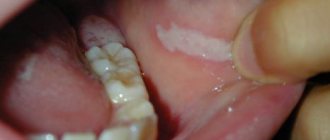
Rice. 5. Syphilis of the tongue in the primary period - chancre. Syphilide is an erosion or ulcer with a dense infiltrate at the base.
Rice. 6. The photo shows a hard chancre on the tip of the tongue.
Symptoms and stages (+photos)
The formation of syphiloma is possible in any area of the oral cavity. Often a cause of concern is a hard formation that appears in the gum area or on the mucous membrane of the lips. It is painless, but causes discomfort during chewing and articulation movements and does not look aesthetically pleasing. The size of the rash ranges from 0.1 to 2.0 cm. Their location can be single or group.
External signs of syphilis in the mouth are characterized by visual manifestations, each of which is characteristic of the stage of the disease.
| Stage | Symptoms |
| First | In the mouth, at the site of the affected mucous membrane, a round ulcer with a hard consistency is observed. Characterized by the absence of pain. A single ulcer may form, or several may form at once in different parts of the oral cavity. The main localization sites for chancre are the tonsils, tongue, periodontal tissue and the inner cheek area. |
| Second | Characterized by the disappearance of chancre. The oral mucosa becomes covered with rashes. As the pathology progresses, they develop into red roseola, which, as the pathology progresses, begin to merge and rapidly increase in size. The main location is the palate, the edging of the lips inside the mouth, and the tongue. Purulent abscesses and papules may appear. |
| Third | Nodules, bumps and abscesses are observed in the affected areas of the oral cavity. At the initial stage, seals form on the tongue, which quickly transform into large ulcers. Gummas are often located singly. It is small in size, with a diameter of no more than 1.5 cm. Subsequently, it opens and turns into an abscess, accompanied by pain. |
Signs of different types of syphilis in the oral cavity
In medical practice, depending on the stage of development of the pathology, the following are distinguished:
- Primary syphilis in the mouth is manifested by a hard ulcerative formation, the so-called chancre. In the initial stage of the disease, an increase in the cervical, submandibular and occipital lymph nodes is noted. The duration of this period is from six to eight weeks. But even in the presence of the first symptoms of syphilis, it is not possible to determine the causative agent of the disease in a laboratory. In such cases, a polymerase chain reaction blood test is prescribed.
- Secondary syphilis in the mouth is characterized by the appearance of rashes of various types and more pronounced symptoms. After the end of the second phase of the disease, remission occurs, which can be periodically interrupted by exacerbation of the pathology. This action can happen up to ten times. The duration of the period reaches an average of five years.
- Tertiary syphilis in the mouth is the last stage of the disease, manifested by irreversible damage to organs of various body systems. The central nervous system especially suffers in the form of paresis, cerebral disorders, dementia and other pathologies.
Routes of infection
The development of syphilis in the mouth involves the following routes of infection:
- Intrauterine , when a pregnant woman with syphilis has not received a therapeutic course, there is a risk of infection of the fetus. It depends on the degree of the disease, and then the child can be born with congenital syphilis, with deformities, with pathologies leading to death, or maybe healthy.
- The everyday route of infection occurs when basic rules of personal hygiene are ignored and the use of household items and medical instruments contaminated with syphilis pathogens that are not disinfected and washed in the proper way.
- The sexual route of infection with oral syphilis occurs from an infected person to a healthy partner during oral sex and kissing.
Intrauterine infection with syphilis
Treponema pallidum enters the human body due to abrasions, scratches of the epidermis and mucous membrane, as well as through the blood. Having penetrated the body, the spirochete is localized in the tissue base. The incubation stage lasts from two weeks to six months. During this period, the microbe has penetrated the mucous membrane, but does not yet cause the degree of damage that would have a clinical picture.
Congenital oral syphilis can result from intrauterine transmission of triponema. When an infected woman gives birth who has not completed the appropriate therapeutic course, the child is born with signs of syphilis in the oral cavity.