Introduction
Traumatic injuries of the oral mucosa (OM) of a mechanical nature are common in dental practice, especially in the form of prosthetic stomatitis [1, 2], as well as during orthodontic treatment for trauma to the mucous membrane with a brace system [3]. In the latter case, such injuries do not have any clinical manifestations [4] and occur in 38.08% of cases [5].
The high prevalence of traumatic lesions of the oral mucosa during orthodontic treatment with fixed orthodontic equipment dictates the need to improve treatment and preventive measures [5], including the use of new domestic agents for topical use.
The purpose of the study is to assess the incidence of traumatic lesions in people of different ages using fixed orthodontic appliances, and to conduct a comparative assessment of the effectiveness of their treatment with domestic drugs for local use.
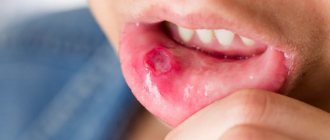
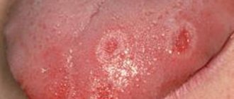
Traumatic stomatitis: before and after photos
In case of one-time, not too serious injuries and the absence of infectious contamination, the consequences of such injuries pass quickly and do not require special drug treatment. But if the impact on the mucous membrane and soft tissue occurs over a long period of time, traumatic stomatitis can become chronic. This is also facilitated by the presence of various diseases that lower the level of immunity, and poor oral hygiene.
Among the symptoms of traumatic stomatitis are:
- the appearance of swelling or redness at the site of injury;
- pain that makes facial expressions, speech and chewing difficult;
- the occurrence of blisters, wounds, ulcers, erosion.
With severe trauma, partial tissue necrosis is possible; some severe cases of the disease are accompanied by fungal infection or suppuration.
Material and methods
We studied the incidence of traumatic lesions of the oral mucosa (traumatic stomatitis - TS) in children 12-17 years old and young adults (18-32 years old) when using brace systems. Frequency T.S. assessed in 52 children and 43 adults for up to 1.5 months immediately after installation of the brace system, as well as in 136 children and 256 adults during orthodontic treatment, from 1.5 months to 1.5 years from the start of using fixed orthodontic equipment .
To evaluate the effectiveness of treatment, 4 groups of patients were formed, taking into account age and the drug used for local treatment of traumatic injuries of the oral mucosa caused by the use of fixed orthodontic appliances (Fig. 1).
Rice. 1. Distribution of patients into study groups.
The 1st group included children who used vinylin for internal use for the treatment of traumatic injuries of the oral mucosa (JSC ", Moscow region, Staraya Kupavna, Russia), in the 2nd group, children used a gum gel with propolis (JSC "VERTEX" ", Saint-Petersburg, Russia). The following groups included adult patients whose TS was treated with topical vinylin (group 3) or gum gel with propolis (group 4). All observed patients (children and adults) underwent orthodontic treatment using vestibular brace systems with correction in order to prevent re-injury of the oral mucosa during treatment.
Control examinations of patients of all groups suffering from TS were carried out on the 3rd, 5th and 10th day from the start of treatment using these drugs. Their local application in children was carried out by parents, and in young people - by the patients themselves, 3 times a day after meals. Along with this, all patients during dynamic observation for up to 10 days before local use of the drug for the treatment of TS, in order to increase the effectiveness of treatment, used the ASEPTA parodontal active mouth rinse (JSC VERTEX, St. Petersburg, Russia).
To ensure the comparability of the results obtained and their reliability, a developed semi-quantitative method was used, which consisted of a visual assessment of various clinical symptoms of TS, each of which was assigned one of three symbols indicating the absence or severity of a specific clinical symptom. Thus, based on an analysis of the symptoms of TS, an index method was proposed for assessing the severity of this pathology, taking into account the following clinical symptoms and their assessment in points:
1. Pathological sensations (pain): absent - 0; moderately expressed - 1; pronounced - 5.
2. The area of the focus of traumatic lesions of the oral mucosa (determined taking into account the damaged organs and
fabrics): TC is not determined - 0; lesion on the mucous membrane in the area of the brace system on one of the jaws - 1; lesion on the mucous membrane in the area of braces on both jaws - 5.
3. Localization of traumatic lesions
: the lesion is not determined - 0; visualized in the lips or cheeks - 1; visualized in the area of lips and cheeks - 5.
4. Presence of hyperemia and edema of the mucous membranes
: no hyperemia, mucous membranes are pale pink, no swelling - 0; moderate (mild) hyperemia and swelling of the mucous membranes - 1; pronounced hyperemia (bright red) and swelling of the mucous membranes - 5.
5. Clinical and morphological characteristics of the focus of traumatic lesions of the oral mucosa
: the lesion is not determined - 0; erosion or erosions are visualized - 1; erosive and ulcerative lesions are diagnosed - 5.
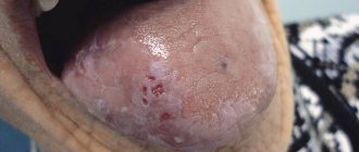
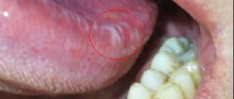
To establish the severity of the clinical picture of TS, the listed clinical symptoms are first diagnosed. After registering the symptoms of the pathology, the sum of points is calculated and the severity of TS is assessed based on the obtained sum of points as follows: 0 - no pathology; 1-4 points - mild severity of the vehicle; 5-9 points - average degree; 10-25 points - severe degree. The data obtained in each group of patients according to symptoms were arithmetically added, and the resulting average values were used for further statistical processing (Fig. 2).
Rice. 2. An erosive-ulcerative traumatic lesion in the area of the mucous membrane of the lower lip on the right of moderate severity, caused by wearing a brace system, in a 14-year-old patient (group 1, subgroup 1). Clinical manifestations: pathological sensations (pain): moderate - 1; area of the focus of traumatic lesion of the oral mucosa (determined taking into account damaged organs and tissues: lesion on the mucous membrane in the area of the brace system on one of the jaws) - 1; localization of traumatic lesions: visualized in the area of the lower lip - 1; presence of hyperemia and swelling of the mucous membranes: moderate (mild) hyperemia and swelling of the mucous membranes - 1; clinical and morphological characteristics of the focus of traumatic lesion of the oral mucosa: erosive and ulcerative lesion was diagnosed - 5 (total 9 points).
To objectify the result of treatment for TS, a method was used that involved determining the effectiveness of the therapy for the specified pathology of the oral mucosa, which was carried out according to the formula:
Efficiency (%)=100·(A–B):A,
where A is the sum of points in the clinical assessment of the severity of TS before the start of therapeutic measures; B - the sum of points for clinical assessment of the severity of the pathology in question on the 3rd, 5th and 10th day from the start of treatment.
The reliability of differences in the average values of independent samples was assessed using the parametric Student's test for normal distribution and the non-parametric Mann-Whitney test for differences from the normal distribution of indicators. Normality of distribution was checked using the Shapiro-Wilk test. For statistical comparison of proportions with an assessment of the significance of differences, the Pearson χ2 test was used, taking into account the Mantel-Haenszel likelihood correction. In all statistical analysis procedures, the achieved level of significance was considered ( p
), the critical significance level was equal to 0.05.
Traumatic lesions of the oral mucosa
- Chronic mechanical injury
Clinical manifestations of a traumatic ulcer depend on the strength of the damaging factor, the general reactivity of the body, the state of the microbiocenosis of the oral cavity and other factors. As a rule, a traumatic ulcer is single. The mucous membrane around the ulcer is hyperemic and swollen. A traumatic ulcer is always painful, has uneven edges, and its bottom is covered with a fibrinous coating that can be easily removed. With the long-term existence of an ulcer, its edges and base become denser (due to the predominance of the phenomenon of proliferation). The edges of the ulcer are hyperemic, painful on palpation, the bottom is often bumpy, covered with necrotic plaque. The depth of the ulcer varies down to the muscle layer. Regional lymph nodes are enlarged, mobile, and painful on palpation.
Traumatic ulcers can be complicated by fusospirochetosis or candidiasis, and with a long course (2-3 months) they can become malignant.
- A traumatic ulcer must be differentiated from:
- ulceration of a cancerous tumor,
- tuberculous ulcer,
- syphilitic chancre,
- chronic ulcerative necrotic gingivostomatitis Vincent,
- trophic ulcer.
Removal of the traumatic factor also serves differential diagnostic purposes. Rapid healing of the ulcer within a few days indicates its traumatic origin.
The bone tissue of the alveolar process is resorbed in places, the alveolar edge becomes soft, mobile (“dangling”). Angular cheilitis often develops simultaneously. In the occurrence of such conditions, in addition to chronic trauma, a certain role is played by the influence of fungi of the genus Candida, which, as a rule, are found in large quantities on prostheses, and less on the mucous membrane of the prosthetic bed. You should also remember about the possibility of an allergic reaction of the oral mucosa to the base material of the prosthesis (usually acrylic plastic).
- Chemical damage
In case of acute injury, sharp pain usually occurs immediately and is localized at the site of contact with the chemical. The clinical picture depends on the nature and amount of the damaging substance and the time of action.
Burns with acids lead to the appearance of coagulative necrosis on the mucous membrane - a dense film of brown (from sulfuric acid), yellow (from nitric acid) or white-gray (from other acids) color. The films are tightly fused to the underlying tissues and are located against a background of pronounced inflammation of the mucous membrane with swelling and hyperemia.
A burn with bases (alkalies) causes liquefaction necrosis of the mucous membrane, while a dense film is not formed, necrotic tissue has a jelly-like consistency. The damage is deeper than with an acid burn. Necrosis from alkali burns can involve all layers of soft tissue, especially on the gums and hard palate. Burns, especially extensive ones, cause severe suffering to the patient. A few days after the necrotic tissue is sloughed off, slowly healing erosive or ulcerative surfaces are exposed. In mild cases, alkali burns can only cause a catarrhal inflammatory process.
Symptoms of “galvanism” should be differentiated from stomalgia, glossalgia, and allergic stomatitis.
Anamnesis (absence of symptoms of “galvanism” before prosthetics), as well as measurement of the magnitude of microcurrents in the oral cavity, are important in the differential diagnosis.
- Radiation sickness
Radiation sickness (morbus radialis) develops as a result of exposure to ionizing radiation on the entire body or its large parts: chest, abdomen, pelvic area.
Radiation damage can be caused by any type of ionizing radiation: X-rays, beams, neutron fluxes, etc. In irradiated tissues, the morphological structure of the walls of blood vessels changes, the barrier function of connective tissue decreases, and regeneration is suppressed. There are acute and chronic forms of radiation sickness.
Acute radiation sickness (morbus gadialis acutus) develops after a single irradiation of the body with massive doses (1 - 10 Gy, or 100 - 1000 rad). In the first period - the period of primary reactions, which begins soon after irradiation (1-2 hours) and lasts up to 2 days, dryness (or salivation) appears in the oral cavity, taste and sensitivity of the mucous membrane decrease. The mucous membrane of the mouth and lips swells, hyperemia appears, and pinpoint hemorrhages may occur. In the second, latent period, which lasts from several hours to 2 weeks, all these phenomena disappear. The period of pronounced clinical phenomena (third period) is the height of the disease. Against the background of a sharp deterioration in the general condition, changes in the oral cavity reach a maximum. A burning sensation appears, the mucous membrane becomes anemic and dry. A picture of radiation stomatitis appears.
Changes in blood vessels and blood composition during this period cause the phenomena of hemorrhagic diathesis: increased bleeding, multiple petechial hemorrhages. Due to a sharp decrease in tissue resistance, a rapidly developing autoinfection, especially putrefactive one, occurs.
Thus, the clinical picture of radiation stomatitis consists mainly of hemorrhagic syndrome and necrotic ulcerative process. The latter is most pronounced in places of injury with overhanging fillings, sharp edges of teeth, dentures, tartar, and where there were accumulations of microflora before the disease, i.e. First, the edge of the gums and tonsils are affected, and then the lateral surfaces of the tongue and the palate. In areas adjacent to the mucous membrane of metal prostheses and fillings, the damage may be more pronounced due to secondary radiation. The mucous membrane of the mouth, lips and face swell. The gingival papillae loosen, bleed, and then the gum edge becomes necrotic. The bone tissue of the alveolar process is resorbed, the teeth become loose and fall out. Multiple necrosis of the mucous membrane does not have sharp boundaries, the inflammatory reaction of surrounding tissues is mild. The bottom of the ulcers is covered with a dirty gray necrotic coating with a putrefactive odor. In severe cases, necrosis can spread from the mucous membrane to the underlying soft tissue and bone, radiation necrosis of the bone occurs with sequestration, and jaw fractures are possible. This is facilitated by the spread of infection from foci of chronic periodontitis and periodontitis.
The tongue swells, becomes covered with a thick coating, cracks, hemorrhages and necrosis appear, most often in the area of the root of the tongue; taste and sensitivity disappear. Severe necrotizing tonsillitis develops. If the patient does not die, then the fourth period begins - recovery: a slow reverse development of the symptoms of the disease occurs. In the oral cavity, everything also returns to relative normal. During this period, relapses of the disease are possible.
Chronic radiation sickness (morbus radialis chronicum) develops as a result of prolonged exposure to low doses of radiation on the entire body or a significant part of it. The oral cavity is sensitive to the effects of ionizing radiation, so at the onset of the disease, changes in the oral cavity can be especially pronounced. Dryness gradually increases due to damage to the salivary glands. Persistent catarrhal gingivitis occurs, which can subsequently develop into ulcerative gingivitis. Initially, erosions and ulcers may appear along the transitional folds on the vestibular side, followed by damage to the gums and red border of the lips.
Glossalgia may develop, and then glossitis with swelling of the tongue, cracks, and coating on the tongue. The long course of chronic radiation sickness usually leads to a picture similar to periodontitis - the so-called radiation periodontitis.
The reaction of the mucous membrane to radiation develops gradually - from hyperemia and swelling to the appearance of erosion. This reaction has its own characteristics in different areas of the mucous membrane. The first clinical signs on the mucous membrane, which does not have a keratinized layer in the epithelium (cheeks, floor of the mouth, soft palate), are manifested by mild hyperemia and swelling, which increase as the absorbed dose increases. Then the mucous membrane becomes cloudy, loses its shine, thickens, wrinkles appear, and when scraped, the surface layer is not removed. This occurs due to increased keratinization of the epithelium. Some affected areas resemble leukoplakia or lichen planus. If the radiation dose increases, the keratinized epithelium in some areas is rejected, desquamated, erosions appear, covered with a sticky necrotic plaque - focal membranous radiomucositis develops. Then the epithelium is rejected over large areas, the erosions merge and focal radiomucositis transforms into confluent membranous radiomucositis. The mucous membrane of the soft palate is highly radiosensitive, there is no keratinization stage when it is irradiated, and the reaction develops faster than in other parts of the oral cavity. In areas of the mucous membrane that are normally subject to keratinization, the radiation reaction proceeds more favorably and leads only to focal desquamation of the epithelium or single erosions.
The course of pathological processes in the oral mucosa is complicated by damage to the salivary glands (with remote irradiation methods). At the beginning (the first 3-5 days), increased salivation may be observed, which is quickly replaced by dry mouth up to complete xerostomia, which is practically impossible to stimulate.
The consequence of the death of the taste buds of the tongue is a violation of taste. At first, sensations in the tongue may manifest themselves as glossalgia, then a perversion of taste is noted, and later - loss of it.
Radiation changes in the oral cavity are largely reversible. After cessation of irradiation or during a break in treatment, the mucous membrane quickly returns to relative normal. This period lasts 2-3 weeks. With a large absorbed dose (more than 50-60 Gy, or 5000-6000 rad), irreversible changes in the salivary glands and mucous membrane (edema, hyperemia, telangiectasia, atrophy, radiation ulcers) may occur.
Radiation therapy should be preceded by sanitation of the oral cavity, since the reaction of the mucous membrane to radiation exposure is more severe in an unsanitized oral cavity.
Due to trophic disorders, the reactivity of the mucous membrane to mechanical trauma and infection is sharply reduced. The edematous mucous membrane with defective epithelial cover is easily injured by the sharp edges of teeth and dentures, which can lead to the appearance of sharply painful, long-term non-healing ulcers.
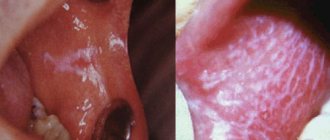
- Leukoplakia
The initial manifestations of leukoplakia are usually invisible, since there are no subjective sensations. Leukoplakia begins with the appearance of areas of clouding of the epithelium (grayish in color, with elements of keratinization on the surface). The lesion occurs against the background of an unchanged mucous membrane. Typical localization of the focus of leukoplakia is the mucous membrane of the cheeks along the line of closure of the teeth in the anterior section, the corners of the mouth and the red border of the lower lip without skin lesions. The dorsum and lateral surfaces of the tongue are somewhat less commonly affected. Smokers are characterized by lesions of the palate, described as “Tappeiner smokers' leukoplakia.”
There are the following forms of leukoplakia: flat or simple, verrucous and erosive. These forms can transform one into another. It is possible to combine different forms of leukoplakia in different areas of the oral mucosa in the same patient. The disease begins with the appearance of a flat shape.
Flat, or simple, leukoplakia (leukoplakia plana) is the most common. This form usually does not cause subjective sensations and is discovered by chance. Sometimes patients have complaints of a feeling of tightness, burning, and an unusual appearance of the mucous membrane. In the presence of extensive lesions on the tongue, taste sensitivity may be reduced. The main morphological element of the lesion in flat leukoplakia is a hyperkeratotic spot, which is an area of turbidity of the epithelium with clear contours. During the examination, foci of hyperkeratosis of various shapes and sizes are revealed, not rising above the level of the mucous membrane, but with clear boundaries of the lesion. Elements of flat leukoplakia resemble a burn of the mucous membrane with lapis, thin tissue paper pasted on, or a white coating that does not come off even with intense scraping. The keratinization can vary in intensity, so the color of the affected areas varies from pale grayish to intensely white.
The surface of the area of flat leukoplakia is usually slightly rough and dry. There is no compaction at the base of the lesion, just as there is no visible inflammatory reaction along its periphery.
There is a direct connection between the shape, color, size of keratinized areas and their location. Thus, with hyperkeratosis of the mucous membrane in the area of the corners of the mouth, symmetry of the lesion is observed in 85% of cases. The shape of the lesion has the shape of a triangle, the base of which faces the corner of the mouth, and the apex faces the retromolar space.
When the focus of leukoplakia is localized on the mucous membrane of the cheeks along the line of closure of the teeth, the elements of the lesion represent an elongated line, the continuity of which may be disrupted in certain areas. Leukoplakia on the red border of the lips looks like sticky tissue paper of irregular shape and grayish-white color (Fig. 11.5). Limited areas of lesions form on the mucous membrane of the tongue, hard palate, and floor of the mouth, sometimes resembling wide stripes or solid spots. More extensive lesions are also noted.
The relief of the mucous membrane and its turgor are also reflected in the appearance of the lesion. So, if leukoplakia develops against the background of folding of the tongue, the protruding areas become more keratinized and the surface of the tongue resembles a cobblestone street. A similar picture can be seen with a decrease in turgor and folding on the cheeks. If there is no pronounced folding on the tongue, flat leukoplakia looks like grayish-white, slightly sunken spots, the papillae of the tongue on them are smoothed.
Tappeiner's leukoplakia (leucoplakia nicotinica Tappeiner, nicotinic leukokeratosis of the palate) occurs in heavy smokers (especially pipe smokers). The mucous membrane of the hard palate and the adjacent part of the soft palate is mainly affected. Sometimes the gum margin is involved. The mucous membrane in the affected area is grayish-white in color, often folded. Against this background, mainly in the posterior half of the hard palate, red dots stand out - the gaping mouths of the excretory ducts of cystically dilated salivary glands, looking like small nodules. They are formed due to blockage of the excretory ducts by hyperkeratotic masses. Damage to the palate in smoker's leukoplakia can be combined with the location of elements on the mucous membrane of the cheeks, corners of the mouth, lower lip, etc. This form of leukoplakia is an easily reversible process: stopping smoking (an irritating factor) leads to the disappearance of the disease.
Verrucous leukoplakia (leucoplakia verrucosa) is the next stage in the development of flat leukoplakia. This is facilitated by local irritants: trauma with sharp edges of teeth and dentures, biting areas of leukoplakia, smoking, eating hot and spicy foods, microcurrents, etc.
The main feature that distinguishes this form of leukoplakia from flat one is more pronounced keratinization, in which a significant thickening of the stratum corneum is detected. The area of leukoplakia rises significantly above the level of the mucous membrane and differs sharply in color. Upon palpation, a superficial compaction may be detected. Patients usually complain of a feeling of roughness and tightness of the mucous membrane, burning and pain when eating, especially spicy food.
There are plaque and warty forms of verrucous leukoplakia. In the plaque form, limited milky white, sometimes straw-yellowish plaques with clear contours are identified, rising above the surrounding mucous membrane. The warty variety of verrucous leukoplakia is characterized by dense grayish-white bumpy or warty formations, rising 2-3 mm above the level of the mucous membrane. The warty form of leukoplakia has a greater potential for malignancy compared to the plaque form. On palpation, the lesions are dense, painless, and not fused with the underlying areas of the mucous membrane. The thickness of the raised areas of hyperkeratosis varies from pronounced to barely noticeable during normal examination. With a slight elevation of the elements, the affected area is almost not determined by palpation. A leukoplakic lesion of considerable thickness becomes dense to the touch, and it is not possible to fold it.
Erosive leukoplakia (leukoplakia erosiva) is actually a complication of simple or verrucous leukoplakia due to trauma. With this form of leukoplakia, patients complain of pain, which intensifies under the influence of all types of stimuli (eating, talking, etc.). Usually, against the background of foci of simple or verrucous leukoplakia, erosions, cracks, and, less often, ulcers occur. Erosions are difficult to epithelialize and often recur. Patients are especially concerned about erosion in the keratinized area of the red border of the lips. Under the influence of insolation and other irritating factors, they increase in size without showing a tendency to heal. The pain intensifies.
Pathohistologically, thickening of the epithelium is revealed due to the proliferation of the horny and granular layers. The stratum corneum reaches considerable thickness, especially with the verrucous form of leukoplakia. In it, foci of hyperkeratosis often alternate with foci of parakeratosis. The granular layer of epithelium in the affected area has varying degrees of severity. Verrucous and erosive forms of leukoplakia are characterized by pronounced hyperkeratosis and acanthosis. Acanthosis in leukoplakia can be significant if the keratinization has the character of parakeratosis; conversely, with severe hyperkeratosis, acanthosis is minimal or completely absent. In the connective tissue stroma of the affected areas of the mucous membrane, diffuse chronic inflammation is determined with pronounced infiltration of the surface layers by lymphocytes and plasma cells, the manifestation of sclerosis. The latter explains poor healing and frequent relapses with the appearance of cracks and erosions, which is most typical for the plaque form of leukoplakia.
Leukoplakia is a precancerous condition, since all its forms can become malignant with varying degrees of probability, transforming into spinocellular cancer.
Flat leukoplakia malignizes in 3-5% of patients, and in some the process of malignancy occurs quickly (1-1.5 years), in others the disease can exist for decades without transforming into cancer. Most often, malignancy occurs in verrucous and erosive forms of leukoplakia (in 20% of cases).
Clinical signs of malignancy are increased keratinization processes; rapid increase in the size and density of the lesion; the appearance of compaction at the base of the plaque, erosion; papillary growths on the surface of erosions, bleeding due to injury; the appearance of non-healing cracks.
- Soft leukoplakia Pashkova
Characterized by the presence of areas of peeling, slightly swollen, pasty mucous membrane with unclear boundaries, without inflammation or compaction. The mucous membrane has a grayish-white color. Most often, soft leukoplakia is localized on the mucous membrane of the cheeks along the line of closure of the teeth and lips, and less often on the tongue and gums. The lesion can be limited or diffuse, when almost the entire mucous membrane, and sometimes the red border of the lips, is involved in the process. In patients with neuropathy, predominantly young, with habitual biting or sucking of the cheeks, lips, tongue, the epithelium along the line of closure of the teeth is unevenly desquamated, has a fringed appearance due to the presence of multiple small flaps, which causes the unevenness of the affected surface (it looks like it is moth-eaten). This altered epithelium is partially removed by scraping. In severe cases, painful erosions may occur from biting the epithelium.
Histologically, pronounced parakeratosis and acanthosis are noted. At all levels of the spinous layer there are so-called “light” vacuolated cells, the cytoplasm of which is almost not stained, and the nuclei are deformed (pyknotized). In the connective tissue layer, expansion of small blood vessels, thickening of collagen, and thinning of elastic fibers are detected.
- White spongy nevus of Cannon
White spongy nevus may appear in early childhood or later, then progresses and reaches its maximum development during puberty. The disease may then regress or remain unchanged.
There are no subjective sensations. Patients may complain of an unusual appearance of the mucous membrane.
The typical location of a white spongy nevus is the buccal mucosa. The lesion is always symmetrical. The mucous membrane of the cheeks is white or grayish-white, somewhat thickened, soft, as if spongy, strongly folded. In some cases, the folding and wrinkling of the mucous membrane is so pronounced that the folds hang into the oral cavity. The surface layers of the epithelium are removed by scraping. At the same time, similar lesions may appear on the mucous membrane of the genital organs and rectum.
White spongy nevus should be differentiated from:
- flat leukoplakia;
- candidiasis;
- lichen planus;
- soft leukoplakia.
There is great clinical similarity between white spongy nevus and soft leukoplakia. Some authors consider white spongy nevus and soft leukoplakia to be different clinical forms of the same disease related to nevi, but there are quite pronounced differences between them. White spongy nevus, unlike soft leukoplakia, occurs mainly in childhood and is often hereditary. The development of mild leukoplakia, as a rule, is associated with various kinds of neurotic reactions or with hormonal changes in the body (puberty, postmenopause).
Patients with Cannon's nevus have congenital defects in the structure and maturation of the epithelium of the oral cavity, and in some cases, the genitals and rectum. With mild leukoplakia, the oral mucosa initially has a normal appearance, and its pathological changes are associated with the action of irritating factors against the background of neurological disorders or hormonal dysfunctions. In addition, damage to the oral mucosa with white spongy nevus is usually generalized.
Results and discussion
A study of the prevalence of TS showed that most often TS occurs in the first 1.5 months from the start of orthodontic treatment ( p
≤0.01), and subsequently the frequency decreases (Fig. 3).
Rice. 3. The incidence of traumatic stomatitis during orthodontic treatment in children and adults at various times during the active treatment period. Thus, according to the data we obtained, the incidence of TS was significantly less common than according to the literature [3–5], and amounted to 6.62–15.38% in children during different periods of the active period of orthodontic treatment with fixed appliances, and 4.69% in adults. -11.63% (see Fig. 2)
.
Before the start of treatment, TS of moderate and severe severity was detected in both children and young adults (Fig. 4, 5).
Rice. 4. The number of patients in the 1st and 2nd study groups depending on the severity of TS at different follow-up periods.
Rice. 5. The number of patients in the ٣-th and ٤-th study groups depending on the severity of traumatic stomatitis at different periods of follow-up. As a rule, the pain syndrome was mild, and the mucous membrane of the cheeks was most often affected, and less often the lips. Typically, in patients, regardless of age, with severe swelling and hyperemia, erosive and ulcerative lesions of varying severity were observed in the area of projection of orthodontic equipment (braces and arches).
The use of topical agents to treat TS has been effective in both children and adults. More pronounced positive dynamics of the reparative process of oral mucosa was noted when using gum gel with propolis in all age groups ( p
≤0.05) and observation period (
p
≤0.05).
At the same time, it should be noted that if in children on the 3rd day from the start of treatment TS of severe severity remained (see Fig. 4), then in young people (see Fig. 5) there was no such degree of TS ( p
≤0.05).
By this time, in adults, regardless of the drug used for the treatment of TS, there were no pathological changes in the mucous membrane in the area of the mucous membrane previously injured by the brace system ( p
≥0.05).
In children of groups 1 and 2, the effectiveness of treatment on the 3rd day was 34.66 and 48.96%, respectively ( p
≤0.01);
on the 5th day - 78.66 and 83.33%, respectively ( p
≤0.05). Similar data on the effectiveness of treatment of TS caused by injury to the mucous membrane of the cheeks and lips by braces and arches were obtained in young adults (Fig. 6).
Rice. 6. The effectiveness of treatment of traumatic stomatitis in patients of different groups at different periods of dynamic observation. Thus, in individuals included in the 3rd and 4th study groups, the effectiveness of treatment on the 3rd day was 60.29 and 68.03%, respectively ( p
≤0.05);
on the 5th day 91.18 and 95.08%, respectively ( p
≤0.05), which also indicated the greater therapeutic effectiveness of the gum gel with propolis for the treatment of TS caused by wearing fixed orthodontic appliances.
On the 10th day, no TS was detected in patients of all four study groups.