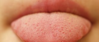
White plaque on the gums can be a symptom of candidiasis, a disease caused by a fungus of the genus Candida. Under the influence of a fungal infection, stomatitis develops. First, multiple cheesy white rashes appear on the surface of the cheeks, tongue, palate and gums. Gradually they begin to grow. As the lesions grow, they merge and a film forms in the mouth.
At the beginning of the disease, plaque can be scraped off. But in the place where it was removed, small ulcers remain. When the disease is advanced, the whitish film can no longer be scraped off. The patient feels serious discomfort. Dryness, burning, pain, and a metallic taste appear in the mouth. In the advanced stage there are systemic symptoms. The head begins to hurt, the patient complains of lethargy and weakness, and the temperature rises.
Candida is an opportunistic microflora. They live in the oral cavity of any person. As long as their number is within normal limits, pathological conditions do not develop. If candida begins to multiply quickly, they provoke candidiasis.
How does the infection manifest and why is it dangerous?
The most striking symptom of oral candidiasis is a white, loose coating on the mucous membrane. It mainly covers the tongue, cheeks, and may affect the gums and palate. Plaque can be easily scraped out; the tissue underneath is prone to redness and may bleed. Other signs of the disease include:
- unpleasant taste;
- dry mouth;
- burning;
- the appearance of cracks in the corners of the lips;
- difficulty or painful swallowing;
- unpleasant sensations with habitual movements of the tongue.
Candidiasis can be acute or chronic. In most cases, it is the acute form that manifests itself; the chronic form is typical for carriers of HIV infections and smokers. Depending on the degree and form of the disease, other symptoms may be observed, so even if one or two appear, you should immediately contact your dentist.
When should you see a doctor if you have a white coating on your tongue?
White coating on the tongue is a symptom, not a disease, so it is necessary to find out the cause of plaque formation from a specialist (especially if it is accompanied by discomfort, pain and burning in the mouth, the plaque persists for more than several weeks, ulcers form, chewing and swallowing are difficult), and undergo diagnostics and treatment of the disease causing such symptom. Depending on the problem, the doctor will prescribe antifungal drugs, local glucocorticosteroid therapy, antibiotic therapy and other treatment methods.
Often, a white coating on the tongue goes away on its own (for example, caused by ARVI) within a few weeks. Don’t forget to take proper care of your oral cavity: brush your teeth regularly with a soft-bristled brush, use a special scraper on your toothbrush to clean your tongue, floss, mouthwash, drink plenty of water, try to eat healthy and balanced, give up bad habits, eat regularly Visit your dentist for preventive examinations (at least once every six months) and treatment.
Causes of oral candidiasis
How does the disease occur? The fungus Candida lives in the body from birth and, under certain circumstances, provokes illness. What becomes the trigger for infection:
- weakened immunity;
- taking antibiotics;
- long-term use of removable dentures, especially if they do not fit tightly;
- insufficient oral hygiene;
- smoking;
- oncology;
- use of inhalers for asthma;
- excess simple carbohydrates in the diet;
- pregnancy period;
- infancy or old age.
It can be seen that there are many reasons, often they act together, less often - individually. In general, oral candidiasis is rare in adults. It mainly affects children in the first years of life and older people. Among these categories, the prevalence of the disease reaches 10%.
Diagnostic search algorithm for diseases of the oral mucosa
I. K. Lutskaya, Doctor of Medical Sciences, Professor BelMAPO (Minsk)
Diagnosis and treatment of diseases localized on the mucous membrane and perioral area present a certain difficulty for the dentist due to the variety of their manifestations in some cases and the striking similarity of the rashes in others.
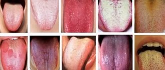
Clinical symptoms change significantly both in the presence of a banal infection and under the influence of treatment. Similar complaints and unclear development of the process often do not allow one to obtain a sufficient impression to determine a possible disease. In such cases, the diagnostic search begins with an objective assessment of the clinical picture, namely the appearance of the lesion elements. The subjective picture (complaints and description of anamnestic data) is reflected in detail in the medical record and occupies an important place in the diagnostic algorithm. As a specific example, consider the presence of white spots localized on the tongue, mucous membranes of the cheeks and lips: whitish areas with uneven borders (Fig. 1). The first step in the doctor’s thinking algorithm is to list diseases that have a similar visual picture. In this case, we should name candidiasis, lichen planus (LP), geographic tongue, desquamative glossitis, secondary syphilis.
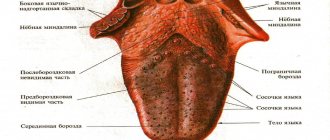
After a thorough examination, returning to the analysis of complaints will allow you to link them to a specific disease. If no subjective sensations, paresthetic or painful, are noted, lichen planus, geographic tongue, or secondary syphilis can be assumed. Candidiasis and desquamative glossitis are characterized by pain when talking and eating food, especially spicy and salty food. Pain may appear with the development of the inflammatory-hyperemic form of LP or with syphilis in the event of a secondary infection. Geographic tongue can be combined with candidiasis, accompanied by pain. Thus, the presence of subjective complaints cannot serve as a pathognomonic sign of a specific disease. Finding out the onset of the disease from the anamnesis will allow you to take the next step in the diagnostic search. Candidiasis has a clear onset and accompanies common diseases or the use of medications that suppress the bacterial flora. Lichen planus can exist for quite a long time (months, years), but it appears in adulthood and is often caused by emotional stress. "Geographic tongue" is detected soon after birth and remains for many years. Desquamative glossitis (stomatitis) occurs and recurs, being associated with general diseases, most often pathology of the gastrointestinal tract (GIT). With syphilis, the rash of secondary elements of the lesion has a clear beginning and can appear against the background of preserved primary affect. The dynamics of the elements of damage have their own characteristic features. Plaque due to candidiasis occupies an increasingly larger area over time, moving to neighboring sections of the oral mucosa, and its quantity increases. With lichen planus, the picture is quite stable, only the clinical picture slowly and gradually increases or changes. “Geographical language” is distinguished by the “migration” of drawings. Desquamation of the mucosal epithelium in general diseases may increase over time, but the administration of anti-inflammatory drugs leads to improvement. With syphilis, the elements of the lesion gradually disappear, but on the tongue the picture of a “mowed meadow” is quite stable. A more detailed examination allows us to identify the features inherent in a particular disease. With candidal stomatitis, changes are observed in the mucous membrane of the entire oral cavity. When individual areas are affected, the disease, depending on the location, is called candidal glossitis, cheilitis, or tonsillitis. Acute candidiasis occurs in infants or weakened people (blood diseases, hypovitaminosis), as well as in persons receiving large doses of corticosteroids, cytostatics, and antibiotics. The patient is concerned about dry mouth and impaired taste. When eating food, especially salty, sour, spicy food, pain is noted. A characteristic sign of candidiasis is foamy saliva that collects in the retromolar region and on the back of the tongue. On the hyperemic, edematous mucous membrane of the cheeks, palate, gums, and tongue, whitish areas appear that merge to form a loose, “curdled” white coating (Fig. 2). After removing the plaque, a smooth, hyperemic mucous membrane is exposed.
Rice. 1. The lesion elements are localized on the mucous membrane of the tongue. Rice. 2. Plaque on the mucous membrane with candidal stomatitis. Rice. 3. Chronic candidal glossitis. Rice. 4. Plaques with lichen planus on the tongue.
Subsequently, the plaque can become saturated with fibrin, taking on the appearance of a dense film of grayish or yellowish color, tightly attached to the surface of the epithelial layer. Pseudomembranous candidiasis, characteristic of HIV infection, develops. The initial symptoms of candidal glossitis may be pinpoint redness of the marginal zones and the tip of the tongue. With chronic candidal glossitis, small furrows with white deposits appear along the edges and at the bottom. The lesions are first detected at the base of the tongue, and then spread to the remaining parts, capturing its lateral surfaces. Against the background of atrophy of the filiform papillae of the tongue, a scant whitish coating is detected, which is not completely removed (Fig. 3). Candidiasis of the red border of the lips is manifested by dryness, hyperemia, swelling, and peeling. Painful erosions, small cracks, and thin gray scales may occur. Subjective sensations include tension and burning. The disease differs in its duration and relapses. The diagnosis of candidiasis is confirmed by the results of complex laboratory studies over time: microscopic, cultural (with determination of the type of fungus), and in some cases, histopathological. Fungi of the genus Candida are found in the form of yeast-like cells and pseudomycelium. The presence of pseudomycelium or chains of round or elongated budding cells upon microscopic examination of the material is considered sufficient to confirm the diagnosis. The predominance of yeast cells with single filaments of pseudomycelium indicates superficial candidiasis. The predominance of the filamentous form over the cellular form indicates the parasitic activity of the fungus and is more often detected in visceral lesions. Lichen planus (LP) can begin unnoticed, last for years, and be discovered accidentally when examining the mucous membrane by a specialist. In some cases, the abundance of rashes attracts the patient's attention. The etiology is not always determined. Often this is psycho-emotional stress, toxic-allergic effects. Elements of the lesion may first appear on the mucous membrane, then spread to the skin, or, conversely, a rash on the body precedes the lesion of the mucous membrane. The main element is always a papule: on the skin it is initially whitish, then pale pink, then reddish, lilac. Papules tend to cluster. The presence of skin rashes makes the diagnosis easier. On the red border of the lips, papules undergo keratinization, are connected by bridges, and form whitish areas in the form of individual raised nodules, bizarre patterns or merging areas of hyperkeratosis with uneven outlines. On the mucous membrane, PL is characterized by the presence of small whitish papules (up to 2 mm in diameter). The last few rise, which causes discomfort, a feeling of tightness or a feeling of roughness. Papules can acquire a grayish or pearlescent tint and tend to group together to form fancy patterns in the form of lace, mesh, or a “frosty pattern.” Favorite localization is the buccal mucosa along the line of closure of the teeth, retromolar region, tongue, gingival margin. On the tongue, the lesion elements can merge into plaques or be located in the form of circles, semi-arcs, or wavy lines (Fig. 4, 5). Papules in LP are painless; scraping does not remove the whitish surface. The exudative-hyperemic form is characterized by hyperemia, swelling of the mucous membrane, on which papules are located, forming a typical picture in the form of patterns, networks, arcs. Subjective sensations in the form of pain when eating food (hot, spicy, hard) are added. The bluish glow in Wood's rays of the lesion elements on the red border and the white glow on the mucous membrane makes it possible to differentiate the lesion elements in LP. The etiology of such a condition as “geographic tongue” has not been fully elucidated: neurotrophic disorders and diseases of the gastrointestinal tract are possible. In a number of cases, “geographical language” acts as a variant of the normal structure and is detected already in childhood. There may be no subjective sensations, and then the peculiarities of the appearance of the tongue are revealed during examination. Rarely, patients complain of tingling, burning, and paresthesia. Symptoms intensify with injuries to the mucous membrane, the development of Candida fungus, and the addition of a secondary infection. With the development of the “geographic tongue” picture against the background of a general disease, the process of desquamation begins with the appearance of a small area of turbidity of the epithelium; in the center, the upper layers of the keratinized epithelium are peeled off, revealing a pink, smooth area that quickly grows along the periphery. Multiple or single lesions reach a diameter of 1-2 cm, having the shape of spots, rings, half-rings, the boundaries merge with the surrounding mucous membrane. In the center, normal keratinization of the filiform papillae begins. The lesions are layered, new ones appear against the background of the old ones, which gives the surface of the tongue the appearance of a geographical map (Fig. 1). Minor keratosis may appear in the form of whitish stripes. In some cases, pigmentation of these areas occurs (Fig. 6). The process usually does not extend to the lower surface of the tongue. A similar picture rarely occurs simultaneously on the lips, cheeks, and gums. A constant feature is the preservation of filiform papillae of the tongue.
Rice. 5. Lichen planus in the form of a lace pattern. Rice. 6. “Geographic tongue” is combined with a deep median sulcus. Rice. 7. Desquamation of the epithelium is combined with plaque during gastritis. Rice. 8. Picture of a “mowed meadow” with syphilis.
Cytological examination reveals epithelial cells with keratohyalin inclusions. Desquamation of the epithelium, combined with plaque, is characteristic of diseases of the gastrointestinal tract (GIT). In such cases, discomfort and pain appear in the oral cavity, especially during meals. The tongue may be swollen, and then teeth marks can be identified on it. Cracks, erosions, and areas of increased desquamation form on the tongue, cheeks, and lips (Fig. 7). Gastritis with reduced secretion is characterized by dry mouth, complaints of burning, pain in the tongue, especially from spicy, hot foods, and a metallic taste. Against the background of atrophy of the filiform papillae of the tongue, desquamation of the epithelium is characteristic. The filiform papillae are smoothed, the mushroom-shaped papillae appear hypertrophied. With atrophic gastritis, atrophic glossitis develops. Characterized by burning and pain in the tongue. The tongue is pale pink, partially coated, the filiform papillae are atrophied. With a gastric ulcer, plaque on the tongue may be more or less abundant and pigmented, but can be easily removed. There may be complaints of a burning sensation (the tongue feels as if it is scalded or sprinkled with pepper), pain and an increase in the size of the tongue due to swelling. There are teeth imprints on the side surfaces. Independent glossitis may develop: “geographical”, “black hairy” tongue. Fungal stomatitis is often associated. A course of treatment for a general disease leads to an improvement in the condition of the oral cavity, but does not exclude relapses of desquamative stomatitis (glossitis). The secondary period of syphilis in the absence of treatment manifests itself after 6-8 weeks as a fresh secondary one, and then its relapses can be repeated for 3-5 or more years, lasting 1.5-2 months and characterized by more or less profuse rashes. About 50% of patients have manifestations in the oral cavity, and this is often the only localization of the lesion elements. The most common lesions found are papular rashes. Favorite localizations are tonsils, palatine arches, cheeks (along the line of teeth closure), tongue. Red papules are small at first, then grow to several millimeters and become covered with a peculiar grayish coating. After scraping with a spatula, flesh-red erosion is exposed. Papules tend to spontaneously erode. The addition of a secondary infection leads to pain.
When localized on the back of the tongue, syphilitic papules acquire the characteristic pattern of a “sloping meadow” - clearly defined oval areas with a smooth surface. The latter is formed due to atrophy of the filiform papillae (Fig. 8). Clinical diagnosis of syphilis is based on the characteristic elements of the lesion with mandatory confirmation by the results of laboratory tests to identify the pathogen or specific reactions.
Conclusion Difficulties in diagnosing diseases affecting the oral mucosa are associated with the lack of clear differences between subjective and objective symptoms. In such cases, the dentist can use a diagnostic search algorithm (without violating the rules and procedure for filling out medical documentation). After receiving the entire amount of information, he consistently excludes diseases that do not have characteristic features inherent in the clinical manifestations of a given patient.
How is oral candidiasis treated?
To begin with, let's say that treatment is necessary, otherwise the infection spreads to the pharynx, tonsils, and gums. Then the disease descends into the lungs, gastrointestinal tract, liver and causes irreparable harm to them.
When the first signs of candidiasis of the oral mucosa appear, you should contact your dentist or therapist to confirm the diagnosis. When you see your doctor, describe all your symptoms, even those that are not on the list. For example, you have noticed increased anxiety or a tendency to stress lately. Information about the medications you are taking, recent illnesses, and being around sick people is also important. To confirm the diagnosis, it is often enough to take a swab from the oral cavity and carry out the appropriate analysis. If the disease has already spread to other organs, a more thorough examination may be required, for example, endoscopy - diagnosing the condition of the esophagus and stomach using a flexible tube with a camera at the end.
How to treat oral candidiasis? Treatment can be local or systemic depending on the form and stage of the disease. When local, antifungal agents and antiseptics are prescribed. These can be sprays, rinses, gels, lozenges and more. Treatment on average takes up to 3 weeks, as a general rule - until symptoms disappear and another week. Systemic treatment is used for chronic forms or when the infection spreads to other organs. It usually involves the use of more medications that are intended to treat the directly affected areas.
Under no circumstances resort to self-medication! A fungal infectious disease has nothing to do with inflammation, so your actions are more likely to harm your body. In doing so, you can reduce the harmful effects of the infection and make you feel better. To do this, eat natural yogurt without sugar and take probiotics as recommended by your doctor. But these actions are not a complete treatment for candidiasis in the mouth!
Symptoms and treatment of leukoplakia -
Leukoplakia is characterized by the fact that under the influence of various irritants, keratinization of the epithelium of the oral mucosa begins to occur. This process occurs due to a violation of the desquamation of the epithelium in those areas of the mucous membrane that is exposed to pathological irritants. These irritants can be:
- smoking,
- sharp edges of carious teeth,
- overhanging edges of fillings,
- malocclusion,
- excessive consumption of spices, very hot foods,
- poorly manufactured removable dentures,
- the presence of artificial crowns made of different metals on the teeth, which gives rise to galvanic currents in the oral cavity.
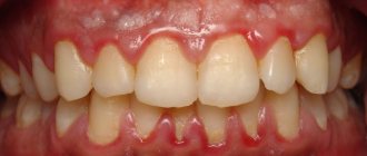
White spots on the gums with leukoplakia: photo
Symptoms of leukoplakia:
The main element of leukoplakia is a white plaque, which may rise above the level of the mucous membrane, but may not rise (as in Fig. 4). In addition, the plaque can have both clear and blurred boundaries. Patients most often call such plaques white spots. As a rule, white spots do not bother their owners and are discovered completely by accident.
In some cases, patients may be concerned about the roughness of the mucous membrane at the site of the white spot, as well as the fact that the mucosa in this place may rise above the level of the mucosa. In some cases, cracks and ulcers may appear on the surface of the stain; in this case, the patient may also be bothered by burning and pain. If leukoplakia develops as a result of smoking, then, as a rule, patients first of all complain of dryness and burning of the oral mucosa (24stoma.ru).
Treatment of leukoplakia will primarily involve eliminating exposure to the irritating factor. Those. it will be necessary to eliminate the impact of the overhanging edge of a filling or crown, replace old low-quality dentures, normalize oral hygiene, and of course, solve something with the effects of nicotine and hot dry air on the mucous membrane (which occurs when smoking).
Only after this are prescribed keratolytic agents for topical use (3-5-10% salicylic acid, celandine infusion), and vitamin therapy. Applying any treatment procedures without eliminating the causative factor will be useless.
The danger of leukoplakia –
Leukoplakia belongs to the category of precancers. Malignancy of leukoplakia, i.e. its transition to cancer occurs in 15-75% of cases (depending on its form). Therefore, if you do not want to develop cancer of the oral mucosa, you need to urgently go to the dentist and look for the cause of its appearance. But in the photo below you can see photos of two clinical cases when leukoplakia transformed into a malignant tumor of the oral mucosa and the red border of the lips.
Cases of cancer associated with leukoplakia –
How to avoid candidiasis?
To prevent the disease, you must follow a few simple rules:
- maintain oral hygiene;
- consult a dentist at least twice a year, even if there are no complaints;
- control nutrition, avoid excess carbohydrates in food;
- refrain from smoking, which can cause a chronic form of the disease.
Treatment of candidiasis of the oral mucosa can take a long time and also cause irreparable harm to health, so we recommend that you take care of yourself in advance by following preventive measures. If symptoms have already begun to appear, consult a doctor as soon as possible so that the infection does not have time to spread to other organs. You can undergo oral cavity diagnostics at Comfort Dentistry. Our specialists will tell you in detail about the prevention of common diseases of teeth and gums, and also, if necessary, provide high-quality treatment.
Why do you need to clean your tongue?
Brushing your tongue twice a day not only cleanses it of bacteria. This cleaning eliminates four problems.
- Prevents the development of caries
by preventing microorganisms from producing acid that destroys enamel. - Removes bad breath
because bacteria do not produce odorous substances. - Removes plaque
- a breeding ground for bacteria. - Increases the sensitivity
of taste buds - a clean tongue senses the flavors of food and drinks more clearly.
When caring for your tongue, it is worth remembering that not every coating is a sign of some problems. There is also a healthy plaque that can be easily removed with proper hygiene.
Healthy plaque
can be easily distinguished by several signs.
- It is formed
in the morning, after eating or during thirst; - disappears
after cleansing; - changes color
due to coloring by products; - through it you can see
the surface of the tongue; - it doesn't smell
.
Unhealthy plaque can be a symptom of many health problems: diseases of the stomach and intestines, dysfunction of the liver and kidneys, infections in the body (whooping cough, foot and mouth disease, scarlet fever, pityriasis rosea), as well as a consequence of taking antibiotics.
This is what characterizes an unhealthy plaque
.
- It remains
even after thorough cleaning; - It smells
bad ; - very dense and quickly restored
after cleaning; - regardless of food, the color ranges
from yellow to brown; - accompanied by
redness, swelling and cracks of the tongue, nausea, bitterness in the mouth, gastrointestinal upset, etc.
If the plaque is deep yellow or gray-yellow, you should pay attention to the gastrointestinal tract - you may have gastritis, ulcers, pancreatitis. And if frequent and severe heartburn occurs, the plaque will turn yellow-orange.
But we must remember that harmful plaque is not always caused by some disease. It can appear due to an unhealthy lifestyle: smoking, a large amount of dyes in foods, poor hygiene, even due to caries and bleeding gums. Brushing your tongue with a toothbrush will not solve this problem - you need to contact a specialist to understand how to proceed.