Anatomical structure of the tongue
The structure of human language corresponds to its multifunctionality, which lies in the fact that it participates in the processes:
- chewing;
- salivation;
- taste perception;
- speech.
The body of the tongue consists of striated muscle tissue, which is covered by a membrane of mucous tissue. Its surface, called the back, is conventionally divided into three parts:
- the last third, located near the pharynx, is called the root;
- the first two thirds are the body of the tongue.
A longitudinal groove runs in the middle, which is an external manifestation of the internal septum; it is, in fact, a reduced thyroglossal duct.
The mucous membrane, tightly adjacent to the muscle tissue, is covered on the outside with stratified squamous epithelium. It contains:
- salivary glands;
- taste buds;
- lymphatic ducts.
The mucous membrane of the posterior part forms three supraglottic folds, with the help of which the tongue is attached to the larynx:
- median;
- two lateral.
The tongue is abundantly covered with papillae, including:
- filamentous - act as organs of touch and, thanks to the rough surface, hold food on the tongue;
- cone-shaped – responsible for sensitivity to temperature and pain;
- mushroom-shaped - equipped with taste buds, thanks to them we distinguish many taste sensations;
- groove-shaped - located near the root, have serous glands and are also responsible for the sense of taste;
- leaf-shaped - equipped with lingual glands that secrete a mucous secretion.
The tongue is attached to the oral cavity by a fold of mucous membrane called the frenulum.
content .. 100 101 102 103 104 105 106 ..
Tongue (human anatomy)
Language
, lingua (in phrases the Latin synonym glossus is used), is a muscular organ consisting of striated muscles. The tongue is covered with a mucous membrane that has a special structure. By function, the tongue is an organ of speech, a taste organ, and also an organ involved in the acts of sucking, chewing and salivation.
The shape and position of the tongue are variable and depend on its functional state. At rest, the tongue has a spade shape, almost completely filling the oral cavity. The tip of the tongue is adjacent to the back surface of the front teeth.
In the tongue, there is a tip or its apex, apex, a body, corpus, and a root, radix (Fig. 85). Its upper surface is convex and is called the back of the tongue, dorsum linguae. The lower surface, fades inferior, is smaller than the upper, since most of it is covered by the root of the tongue. Both surfaces are connected by the lateral edges of the tongue, margo linguae. The upper surface is divided into two sections - the anterior, larger - oral, pars oralis, lying in the horizontal plane, and the posterior, pharyngeal, pars pharyngea, facing the pharynx and running almost vertically. At the border between these sections along the midline, a small depression is visible leading into the blind foramen, foramen coecum, the remnant of the reduced thyroglossal duct of the thyroid gland, ductus thyreoglossus. In some people, this embryonic duct may not be reduced completely or partially, which causes the formation of median cysts and fistulas of the neck.
Rice. 85. Tongue and entrance to the larynx. a - general view; b - fungiform papilla; c - filiform papilla; d — papilla surrounded by a shaft (top view); d - papilla surrounded by a shaft (in section). 1 - glottis; 2 - interarytenoid notch; 3—horn-shaped tubercle; 4 - wedge-shaped tubercle; 5 - aryepiglottic fold; 6 - pear-shaped pocket; 7 - lingual-epiglottic fold (median); 8 - lateral lingual-epiglottic fold; 9 - left palatine tonsil; 10 - lingual follicles; 11 - conical papillae;; 14 - median lingual groove; 15 - tip of the tongue; 12, 16 - filiform papillae (see Fig. c); 17 - mushroom-shaped papillae (see Fig. b); 13, 18 - leaf-shaped papillae; 19, 20 - papillae surrounded by a shaft; 21 - blind hole; 22 - right palatine tonsil; 23 - epiglottis; 24 - greater horn of the hyoid bone
As mentioned above (see section Development of the digestive organs, this edition), the language develops from three rudiments. As a trace of the fusion of these rudiments, two furrows are visible on the tongue. One of them, the median groove of the tongue, sulcus medianus linguae, is located longitudinally on the back of the tongue along the midline from the apex of the tongue to the foramen cecum. The second, border groove, sulcus terminalis, runs transversely from the blind foramen to the right and left.
The bulk of the tongue consists of muscles and their connective tissue apparatus. It consists of a dense fibrous septum of the tongue, septum linguae, which lies longitudinally in the thickness of the tongue. On the back of the tongue, the septum projects onto the median sulcus, and below it passes into the raphe tendinea m. mylohyoidei. The septum divides the muscles of the tongue into two more or less symmetrical halves. In addition, the muscles of the tongue are covered with a strong lingual aponeurosis, aponeurosis linguae, which contains a lot of collagen and elastic fibers intertwined in the form of bundles. The aponeurosis contains numerous openings through which small tendons of the tongue muscles pass into the mucous membrane of the tongue.
The striated muscles of the tongue consist of bundles of muscle fibers running in three mutually perpendicular directions: longitudinal, transverse, and vertical. Depending on the position, there are two groups of muscles of the tongue: internal and external. Internal, or intrinsic, muscles lie only in the thickness of the tongue and do not extend beyond its limits. They change the shape of the tongue. External muscles begin on the nearest bones, enter the thickness of the tongue and, when contracted, change its position. In addition, the muscles of the tongue are grouped depending on their function: 1) muscles that lengthen and flatten the tongue (m. genioglossus, m. verticalis); 2) muscles that shorten the tongue and move it upward (m. styloglossus, m. longitudinahis superior, m. longitudinalis inferior); 3) muscles that shorten the tongue and shift it downwards, and with unilateral action - to the side (m. hyoglossus, m. transversa linguae). The first group of muscles originates on the derivatives of the 1st branchial arch, the second on the derivatives of the 2nd branchial arch, and the third on the derivatives of the 3rd branchial arch (Fig. 86). The internal muscle group of the tongue includes the following muscles.
1. Superior longitudinal muscle of the tongue
, m. longitudinalis superior, steam room, a thin muscle layer lying directly under the lingual fascia, from which it originates in the region of the root of the tongue. It lies on the sides of the septum linguae above all the others, runs along the entire tongue and is attached to the fascia of the tongue at its apex.
Function: shortens and somewhat thickens the tongue. With unilateral contraction, it takes it to the side.
2. Inferior longitudinal muscle
, m. longitudinaHs inferior, steam room, begins from connective tissue bridges in the area of the root of the tongue, separating individual muscles. Goes between m. genioglossus and m. hyoglossus near the lower surface of the tongue to its apex, intertwining with the fibers of these muscles and the transverse muscle of the tongue. Attaches to the fascia of the tongue at its apex.
Function: shortens the tongue and bends its back upward; with unilateral contraction, it takes it to the side,
3. Transverse tongue muscle
, m. transversus linguae, steam room, originates from the septum linguae and spreads laterally between the upper and lower longitudinal and genioglossus muscles. The upper muscle bundles are attached to the lingual fascia in the lateral parts of the back of the tongue, and the rest are attached to its edges, where the m. hyoglossus. The posterior sections of the muscle give off bundles of fibers that go along with m. palatoglossus and in small quantities with m. palatopharyngeus, reaching the soft palate and the wall of the pharynx.
Function: narrows the tongue and lengthens it, participates in compression of the pharynx and pharynx.
4. Perpendicular muscle of the tongue
, m. verticalis, steam room, starts from the lingual fascia in the region of the back of the tongue and goes between the fibers of other muscles down to the fascia of the lower surface of the tongue.
Function: flattens and lengthens the tongue, forms a longitudinal groove on its back.
The external muscle group of the tongue consists of 4 muscles:
1. Genioglossus muscle
, m. genioglossus, steam room, departs from the spina mentalis and, diverging in a fan-shaped manner, goes to the septum and attaches to the lingual fascia on the back of the tongue. The bundles of muscle fibers of this muscle partially merge with the bundles of the vertical and longitudinal muscles of the tongue.
Function: pushes the tongue forward, and with unilateral contraction, deflects it to the side.
2. Hyoglossus muscle
, m. hyoglossus, steam room, originates on the large horns and superolateral parts of the body of the hyoid bone and goes forward and upward, intertwining with the vertical muscle. The fibers of the muscle reach the lingual fascia along the edges of the tongue, and the fibers of the posterior sections of the muscle reach the back of the tongue. Quite often in m. hyoglossus there is an independent, variable in size, bundle of muscle fibers, starting on the lesser horn of the hyoid bone and going to the back of the tongue, which is designated as the cartilaginous muscle, m. chondroglossus.
Function: flattens the tongue and pulls it down and back.
Rice. 86. Muscles of the tongue. 1 - lower longitudinal muscle; 2 - genioglossus muscle; 3 - geniohyoid muscle; 4 - thyroid cartilage; 5 - hyoid bone; 6 - hypoglossus muscle; 7 - styloglossus muscle
3. Styloglossus muscle
, m. styloglossus, steam room, begins on the styloid process of the temporal bone and from lig. stylomandibulare and passes down and medially, fanning out in the lateral parts of the tongue outward from m. hyoglossus, where it is intertwined with bundles of the transverse muscle.
Function: pulls the tongue upward and backward.
4. Palatoglossus muscle of the tongue
, m. palatoglossus (see section The oral cavity itself, this edition).
In general, the listed internal and external muscles of the tongue form a complex interweaving of bundles, which explains the exceptional mobility of the tongue and the variability of its shape.
The mucous membrane of the tongue is tightly fused with the lingual fascia and intermuscular connective tissue. There is no submucosal layer in the tongue, so the mucous membrane is motionless and does not fold. On the outside, this membrane is covered with stratified squamous epithelium. It contains glands, taste organs and lymphatic formations. In the area of the tip, back, root and edges of the tongue, the mucous membrane is rough. Posterior to the sulcus terminalis it is thicker than in front, and has nodular elevations due to the presence of lymphatic follicles, and on the lower surface it is smooth. Along the midline, the mucous membrane forms the frenulum of the tongue (see section The oral cavity itself, this edition), and on the sides of it there are fringed folds converging anteriorly, plicae fimbriatae, more clearly expressed in children. The mucous membrane of the posterior part of the tongue forms three lingual-epiglottic folds going to the epiglottis: unpaired - median, plica glossoepiglottica mediana, and paired - lateral, plica glossoepiglotticae laterales. Between them there are supraglottic recesses, valleculae epiglotticae. On the upper surface and along the edges of the tongue anterior to the sulcus terminalis there are numerous different devices of protrusion of the mucous membrane - papillae of the tongue, papillae linguales. Some of them contain fibers of taste nerves. There are five types of papillae: filiform, conical, mushroom-shaped, papillae surrounded by a shaft (vallate) and leaf-shaped (see Fig. 85).
1. Filiform papillae
, papillae filiformes, are the most numerous, scattered throughout the wall and along the edges of the tongue. Their length is from 0.6 to 2.5 mm, thickness 0.1-0.6 mm. In front they are longer than in the posterior sections of the dorsum of the tongue. The basis of the papilla is a protrusion of the lamina propria of the mucous membrane, which is covered with keratinizing stratified squamous epithelium. The peeling horny scales have a whitish color, as a result of which the tongue is whitish-pink. In case of digestive disorders, the rejection of keratinized epithelial cells is delayed, as a result of which a white coating (“coated” tongue) forms on the tongue. Filiform papillae are not taste organs. They function as organs of touch and help keep food on the tongue.
2. Conical papillae
, papillae conicae, are among the filamentous ones and are very close to them in their structure and function. They are considered as a transitional form to taste buds.
3. Fungiform papillae
, papillae fungiformes, are less numerous than filiforms (150-200), distributed over the upper surface of the tongue without any order. There are slightly more of them on the tip of the tongue. The mushroom-shaped papillae are 0.5-1.5 mm long and 0.5-1 mm thick. The surface of these papillae is covered with non-keratinizing stratified squamous epithelium. They are clearly visible to the eye in the form of reddish dots, since the capillaries of the papilla are visible through the epithelium. The epithelium of the fungiform papillae contains taste buds, which are taste organs.
4. Papillae surrounded by a shaft
, or
grooved
, papillae vallatae, are the largest papillae of the tongue. Their length reaches 3-6 mm and width 1-2 mm. The papillae, surrounded by a shaft, are concentrated in a row in the form of an angle, whose apex is directed towards the blind foramen. At the bottom of the opening there is a separate papilla, papilla solitaria. There are few papillae - from 7 to 18, more often there are 7-12. The papillae, surrounded by a shaft, do not protrude above the surface of the mucous membrane of the tongue. They are immersed in it due to the deep groove surrounding the papilla. The mucous membrane around the papilla forms an elevation - a roller. At the base of the papillae, the intermuscular connective tissue contains small serous glands, which open in the grooves surrounding the grooved papillae. The epithelium lining the lateral surfaces of the grooved papillae and the ridges surrounding them contains a large number of taste buds (from 40 to 150 bulbs in one papilla).
5. Leaf-shaped papillae
, papillae foliatae, lie along the edges of the tongue, in its posterior sections, slightly anterior to the papillae, surrounded by a shaft, 15-20 on each side, forming several small folds or wrinkles. The height of the folds can reach 7 mm, and the thickness - 2-3 mm. The epithelium of the leaf-shaped papillae contains taste buds.
Taste buds, or buds, contain the terminal receptor apparatus of the taste analyzer and are located in the papillae of the tongue, as well as on the posterior edge of the palate, in the epithelium of the pharynx and epiglottis. It is believed that the mushroom-shaped and leaf-shaped papillae perceive the taste of sour, sweet and salty, and surrounded by a shaft - bitter.
Between the muscle bundles under the mucous membrane lie glands, the excretory ducts of which open on the tongue, and accumulations of lymphoid tissue. The glands of the tongue are serous, mucous and mixed. The following glands of the tongue are distinguished:
1. Anterior gland
, glandula lingualis anterior, paired, mixed, located under the lower longitudinal muscle near the tip of the tongue. The excretory ducts of the gland (up to 7) open on the lower surface of the tongue.
2. Posterior glands
, glandulae linguales posteriores, numerous small glands of mixed, serous and mucous types. They lie between bundles of muscle fibers in the posterior half of the tongue. Their ducts open in the grooves of the grooved papillae, as well as in other areas of the mucous membrane.
In the mucous membrane of the root of the tongue, posterior to the groove, there are accumulations of lymphoid tissue in the form of follicles of various sizes. The collection of lingual lymphatic follicles is called the lingual tonsil, tonsilla lingualis. In the area of accumulation of follicles, the mucous membrane forms a noticeable elevation, in the center of which there is a depression - the crypt. The set of palatine, glossopharyngeal, tubal and laryngeal tonsils located at the border of the oral cavity and pharynx is called the lymphoepithelial pharyngeal ring.
The blood supply to the tongue is provided by a. lingualis, the branches of which form the intraorgan vascular bed. The outflow of blood occurs through v. lingualis, which flows into the internal jugular vein.
Lymph flows into the mental, submandibular and retropharyngeal lymph nodes.
Innervation of the muscles of the tongue is carried out by n. hypoglossus, mucous membrane in the anterior two-thirds - n. lingualis (from n. mandibularis), in the posterior third - n. glossopharyngeus, part of the root near the epiglottis - n. laryngeus superior (from n. vagus). Nerve fibers to the taste buds go as part of the chorda tympani (n. intermedius) to the mushroom-shaped and leaf-shaped, n. glossopharyngeus - to the grooved papillae.
content .. 100 101 102 103 104 105 106 ..
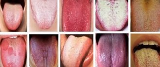
What does a healthy tongue look like?
A white-pink color of the tongue is considered normal. There are a number of other accompanying signs of his health:
- the longitudinal fold of the tongue is clearly visible;
- the papillae are clearly visible, but not hypertrophied;
- the edges are smooth.
The surface must be clean, although a slight coating on the tongue is acceptable.
For an adult, seasonal changes in the color of the tongue are possible:
- in winter, a slight yellowish coating on the tongue in adults can be considered normal if there are no other abnormalities - pain, increase in size, lack of taste;
- in summer, a light white coating, not localized, but over the entire surface, is also not considered a pathology.
A healthy child’s tongue is not much different from an adult’s tongue. One significant feature: it reacts to the slightest changes in the condition of the baby’s body - teething, the introduction of complementary foods, even a change in the brand of baby food. Therefore, plaque on a child’s tongue requires close attention.
Diseases that can be diagnosed by changes in tongue color
Not only the appearance of plaque, but also a change in the color of the tongue can tell a lot about the condition of the body as a whole. As mentioned earlier, a whitish coating in the middle part of the tongue will indicate diseases such as stomach ulcers, gastritis and duodenal ulcers.
The tongue will become dry due to dehydration, which can be caused by an intestinal infection, diarrhea or acute abdomen - peritonitis, appendicitis, internal bleeding, etc. Dryness of the tongue may well be combined with a bitter taste in the morning - this is quite likely due to disruption of bile secretion processes. In addition, a feeling of dryness of the tongue can signal both a disturbance in sugar metabolism and problems with the thyroid gland.
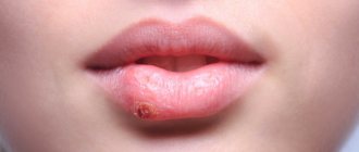
White plaques may often appear on the tongue, which can be easily removed if desired. This is a clear sign of dysbacteriosis, stomatitis or thrush. The cause of these diseases may well be improper treatment with antibiotics. But white or grayish-yellow ulcers, quite painful for a person, can occur due to injuries to the tongue or increased acidity of gastric juice.
Thus, it makes sense to look at your tongue from time to time - after all, its condition may well signal some problems in the body. And as doctors assure, a timely diagnosed disease is a serious step towards a healthy body.
What is plaque on the tongue?
The most numerous - filiform papillae - form a white coating on the tongue due to their structure:
- the lamina of the mucous membrane of the filiform papilla is covered with stratified squamous epithelium;
- this is a keratinizing epithelium that periodically exfoliates, covering the entire tongue with a light white coating;
- In case of any malfunctions in the human body, desquamation slows down and a layer of keratinized cells grows, which acquire different colors depending on what pathology led to the malfunction.
By the color of the plaque and where it is localized, diseases that have led to pathological changes in the tongue are judged.
Taste buds
On the surface of the tongue in its upper part, from the base of the root to the very end, there are many papillae and bulbs scattered, which help to feel the taste in its entirety.
The main types of papillae include:
- mushroom-shaped;
- leaf-shaped;
- groove-shaped;
- threadlike;
- cone-shaped.
But only the first 3 of them help to feel the taste sensations from food and foreign objects. The mushroom-shaped papillae are responsible for determining sweet taste, and the leaf-shaped papillae are responsible for determining sour taste.
Bulbs also have the ability to sense taste. It is in them that the glands pass and there are receptors that are able to distinguish between bitter and salty. Certain types of papillae and bulbs are densely located in different parts of the tongue.
The structure of the tongue, the location of the root.
Therefore, it is believed that the tip of the tongue detects sweet faster, and the root detects bitter. On the sides of the tongue, closer to its root, there are sections that allow you to identify sour, and closer to the end - salty. The central part of the tongue contains a minimal number of taste receptors and is neutral.
It is important to remember that the tongue is involved in stirring food while eating. The sensation of taste while chewing food causes a feeling of euphoria for individual foods in each person differently, depending on his preferences. Different pieces of food, falling on separate parts of the tongue, cause the perception of food in a new way, combining it into a complex combination.
Why does plaque appear on the tongue?
The tongue is a muscular organ that can tell a lot about the state of the body. It is soft and easily mobile, and has a pale pink color if the person is healthy. From time to time, plaque may appear on the mucous membrane, the density of which is often seasonal. This is explained by the fact that at different times of the year the body needs certain vitamins. For example, in summer the deposits are thicker and more saturated. At this time they may acquire a yellow tint.
Bacteria constantly accumulate on the mucous membrane of the tongue. These microorganisms are the cause of plaque and bad breath.
The following factors contribute to their intensive reproduction:
- excessive alcohol consumption;
- smoking;
- poor nutrition;
- infections and inflammatory processes;
- taking medications;
- chronic diseases;
- poor oral hygiene.
Since the tip of the tongue is mobile, it is cleaned more and, accordingly, there is less plaque here. At the root, its density is higher, since in this place there is contact only with the sky. Such manifestations are also possible with dysbacteriosis, vitamin deficiency, and improper hygiene.
Taste buds
The most interesting thing that is found in the mucous membrane of the tongue is the taste buds, which perceive sweet, bitter, sour and salty tastes (Fig. 5). The total number of taste buds in humans is more than 2,000[1]. On the tongue they are concentrated in the grooved, mushroom-shaped and foliate papillae, but are also present in the mucous membrane of the palate, epiglottis, and posterior pharyngeal wall. More than 50% of taste buds are located in the circumvallate papillae. With age, their number decreases. It has been experimentally established that the taste buds of the tip of the tongue perceive predominantly sweet, the side sections - sour and salty, and the root - bitter (see Fig. 1).
Taste buds are ellipsoidal bodies about 70 microns long. They have an opening - a taste pore, through which they open into the oral cavity. Each taste bud contains several sensory taste cells surrounded by supporting cells. Saliva with chemicals dissolved in it enters the taste pores and irritates the taste cells. Taste cells are equipped with nerve endings of the cranial nerves, through which information about the taste of food is transmitted to the brain. As a result of the analysis of the received information, a complex chain of reactions is launched in different parts of the brain, leading to different functioning of the digestive organs or the removal of substances harmful to the body that come from food.
The lifespan of taste cells is on average 250 hours; after such an interval, each cell is replaced by a young one, moving towards the center of the taste bud from its periphery. At the end of the taste cell, facing the lumen of the taste pore, there are 30-40 tiny microvilli - it is believed that they selectively perceive certain chemical substances.
Different people have significantly different taste sensitivity to different substances. The threshold of taste sensitivity can depend on the state of the body and change significantly during pregnancy, fasting, and various diseases. Sometimes about certain substances. With prolonged exposure to flavoring substances, the intensity of the taste sensation decreases (adaptation). Adaptation to sweet and salty foods develops faster than to bitter and sour foods. At the same time, adaptation to bitter increases sensitivity to sour and salty, and adaptation to sweet sharpens the perception of all other taste sensations.
In the process of evolution, taste was formed as a mechanism for selecting or rejecting food. In modern humans, taste sensations are combined with olfactory, tactile and thermal sensations, also created by food. It is worth noting that the “taste” of tobacco, tea, wine, and spices is mainly the result of olfactory sensations. This is confirmed by the “loss of taste” with a runny nose.
Types of plaque on the tongue
White plaque
A thin white coating is a common occurrence. You can especially notice it in the morning, when your teeth have not yet been brushed. Homogeneous white deposits occur in infants after feeding. This is also considered normal in older children.
The presence of other features may indicate certain ailments:
- an increase in plaque thickness is a symptom of prolonged constipation;
- elevated temperature and symptoms of intoxication - indicate infectious processes in the body;
- localization on the root of the tongue, its back – gastrointestinal diseases;
- placement on the sides of the tip of the tongue - pay attention to the kidneys.
A white coating with a cheesy structure, as well as dry mouth, indicate candidiasis (popularly called thrush). It often affects infants. The cause of the disease is weak immunity. To prevent thrush from spreading to the cheeks and gums, you should consult a doctor. He will prescribe antifungal medications. This disease is also possible in older children, but more often these are asthmatics or children with weakened immune systems. The disease may be accompanied by an unpleasant taste in the mouth.
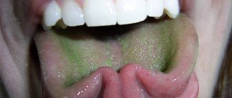
Gray, green and brown plaque
And if the deposits are not white, but of a different color - what is it? Each shade has its own characteristics:
- Grayish
is a common symptom of gastrointestinal diseases. This could be, for example, a stomach ulcer. But a grayish-white coating is not a deviation from the norm. - Brown
. Such deposits on the root of the tongue appear in chronic alcoholism. It also occurs in smokers and with lung diseases. - Green
is a rare occurrence. Occurs with different types of glossitis. The disease can develop as a result of taking antibiotics, steroids and other substances that reduce the body's immune strength.
Please note that the tongue often changes color after eating and drinking. For example, strong tea can turn it brown.
Yellow plaque
As already mentioned, a yellowish coating appears in the summer. You need to worry if its shade becomes saturated. You should pay attention to the following signs:
- Bright yellow color - the liver and bile ducts may be affected.
- Yellowness of the lower part of the tongue is a symptom of incipient jaundice.
- A thick yellow-green coating is a sign of improper functioning of the digestive organs and stagnation of bile. These disturbances in the body may be accompanied by the formation of a red plaque.
Sometimes yellow deposits indicate an increased amount of bile.
Black plaque
Black deposits on the tongue are very rare. More often this is one of the signs of a serious illness:
- serious disruption of the pancreas, gallbladder, and gastrointestinal tract;
- high blood acidity resulting from dehydration;
- cholera.
- There is such a thing as a “villous” tongue, when the papillae on it turn black and become hard. Such manifestations can be observed in smokers and people who abuse alcohol, as well as when exposed to certain organisms and medications.
Spotted plaque
Geographic tongue, when its mucous membrane is covered with uneven red spots, scares many. This condition occurs in people of all ages. There is no danger in it, and often it goes away on its own.
Today, science does not fully know what the appearance of such spots means. Each case is individual, so it is important to monitor your condition. For example, spots may occur due to an allergic reaction. But in most cases they are then present on the skin.
Functions and properties
The root of the tongue is located at the beginning of the pharynx and forms its basis. The root of the tongue performs many functions independently and as a single organ.
The main functions of the tongue root include:
- mixing food and pushing it down the throat;
- participation in swallowing, sucking food;
- perception of 5 basic tastes;
- sensitivity to the temperature of food consumed and any objects;
- participation in speech by determining the purity of individual sounds;
- tactile functions for the perception of various foods and objects;
- supporting the quality of work of the entire body;
- participation in the articulation and expression of feelings.
When chewing food, the tongue triggers the process of salivation and determines the beginning of digestion, so it is believed that it is also involved in the digestion process. The tongue can perform several functions simultaneously, and any disruption in its functioning affects the performance of its individual functions.
Determining the disease by the color of plaque
The first diagnostician on the condition of the tongue was the Russian doctor M. A. Nechaev, who in 1833 published the book “Recognition of diseases by changes in the language” in the printing house of Kazan University. Several generations of Russian doctors were grateful to him for this unique work, which helped to carry out diagnostics without additional instruments.
Today, the technique is widely used not only among traditional healers, but also among practitioners of traditional medicine. However, the diagnosis must be confirmed after a comprehensive examination carried out in a laboratory, or using ultrasound, CT, MRI, fluoroscopy, etc.
What do you pay attention to during this diagnosis:
- plaque color;
- its consistency.
As for the color of plaque, it can be:
- white;
- grey;
- yellow;
- greenish;
- bluish;
- brown;
- even black.
And the consistency can be:
- almost transparent;
- flaky;
- viscous.
All signs are compared, and a certain diagnostic verdict is made.
Organ characteristics
The root of the tongue is located deep in the mouth, namely in the recess of the lower part of the palate between the teeth. It, like the rest of the tongue, is covered with a mucous membrane. The root of the tongue is its base and consists of several muscles that continue into the body of the tongue. The tongue is considered the strongest muscle of the body and combines 8 different muscles that are woven together and complement each other.
On the outside, the tongue is covered with a mucous membrane, which consists of papillae of various types. It is the presence of papillae that helps to sense taste and starts the digestion process.
The papillary layer forms a rough surface on the tongue, on which plaque from food or bacterial activity is quickly deposited. It is the quality and color of plaque that signals disturbances in the functioning of the body.
Often, based on the condition of the tongue, it is possible to determine the presence of diseases of individual organs for further examination and diagnosis, therefore, during the examination, the therapist must note the color of the tongue.
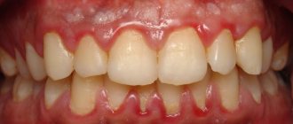
Diseases of the oral cavity and plaque on the tongue
Most often, the condition of the tongue depends on the conditions in the oral cavity. The presence of plaque may be due to:
- caries;
- stomatitis with fungal and bacterial etiology;
- periodontal disease – systemic damage to periodontal tissue (gums, bone and tooth ligament);
- glossitis - inflammation of the tongue that occurs as a result of mechanical damage to the organ, or as a condition accompanying other diseases;
- gingivitis - inflammation of the gums without damage to bone tissue.
The mucous membrane of the tongue reacts very sensitively to any problems in the oral cavity caused by inflammation, caused by bacterial or fungal infections.
They are diagnosed quite easily:
- A loose white coating indicates that a yeast-like fungus of the genus Candida has settled in the mouth.
- The presence of periodontal disease and gingivitis is determined by the condition of the gums.
- The presence of caries is accompanied by an unpleasant odor and putrefactive damage to the bone tissue of the tooth.
- Glossitis is accompanied by a burning sensation, salivation, pain and inflammation.
If everything is more or less clear with these signs, then plaque caused by systemic diseases is not so easy to recognize without special knowledge.
What diseases does plaque on the tongue foreshadow?
It is believed that the nature of the disease and its location can be determined by the color of the plaque:
- White plaque is quite acceptable if it is easily removed after hygiene procedures. If it lies in a thick layer and has a cheesy consistency, then this is a sign of fungal infection, intoxication, the presence of foreign bodies in the oral cavity - implants or dentures - and the allergic reactions they cause.
- A gray coating may indicate that a course of antibiotic treatment has been carried out or there are problems with the gastrointestinal tract. Most often these are ulcerative lesions of the stomach or duodenum. A decrease in general immunity can also be the cause of plaque of this color.
- A yellow coating indicates stagnation of bile or problems with the liver. It can also be observed with kidney damage, then its localization is at the edges of the tongue. Constipation also causes such plaque, in which case bad breath also appears.
- The green color of plaque occurs from excess bilirubin during hepatitis of various etiologies. This may also be a consequence of a viral infection.
- Brown plaque can be a consequence of gastronomic preferences - among lovers of brewed coffee and strong black tea. Heavy smokers also often have a brown coated tongue. Inflammatory processes in the gastrointestinal mucosa can cause such plaque.
- A bluish coating is a consequence of problems with the cardiovascular system. This may be coronary heart disease or chronic hypotension.
- A dark, almost black coating should alert you. This is a consequence of a serious pathology in the body - oncology, severe dehydration, rare Crohn's disease or cholera infection.
For adults, a constant coating on the tongue may mean that the person is a heavy smoker. It is difficult to find among smokers those whose organs have not been damaged by nicotine tar. This means that they are no longer healthy.
Innervation of the tongue
All the variety of functions of the tongue is provided by a finely structured nervous apparatus. 5 out of 12 cranial nerves are involved in the innervation of the tongue! One of them, the hypoglossal nerve, controls the activity of all the muscles of the tongue, and the rest carry information from the mucous membrane of the tongue to the brain.
In conclusion, it should be noted that the state of the tongue and its appearance reflect the state of the organism as a whole. Examine your tongue more often and you will see that this conclusion is correct.
A smooth, whitish-pink tongue without cracks or plaque indicates good health. “Coated tongue”, changes in its color and relief are signs of trouble in the body. A thick white, gray or yellow coating is observed in diseases of the digestive system. The red color of the tongue carries information about disruption of the cardiovascular and respiratory systems and some infectious diseases. With heart defects, the color of the tongue changes to bluish. Delirium tremens paints the tongue with dark stripes, making it “tiger-like.” Weak or, conversely, increased development of the papillae of the tongue indicates a violation of secretion
Author: Olga Gurova, Candidate of Biological Sciences, Senior Researcher, Associate Professor of the Department of Human Anatomy of the RUDN University
[1] Other mammals have a much larger number of taste buds. For example, in ruminants it reaches several tens of thousands.
Plaque in diseases of the gastrointestinal tract
The gastrointestinal tract, or digestive system, includes:
- oral cavity;
- esophagus;
- stomach;
- liver;
- gallbladder;
- pancreas;
- duodenum;
- small and large intestines;
- rectum and anal sphincter.
Any malfunctions in the organs of the digestive system cause plaque on the tongue:
- if it is concentrated in the area of the root of the tongue and has a gray tint, this means that the large intestine and rectum are affected;
- a thin yellow coating in the middle of the tongue indicates the presence of gastritis or gastroduodenitis, and a thick layer localized in the middle indicates its exacerbation;
- with cholecystitis - inflammation of the gallbladder - a yellow-brown coating appears, while the tongue itself is dry, bitterness and dryness in the mouth are felt;
- if there is a problem with the biliary tract, the plaque can take on a color from yellow to green; it is the greenish shade that indicates that not everything is in order with the biliary system;
- a bluish coating indicates an intestinal infection;
- a thick yellow coating in combination with heartburn, belching and a burning sensation indicates that pancreatitis has worsened - inflammation of the pancreas;
- a reddish-brown, and sometimes even black, coating may indicate oncological processes or abscess inflammation in the gastrointestinal tract.
In any case, plaque on the tongue is not the main sign of diseases of the digestive system. Only a doctor, having collected an anamnesis, can make the correct diagnosis.
Plaque for bronchitis and pneumonia
The area of the tongue immediately following its tip is an indicator of the health or disease of the respiratory system (bronchi and lungs). Based on the condition of this area, one can judge the presence of bronchitis or pneumonia:
- Red spots indicate that pneumonia or bronchial asthma is possible.
- A light film on the front of the tongue indicates the presence of a respiratory allergy or congestion in the lungs.
Plaque on the tongue caused by inflammatory processes of the upper and lower respiratory tract is not decisive in the diagnosis of these diseases.
Human tongue as an indicator of disease
Among other organs of the human body, the tongue is the most accurate sensor of the state of the digestive tract and an indicator of diseases of some internal organs.
Thus, the back of the tongue reacts to pathological processes occurring in the body, such as blood diseases, various types of infections, etc. As a result, a clearly visible coating appears on the tongue. But on the other hand, poor oral hygiene can also lead to plaque, decreased sensory function, and even inflammation.
A healthy tongue should have a pleasant pink color, normal size, moderate moisture and normal sensitivity. Any coating present on the tongue indicates a problem in the body. Therefore, you need to get rid of this phenomenon in any case, regardless of whether it becomes a solution to the problem or not. Ultimately, this will improve your oral health and reduce the rate of plaque formation.
Plaque due to oral chlamydia and thrush
There are two types of infectious diseases that affect the urogenital organs and the oral cavity. These are chlamydia and thrush. For candidiasis caused by a fungus of the genus Candida:
- a dirty white cheesy coating forms on the walls of the mouth and on the tongue;
- when mechanically cleaning the tongue from plaque, bloody discharge appears;
- an unpleasant putrid odor and taste appear in the mouth;
- treatment with special antifungal drugs is necessary.
Oral chlamydia shows a slightly different picture:
- thick sticky mucus forms in the nasopharynx;
- then it migrates to the upper and lower palate;
- only after this does it appear on the tongue, first in the form of spots, and later covering the entire tongue with a white pasty coating;
- At the same time, it has the smell of rotten fish.
It is diagnosed both by visual examination and by laboratory analysis of scrapings from the tongue and palate.
Taste analyzer and taste organ
As you remember, the analyzer of a feeling and the organ of this feeling are not the same thing. The sense organ is a peripheral section of the analyzer, and there is also a central section - a section of the brain in which information received from the periphery is processed. The center and periphery are connected by a conductive path. In our case, the cortical analysis of the sense of taste is performed in the temporal gyrus - this is the 43rd field on the Brodmann map.
So, the analyzer of any feeling is a sensory organ that conducts the path and area of the brain in which information from the sensory organ is processed.
What else can cause plaque on the tongue?
There are many other reasons that cause plaque on the tongue:
- Chronic alcoholism leads to the development of fatty hepatosis, and later cirrhosis of the liver. As you know, a dirty yellow or even greenish coating on the tongue is characteristic of people suffering from liver diseases. In addition, alcoholics are rarely concerned about body hygiene, much less oral hygiene. This further enhances the coating and odor on the tongue of a person suffering from alcoholism.
- Plaque can occur as a side effect of taking medications, mainly antibiotics. Taken orally, they kill the beneficial microflora of the small and large intestines, causing dysbiosis, accompanied by poor digestion and absorption of food. And this, in turn, leads to the formation of plaque on the tongue.
- Intoxication of any origin necessarily causes a coating on the tongue. Thus, cancer patients after a course of chemotherapy all suffer from a dirty-brown coating on the tongue, which is caused by toxic chemotherapy, as well as tissue breakdown products destroyed by cancer cells.
- Impaired immunity, especially if failures occur in that part of the immune system that is located in the intestines, also leads to the formation of plaque, because T-lymphocytes die, settling in the form of a yellowish coating on the tongue and intestinal walls.
In these cases, consultation with a specialized specialist is necessary.
Plaque on the tongue in children
In children from birth to 5 years of age, the immune system is so imperfect that a slight coating on the tongue is considered normal. Moreover, a rare baby has avoided thrush, which affects the oral cavity and tongue from the first days of life. But you need to know and be able to differentiate plaque on a child’s tongue in order to recognize dangerous infectious and autoimmune diseases in time and seek medical help:
- Thrush is characterized by a loose, cheesy coating on the tongue and oral mucosa. Cleansing causes the baby to cry because the papillae are hypertrophied and react painfully to touch.
- A dirty gray coating on a child's tongue may be an indicator of scarlet fever. This is an infectious disease that must be treated under the supervision of a doctor. With scarlet fever, the tongue gradually turns from dirty gray to scarlet, similar to strawberries, with characteristic dots along the entire surface of the tongue.
- A filmy coating covering the root of a child’s tongue indicates that he has diphtheria. This sign requires urgent hospitalization, because the disease develops rapidly and leads to suffocation.
- Black or dark brown plaque in babies can be caused by a latent form of diabetes, bacterial sore throat, or taking strong antibiotics.
- There is also such a thing as “geographical language”. It is also typical for young children. These are red spots scattered across the entire surface of the tongue against the background of a light white coating, making the picture resemble a map of the world. In this case, benign migratory glossitis is diagnosed. It occurs against the background of helminthic infestation, vitamin deficiency, acute infectious diseases, and exudative diathesis. Only a doctor can identify the cause of the disease, so you should contact him immediately.
All other causes of plaque on the tongue in children are not much different from adults. These are the same dysbacteriosis, gastritis and even stomach ulcers, problems with the liver and gall bladder.
Should you see a doctor?
Having a general idea of what type of plaque may be associated with a serious illness, it is worthwhile to be guided by it. It should also be taken into account that all serious systemic diseases, in addition to plaque on the tongue, have a number of formidable symptoms that cannot be ignored. It can be:
- pain;
- vomit;
- diarrhea;
- constipation;
- skin rashes;
- increase in body temperature, etc.
Plaque on the tongue must be taken into account along with other signs and contact a specialist for diagnostic measures.
Prevention and elimination of plaque
The main “commandment” for the prevention of this unpleasant phenomenon is compliance with hygiene rules and regular sanitation of the oral cavity. This concept includes:
- mandatory brushing of teeth in the morning and evening;
- using floss to clean the space between teeth;
- using a toothbrush with a grooved surface, which cleans the surface of the tongue in the presence of plaque;
- the use of mouth rinses, which help get rid of unfriendly bacterial microflora present in the mouth even in absolutely healthy people.
There are many more ways to sanitize the oral cavity in order to prevent unhealthy plaque on the tongue, which you should familiarize yourself with in more detail.
Internal muscles of the tongue[edit | edit code]
Internal muscles of the tongue
So-called internal muscles
tongues change the shape of the tongue. Their action depends on the combination of contractions of specific muscles. The antagonist of these muscles is tissue pressure, which increases with contraction of the internal muscles (water cushion principle).
- The superior longitudinal muscle
(
m. longitudinalis superior
) shortens and widens the tongue and raises its apex.
- The lower longitudinal muscle
(
m. longitudinalis inferior
) shortens and widens the tongue and lowers its apex.
- The transverse muscle of the tongue
(
m. transversus linguae
) narrows the tongue, stretches it and lifts its edges up.
- The vertical muscle of the tongue
(
m. verticalis linguae
) flattens the tongue, widens and shortens it.
Home[edit | edit code]
- Superior longitudinal muscle: root of tongue
- Inferior longitudinal muscle: root of tongue
- Transverse tongue muscle: edges of the tongue
- Vertical muscle of the tongue: lingual aponeurosis
Attachment[edit | edit code]
- Superior longitudinal muscle: tip of tongue
- Inferior longitudinal muscle: tip of tongue
- Transverse tongue muscle: edges of the tongue
- Vertical tongue muscle: inferior surface of the tongue
Innervation[edit | edit code]
- Hypoglossal nerve (XII pair of cranial nerves)
Movements of the tongue. Functional muscle tests[edit | edit code]
*
Clinical relevance
- When the hypoglossal nerve (XII pair of cranial nerves) is damaged, the tip of the tongue deviates towards the lesion.
- Tongue mobility may be limited by thickening and shortening of the lingual frenulum in systemic scleroderma.
Issues and comments
- The patient cannot perform some movements “on orders.” However, the function of the intrinsic muscles of the tongue can be assessed by changes in tongue movements.
You can test the function of these muscles in more detail by grasping the tip of the tongue with your fingers and resisting the test movement.
How to properly brush your teeth and tongue
At first glance, the simple procedure of brushing your teeth is so familiar that there is nothing to add. In fact, proper cleansing can protect you from a host of oral diseases and more. After all, the mouth is the “gate” for any viral and bacterial infection. Therefore, it would be useful to recall that:
- You must brush your teeth twice a day – morning and evening;
- Cleaning your teeth should be done from top to bottom for at least 3 minutes;
- Using the corrugated surface of the toothbrush, use careful movements without much pressure to clean the tongue in the direction from root to tip, after each movement the brush is rinsed with running water;
- the evening procedure includes cleaning the space between the teeth with a special dental floss;
- Finally, use an antibacterial rinse, rinsing your mouth thoroughly.
Ideally, you should brush your teeth after every meal.
Professional cleaning at the dentist
Even such thorough self-cleaning of the oral cavity is not enough to be sure that you will be free from periodontal disease or caries. From time to time it is necessary to contact a dentist so that he can carry out professional sanitation. Typically it includes:
- preventive examination and assessment of the condition of gums and teeth;
- removal of tartar mechanically or using ultrasonic devices;
- treatment with a special powder mixture to get rid of food pigmentation of teeth, typical for smokers, lovers of strong coffee and tea;
- final flossing to remove tartar fragments from the most difficult to reach places;
- polishing using a special paste to create the most even surface relief of the teeth.
It is recommended to carry out such cleaning every six months, in case of predisposition to caries and periodontal disease - once every 3 months.
Cleaning your tongue with a home irrigator
As an alternative to going to the dentist for professional oral cleaning, you can consider a home irrigator. This is a special device equipped with replaceable nozzles and a reservoir that supplies liquid under pressure to clean the space between the teeth. When choosing an irrigator, you must be guided by the following requirements:
- the number of attachments should be a multiple of the number of family members who will use it, because this is a means of individual use;
- It is highly desirable that the kit include devices for cleaning the tongue and dentures of any configuration;
- it is necessary that the device be equipped with a pressure regulator when supplying liquid, because everyone has an individual level of tooth sensitivity;
- It is better to choose a larger tank volume, this will allow cleaning more efficiently;
- It would be great if it was also equipped with a water supply regulator, that is, it could be a stream of water or a spray.
This device will save you the time and money needed to visit the dentist's office.
Using rinse aids
You can use rinses to clean your tongue only in conjunction with all other hygiene procedures. The choice depends on the condition of the gums and teeth:
- For loose, bleeding gums, you need to choose a rinse with a high content of fluoride and oak bark extracts.
- Coniferous tree extracts included in the mouthwash thoroughly sanitize the oral cavity, destroying bacteria.
- Zinc chloride, which is part of the mouthwash, helps keep teeth white and prevent the formation of tartar.
Using mouthwash ensures fresh breath.
Colloidal silver is a natural antiseptic
It is advisable to have colloidal silver in your home medicine cabinet, which is an excellent antiseptic and antibiotic. It destroys bacteria, fungi, and viral infections.
It can be used to treat your hands, mouth, and even be taken orally. The product is a suspension of silver microparticles in distilled water. They treat wounds with it, and rinse the mouth with the solution for any problems with the oral cavity, including plaque on the tongue.
Propolis tincture to cleanse the tongue
Propolis tincture, which can be purchased at any pharmacy, does an excellent job of sanitizing the oral cavity. It is used:
- for rinsing - prepare a solution at the rate of 15 ml of tincture per 100 ml of water and rinse your mouth after each meal;
- to clean the tongue - use undiluted tincture, apply to a tampon and clean the tongue from root to tip, changing the tampon each time.
Before you start cleaning with propolis tincture, you should test for an allergic reaction. Use a cotton swab soaked in the solution to clean a very small area of your tongue. Wait at least 12 hours for the reaction. If no manifestations of allergies occur, then you can clean the surface of the entire tongue.
Herbal decoctions to get rid of plaque on the tongue
Using herbal decoctions to sanitize the oral cavity is a great idea. But it is unreasonable to expect that simply rinsing will get rid of plaque on the tongue. Decoctions of medicinal herbs should only be used in combination with other cleaning methods. Herbal decoctions are ideal for rinsing the mouth:
- from oak bark;
- calendula;
- sage;
- chamomile;
- peppermint;
- lemon balm.
It is not difficult to prepare such a decoction:
- Buy a herbal mixture or use a monocomposition at the rate of 1 teaspoon of herbs or herbal mixture per 100 ml of water.
- Pour boiling water over it and leave over low heat, avoiding boiling, for 10–15 minutes.
- Cool and strain.
The decoction can be used to rinse your mouth after mechanical sanitation.
Method of mechanical tongue cleaning
Mechanical methods of getting rid of plaque on the tongue include:
- cleaning with the grooved side of a toothbrush or a special brush;
- the same action using a special scraper in the form of a plastic ring;
- cleansing with a teaspoon or a special scraper that resembles one.
The method of application is simple - you need to scrape off the plaque from root to tip, each time rinsing the scraper under running water.
The main thing here is not to overdo it. Do not press too hard on the tongue to avoid damaging the papillae and causing bleeding. After mechanical cleaning, be sure to rinse your mouth with a decoction of herbs, mouthwash or colloidal silver solution.
Cleansing the mouth with vegetable oil
This method of cleansing the oral cavity from any infectious lesions was known to our forefathers. It will help not only get rid of plaque, but also solve problems with caries, periodontal disease, and gingivitis. There are no contraindications for it, and the benefits will be obvious after the first procedure. The essence of the method is as follows:
- In the morning on an empty stomach or in the evening, 3 hours after the last meal, take 1 tbsp into your mouth. a spoonful of unrefined vegetable oil.
- Next, for 10 minutes, you need to rinse your mouth with this oil through closed teeth, without swallowing it.
- Ideally, the oil should turn white or dirty gray depending on your health.
- The oil is spat out, and the mouth is rinsed with water or herbal decoctions.
This procedure brings tangible results - bad breath disappears, plaque disappears, gums become stronger, and teeth become healthy and shiny. General well-being improves.
How to use saline or soda solution
You can also use a saline or soda solution only in combination. This procedure alone will not bring the desired result. But this solution is quite suitable as a rinse.
It is enough to dissolve 1 teaspoon of soda or ½ teaspoon of salt in 200 ml of boiled water, cool to room temperature and rinse the mouth after mechanical cleansing of plaque.
You shouldn't self-diagnose. If you have any suspicious symptoms, consult a specialist.