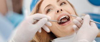
A number of dental procedures are impossible without laboratory diagnostic results.
Sometimes it is difficult to establish a diagnosis through examination of the patient by a dentist and using hardware diagnostic tools. In case of planned surgery, tests are also mandatory.
Laboratory diagnostics in dentistry can include both general clinical tests and complex morphological and biochemical screening.
This allows you to assess the work and condition of individual systems of the patient’s body, as well as predict the effectiveness of therapy, the approximate time of treatment and recovery.
At the first stage of making a diagnosis, the dentist examines and interviews the patient about problems with the dental system, listens to his complaints and the history of the development of the disease. In some cases, this is enough to make a diagnosis. But most often, the dentist refers the patient for an additional X-ray examination or orthopantomogram (second stage).
In 90% of cases, the data obtained is enough for a correct diagnosis and development of a treatment plan. In difficult cases, the doctor prescribes laboratory diagnostics, as well as hardware examination methods - MRI, CT, ultrasound. Popular diagnostic measures are general clinical blood and urine tests, as well as serological and microscopic examination.
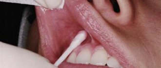
Saliva examination
To make a diagnosis, it is sometimes necessary to determine the amount of saliva and the rate of its secretion, calculate the pH value, buffer capacity, and check the mineral composition. For example, a deficiency or excess of one or another element can provoke unfavorable conditions for enamel.
No less important are the biochemical parameters of the oral fluid:
- indicators of anti- and pro-oxidant systems;
- free radical oxidation values;
- the amount of enzymes in saliva, etc.
How complex digital diagnostics are carried out
The most popular area for digital diagnostics is complex dental implantation with the installation of a complete fixed bridge prosthesis 1-3 days after surgery. Getting teeth back so quickly is quite a difficult task. After all, as a rule, the remains of patients’ own teeth are in poor condition or missing, and the bite is disturbed. The implant surgeon and orthopedist need to re-create the balance of the entire jaw system. But it is impossible to do this right away; muscles and joints need time to rebuild. That is why there is a so-called adaptation period or adaptation prosthesis, through which orthopedists set the vectors of change in combination with the necessary balance of load on the implants. As a result, in addition to aesthetics, prostheses must normalize the position of the joint and the functioning of the facial muscles - and such a large-scale task cannot be solved without careful functional diagnostics and planning.
Comprehensive functional diagnostics is important not only when installing an adaptive fixed prosthesis, but especially when replacing prosthetics. A permanent prosthesis on implants lasts for decades, but only if it functions without overload.
However, a detailed analysis of the initial situation is useful in any orthopedic treatment: when installing veneers, crowns, bridges, etc. Any intervention in the shape of the teeth shifts the balance - the chewing load is distributed slightly differently, the facial muscles are tensed differently, and the movement of the joints changes. Will these changes be for the better or will they lead to structural failure and complications? This can be done
Types of laboratory tests
Cytological
They help determine the nature of the cellular material, detect caries pathogens or specific cells that cause a pathological process in the oral cavity. Exfoliative material is obtained from the surfaces of ulcers, areas of leukoplakia, and from surgical discharge. Aspiration – from tumors, lymph nodes, foci of inflammation. In addition, swabs are used for research. The material is fixed, stained and examined under a light microscope for its constituent components.
Mycological and microbiological
Biomaterial is taken from areas of damage to the tooth or mucous membrane. The objective of the study is to obtain the most detailed information about the microecology of the oral cavity, salivary gland duct, periodontal pocket or other biological niche. These methods help to identify the causative agents of the pathological process and determine the sensitivity of microorganisms to antibacterial agents.
Virological
This group of methods is aimed at identifying traditional pathogens that cause the most common diseases of the oral mucosa. The presence of specific IgG and IgM antibodies in certain reactions will indicate that the disease is viral in nature. During acute infection and during relapse, they manifest themselves differently, which makes it possible to differentiate the stage of the pathological process.
Immunochemical
This includes various research methods, including the indirect enzyme immunoassay method. It is used to determine the titers of specific antibodies to fungi, viruses and bacteria in saliva and blood serum.
DNA polymerase
The material being studied is tested using high-precision methods for the presence of DNA pathogens.
Immunological
The methods are focused on checking the level of general (systemic) and local immunity of the patient and allow further adjustment, for example, of the treatment of pulpitis of primary teeth depending on the immune response. Instead of skin tests, today laboratories often resort to enzyme immunoassay methods.
General clinical laboratory tests are also carried out to diagnose dental diseases. They study the physicochemical and microscopic properties of blood, urine, feces and other fluids and separated substrates from the body.
Diagnostics in dentistry
The primary task for a dentist is to make the correct diagnosis for the patient. The diagnosis is the starting point for planning the treatment process, and the achievement of a successful result depends on its accuracy.
Why is accurate comprehensive diagnosis so important?
Nowadays, some patients perceive dentistry in a distorted way. Among the reasons we can highlight competition between clinics that, in search of patients, are ready to promise everything at once. In this case, insufficient time is devoted to diagnosis, and the correctness of the diagnosis remains in question.
Doctors are also contributing to this negative trend. Some of them quickly make a diagnosis based only on images or a simple examination, and are ready to immediately begin treatment. This practice is common not only in therapeutic dentistry, but also in prosthetics and implantation. One can only imagine what risks exist for the patient without a full diagnosis, and what complications may arise as a result of such an approach.
When contacting a general practitioner at a clinic, we all understand the appointment of many tests and images, as well as referrals to other specialists. Why shouldn’t dentists adopt this approach? We know well that everything in the human body is interconnected. The dental system also interacts with other systems of the body, so simple dental treatment can affect them as well. It can be difficult to establish the root cause of dental diseases without additional diagnostics, because by the time the patient comes to the clinic, his condition may worsen under the influence of psychosomatics.
Thus, thanks to the diagnostics carried out in each specific case, the correct diagnosis can be made. This is the only way to confidently exclude diseases of the respiratory system and spine, as well as neurological diseases. An individual approach to each patient includes personal communication with the dentist to establish contact and clarify the medical history. Thanks to this, the upcoming treatment will take place in an atmosphere of mutual trust and cooperation.
Palpation
One of the standard diagnostic methods is muscle palpation. Using this method, you can analyze the work of the muscles of the head and neck. When comparing muscle function with bilateral palpation, dysfunctions in the functionality of the muscular system can be identified. Thus, having basic anatomical knowledge, the dentist diagnoses problems of the dentofacial apparatus as a whole.
Bite analysis
Wax plates are used to record occlusion and articulation indicators in dentistry. They are used to determine the movements of the lower jaw and the contact of teeth at rest. Using plates is a simple and effective way to evaluate the closure process. However, in addition to this, a general examination is also important, during which you can find out about the condition of the periodontium and teeth, the presence of removable structures and restorations. Thanks to this, the dentist can draw conclusions about oral hygiene and the condition of the mucous membrane. This information is extremely important because malocclusions are often accompanied by periodontal problems.
Photo protocol
Currently, photography is widely used in diagnostics. To optimize the process, a protocol has been developed that the specialist follows.
For a complete diagnosis, it is necessary to take two groups of images – extraoral and intraoral.
Extraoral photographs record the position of the patient's head in individual positions. Based on these images, one can judge the characteristic features of appearance, asymmetry and facial features, muscle tension in the neck and head.
Intraoral photographs can reveal the position of the jaws, the condition of the dentition and other features.
If you have a problem similar to that described in this article, be sure to contact our specialists. Don't diagnose yourself!
Why you should call us now:
- We will answer all your questions in 3 minutes
- Free consultation
- The average work experience of doctors is 12 years
- Convenient location of clinics
Single contact phone number: +7
Make an appointment
Diagnostic models
One of the most important components of the diagnostic process is the production of models based on casts. At this stage, an articulator is used, which shows the position of the upper and lower jaws with maximum accuracy. The articulator is adjusted individually in each specific case, thanks to which the dentist receives comprehensive information about the dental system.
When diagnosing, an important role is played by the analysis of information about the patient’s bite. Maximum contact between teeth is not informative enough for the doctor. The primary task is to calculate the unforced posterior position of the mandible. Thanks to this, it is possible to determine the real relationship of the jaws and identify the source of the detected pathology.
Condylography
An effective method for diagnosing TMJ is condylography. It allows you to calculate the characteristics of movement in a joint. By analyzing the information obtained during condylography, the dentist can determine the presence of pathologies and make the correct diagnosis. Of course, data from other diagnostic methods are also taken into account.
Due to its non-invasiveness, condylography is an easy-to-use method. However, it requires special knowledge and skills from the dentist, without which it is impossible to obtain accurate data.
It is important to mention that condylography data is used both to clarify the diagnosis and for subsequent orthopedic work.
Other diagnostic methods
Currently, the following types of diagnostic methods are used in dental practice:
- Computed tomography;
- Magnetic resonance imaging of the central nervous system;
- Orthopantomogram;
- Teleradiogram, etc.
Let's talk briefly about some of these methods.
Teleradiogram
This method allows you to obtain accurate data on the structure of the skeleton and soft tissues. It is carried out according to a standard scheme, which increases the efficiency and effectiveness of treatment. In reconstructive dentistry, the importance of TRG analysis cannot be overestimated.
Magnetic resonance imaging of the HFNS
MRI is one of the most important methods for diagnosing dysfunctions of the HFNS. It helps to analyze the condition of the joint and soft tissues with maximum accuracy. However, the effectiveness of tomography directly depends on the qualifications of the doctor and his ability to interpret the image.
CT scan
Computed tomography is another common diagnostic method. It is used when making an initial diagnosis and in preparation for dental implantation.
Scanning facial parameters using the Proface scanner
The final stage with the participation of the patient is scanning of facial parameters. There are two options here - use a facial scanner and/or a special application for Mac OS on the iPad.
The Proface scanner or program creates a three-dimensional model of the patient's appearance. For this purpose, a specially developed craniometric analysis algorithm is used (craniometry is a technique for measuring the skull). That is, many points of the skull are calculated - due to this, a copy of the patient’s face and all the bones of the head is transferred to the computer.
In addition, photometry is carried out - photographing the face is necessary to evaluate the result “live” and maintain a digital photo record.
An example of photometry is photographing the face from all angles before and after dental implantation with the installation of a fixed prosthesis immediately
We approach diagnostics wisely! We use the latest developments and artificial intelligence! We make an accurate diagnosis and find the best solution to your dental problems.
Sign up for a consultation
Photogrammetry
This technology stands somewhat apart from optical scanning. It offers high performance because it measures the image, not the object. Other benefits include:
- high accuracy;
- strict methods of processing measurement results;
- the ability to study stationary and moving objects;
- remote nature of measurements.
Monophotogrammetry with structured illumination is most often used. The latter represents a certain known pattern projected onto an object. To build a three-dimensional model, they take the distortion when projecting onto the object.
Study of muscle activity using a myograph
A myograph is a device that helps to study the bioelectrical activity of the muscular system. We use a Protens device, which is both a myograph and a tens device. That is, it allows not only to obtain data on the state of certain muscle groups in the facial area, but also to adjust activity and reduce tension.
The device is very miniature - it is fixed on the neck and has sensors that are located in the area of the temples and temporomandibular joints. To take data, you just need to move your lower jaw for a few minutes and just talk to the doctor. The device reads the data and transfers it to the computer, loading it into a special program.
Are there any disadvantages to digital diagnostics?
The disadvantages of high-precision digital solutions include their low prevalence due to the high cost of the equipment, as well as the time spent by the doctor. The technologies are relatively new and are just beginning to come into practice. There are still few doctors who have a full range of equipment and can carry out the analysis inside and out. Most are just learning new products and using only individual devices - for example, myographs or face arcs. That's why you won't see them in every clinic.
Smile-at-Once is so far the only clinic in Moscow with a full diagnostic range of devices and programs for planning orthopedic treatment and creating prosthetics.
How to identify “hidden caries”
Modern diagnostic methods in dentistry make it possible to identify even hidden forms of dental caries. It is often almost impossible to diagnose caries in the early stages of development without using auxiliary equipment. A breakthrough in diagnosing this disease was the creation of the German company KAVO - the Diagnodent device.
The advantage of the device is its safety; the device can be used to detect caries in both adult patients and children.
Additional Information! Diagnodent allows you to diagnose pathology with an accuracy of up to 90%
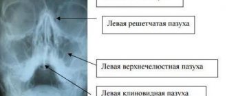
CT scan
This method, which is based on the exponential law of attenuation of radiation for absorbing media, allows one to obtain many points with different optical densities. This is necessary to assess the condition of hard tissues that cannot be examined visually. During CT scanning, teeth, bone and skin surfaces act as a regulator of optical density.
An even more accurate method, compared to CT, is optical scanning (up to 5 microns, which is much more accurate than 300 microns in tomography). This technology is necessary for visualizing soft tissues, including the gingival surface, in a 3D scene of the dentofacial system.
Optical strip scanning
This technology shows better results compared to the above. A special grid or stripes are projected onto the object. Based on their distortions, the relief of the scanned surface is determined. In this case, it is enough to determine the coordinates of the object’s surface using one camera and additional structured lighting. This approach is most often used in modern orthopedic dentistry.
Structured light can be different - laser, infrared, visible. The main condition is automatic recognition of these lines by software. The known distance between the projected stripes allows you to calculate the coordinates of the surface points. The more lines, the more accurate the result will be. The more cameras are used, the higher the scanning speed will be.
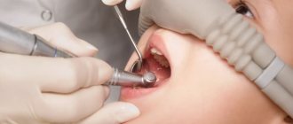
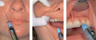
Spot X-ray
An X-ray picture of a separate tooth is most often necessary in order to monitor the treatment process when an accurate diagnosis has already been established.
In our clinic, targeted images are performed using a radiovisiograph - a modern digital X-ray machine, which differs from its predecessors in a much lower radiation dose and the ability to display the obtained data directly on the monitor screen. This allows X-ray examinations to be performed as many times as necessary (up to 60 times a year).
The results of the study are saved in the patient’s electronic record - this allows you to monitor the dynamics of treatment of the disease, and they can also be copied to any digital medium.
The study is carried out as follows: a miniature sensor is placed in the patient's mouth, which looks like a tiny plate and is connected to a computer using a wire. The research process takes only a few seconds. The data instantly appears on the monitor screen, and if necessary, you can immediately take a second photo. Thanks to computer processing, it is possible to enlarge or highlight individual fragments of the image and determine the density of bone tissue.