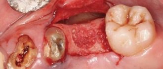
A human tooth is a fairly small part compared to all the other bones in the skeleton. However, it can be considered the most complex structure of all the solid parts of the human body, because each tooth, unlike other bone tissues, protrudes outward and is in direct contact with the external environment, while also bearing the chewing load. And when patients come to the VivaDent clinic with certain problems, dentists and orthodontists try to preserve (or reconstruct) the teeth as much as possible as they should be in their natural healthy state.
If you compare a tooth to an iceberg, then it also has two parts: visible in the mouth and invisible, deepened in the gum tissue. The upper part is called the coronal part (or simply the crown - by the way, the same name is given to a dental prosthesis without a part inserted into the gum), and the lower part is called the root part.
Central lower incisor
Average age of eruption: 6-7 years
Average age of root formation: 9 years
Average length: 20.7 mm
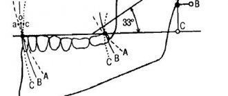
Narrow and flat in the labial-lingual direction, the lower central incisor is the smallest adult tooth. Radiologically it is visible in only one projection and thus appears more accessible than it actually is. The crown, narrow in lingual projection, has a limited area for access. For gentle access formation, a fissure bur and a spherical bur No. 2 are used. The access cavity should be oval and performed on the lingual side.
The lower central incisor often has two canals. One study reported that 41.4% of mandibular central incisors studied had two separate canals, of which only 1.3% had two separate apical foramina [1]. After completing the access formation, the doctor must examine the tooth cavity to identify an additional canal. Endodontic treatment failures in the lower incisor area are most often associated with an unidentified canal, usually in the lingual location. To create conditions for the straight entry of endodontic instruments into the additional canal, access can be expanded in the incisal direction.
There is a great danger of perforation of the vestibular wall, but it can be avoided if the doctor remembers that it is almost impossible to perforate in the lingual direction, since the bur axis is in contact with the incisal edge. The slit-like lumen of the canal is so common that it can be considered a normal variant, and this requires special attention when cleaning and shaping.
Lateral perforations and pulp anatomy will be discussed in an illustration of the lower second molar.
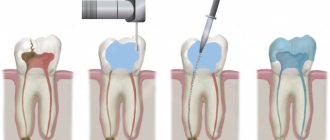
What does the crown of a tooth consist of?
The part of the tooth that rises above the gum is not a monolithic piece made of homogeneous bone material. It is something like a nut in a shell. On top of the tooth is covered with a harder layer designed to protect against various influences - this is tooth enamel. And underneath it lies a softer and more vulnerable layer - it is called “dentin”. And even deeper, under the dentin, going into the root part, lies the “core” of the tooth - a cavity called the pulp.
Let's consider each component separately.
Enamel
Tooth enamel is de facto one of the hardest tissues inside the human body. Even large bones, designed to bear a load of tens of kilograms, are actually looser in structure than tooth enamel - after all, they do not need to constantly encounter various bacteria, constantly grind anything foreign (that is, food), and even collide with antagonist teeth.
Healthy enamel has a light (although not completely snow-white) color and a glossy surface texture; it is very smooth and dense.
Dentine
Dentin is also a type of bone tissue: that is, in an adult, dentin, like enamel, does not grow or change (unless we talk about cases of carious lesions). It is looser and more fragile compared to enamel - but it is still a “non-living” material that does not have nerves or capillaries. Dentin is something like a backup protective layer between the enamel and pulp.
Pulp
Under the dentin layer in the tooth there is a cavity, securely closed on all sides by hard “walls”. But this is not an “air bubble”, but a kind of chamber where the finest blood vessels and nerve fibers are concentrated. The pulp is very necessary during the period when the tooth is growing - all nutrients are transferred to it through this living core.
However, when a person becomes an adult and his teeth are finally formed, the role of the pulp ceases to be so important - it ceases to nourish anything. However, it may still be useful: after all, if the patient feels toothache, it means that it is the nerve fibers in the pulp that make it known about some kind of inflammation. However, when treating pulpitis, dentists often remove the pulp with all its contents - the adult patient’s tooth does not suffer from this, either functionally or aesthetically, and in the future such a pulpless tooth will definitely not cause pain.
Lateral lower incisor
Average age of teething: 7-8 years
Average age of root formation: 10 years
Average length: 21.1 mm
Very similar to the lower central incisor, so access cavity preparation follows the same principles.
Their similarity can cause rare but serious errors. Hasty installation of a rubber dam, identical fillings and carelessness can lead to preparation of the access cavity on the wrong tooth. This error can be prevented by marking the vestibular surface of the tooth with a felt-tip pen before applying the rubber dam.
Trauma, periodontal disease, carious lesions and malocclusion can lead to obliteration of the canal. When moving apically to identify the orifice, great care must be taken to prevent unnecessary destruction of the crown and root. Labial perforations were discussed in Illustration IX. If the bur is not oriented along the long axis of the tooth, there is a risk of lateral perforation. The situation becomes more complicated with the traumatic loss of an anatomical crown. Without anatomical landmarks, a lateral perforation can be easily made when moving in a coronal direction. To prevent this, access is carried out without a rubber dam so that the root can be palpated.
Lateral perforation with endodontic files and Gates-Glidden burs is facilitated by the presence of slit-shaped canals with a narrow, hourglass-shaped cross-section. To avoid vertical fracture along the approximal root wall, minimal expansion and preparation of the space for the abutment pin is indicated.
Apical curves and accessory canals are common in the lower incisors.
Grinding food
The human stomach is capable of fully digesting only finely ground food, which is abundantly moistened with saliva. For this purpose, there are premolars and molars in the mouth, which act like millstones - they grind large pieces of food, while simultaneously mixing it with the secretion of the salivary glands.
The absence or loss of one of these teeth leads to the fact that unprocessed large particles of food enter the gastrointestinal tract, which are difficult to respond to enzymes and can cause various chronic diseases of the stomach and intestines.
Mandibular canine
Average age of teething: 9-10 years
Average age of root formation: 13 years
Average length: 25.6 mm
The canine of the lower jaw is more powerful and significantly wider than the incisors in the mesial-distal direction. It rarely causes treatment problems. The atypical form with two roots can be problematic, but is rare.
The access cavity is oval and can be expanded in the incisal direction to facilitate vestibular-lingual access. In the cervical region the canal is oval, in the middle third it is rounded. To completely clean its walls, directed instrumental action is necessary.
If there are two roots, one of them will always be easier to instrument. The other canal must also be opened and funnel-shaped in accordance with the first to prevent dentine filings from entering it and impairing access. Pre-bending the instruments during the initial approach will allow the clinician to walk along the walls of the buccal or lingual root until the tip of the instrument enters the orifice. Once a difficult-to-reach canal has been identified, every effort must be made to shape and create a funnel-shaped orifice to keep access open.
Indicative
Teeth are an indicator of the general condition of the body. The presence of any metabolic disorders, chronic diseases of the endocrine, digestive and other systems, vitamin deficiencies, unbalanced nutrition - all this negatively affects the condition of the dentition - the teeth change their shade and begin to crumble.
Therefore, caring for your smile includes not only regular hygiene procedures and visits to the dentist’s office, but also treatment of concomitant pathologies.
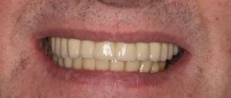
As you can see from the article, teeth are not only about comfortable eating. The loss of at least one element of the dentition is fraught with possible problems with digestion, speech, as well as the development of complexes against the background of a cosmetic defect.
First lower premolar
Average age of teething: 10-12 years
Average age of root formation: 12-13 years
Average length: 21.6 mm
Often considered a mystery to endodontists, the mandibular first premolar, with its two canals separating at different levels of the root, can be very difficult to machine.
The crown consists of a well-developed buccal cusp and a small or almost non-existent lingual enamel protrusion. The approach is performed buccally from the central sulcus and directed along the long axis of the root to the central cervical region. The oval-shaped pulp chamber is opened using fissure burs with a cutting apex and elongated spherical burs No. 4 or 6. In teeth with one canal, the pulp cavity in the neck area has an almost circular cross-section, and in teeth with two canals it is oval.
One study reported that “at least 23% of first mandibular premolars have a second or third canal”[17]. The canals can split almost anywhere in the root. Due to the lack of direct access, cleaning, shaping and filling these teeth can be extremely difficult.
In a recent study, Vertucci [13] showed that the first lower premolar has one canal at the apex in 74.0% of cases, two canals in 25.5% and three canals in the remaining 0.5% of cases.
Classification and frequency (%) of canal types in first and second lower premolars
By Vertucci, F. J. Am. Dent. Assoc. 97:47, 1978.
How to make teething easier
What measures should be taken to relieve toothache and other symptoms accompanying the “birth” of the upper and lower canines? Gentle massage of the gums surrounding the erupting tooth. The procedure is carried out 1-2 times a day, the duration of manipulations should be no more than 2-3 minutes. Auxiliary products: clove or chamomile oil, a piece of ice, special dental gels with analgesic and anti-inflammatory properties.
You can purchase comfortable teethers in pharmacies and specialty stores. These are modern designs filled with distilled water. If you place them in the refrigerator for a while, in addition to their main functions, they will cool and soothe the inflamed gum.
Eruption of the gum is a painful process, often accompanied by hyperemia, swelling, inflammation of the surrounding gums, and increased body temperature
Special anesthetic gels help ease the eruption of front teeth. Among such products, you need to pay attention to Kalgel, Dentinox, Cholisal. The compositions are applied directly to the inflamed gums 1-2 times a day, the anesthetic effect appears a few minutes after the procedure.
The use of homeopathic Viburkol suppositories (sold in pharmacies) allows you to reduce low fever, soothe and anesthetize soft tissues. The suppositories begin to act no earlier than 20 minutes after administration. If the body temperature has exceeded 38 degrees, the baby is given antipyretic drugs (syrups, tablets) or suppositories with paracetamol are used (on the advice of a doctor).
Proven folk remedies can also help cope with the unpleasant sensations associated with teething in babies. In particular, you can relieve pain, stop inflammation, and cope with swelling and hyperemia of the gums with the help of medicinal rinses. The best choice is decoctions of oak bark and chamomile (1 tablespoon of dry plant material per glass of water).
To alleviate the general condition of a child with a runny nose (a symptom accompanying the eruption of a gastrointestinal tract), you can (on the recommendation of a pediatrician) use vasoconstrictor drops - Nazivin, Otrivin, Nazol baby. It is worth remembering that the total duration of use of such formulations should not exceed 7 days.
Second lower premolar
Average age of teething: 11-12 years
Average age of root formation: 13-14 years
Average length: 22.3 mm
The second lower premolar, which is very similar in crown shape to the first premolar, has a less complex root.
Its crown has a well-developed buccal cusp and a much better formed lingual cusp than on the first premolar. The access is made slightly oval, wider in the mesial-distal direction. They begin to form access in the central sulcus with a fissure bur with a cutting apex, and then expand and form the contour of the burr hole with spherical burs No. 4 and 6.
Researchers reported that only 12% of mandibular second premolars studied had a second or third canal [17]. Vertucci [13] also showed that second premolars had one apical foramen in 97.5%, while only 2.5% of the teeth examined had two foramina.
An important circumstance that should not be forgotten is the anatomical location of the mental foramen and the vessels and nerves passing through it. Due to the proximity of these structures, an acute inflammatory process in the area of the lower premolars can cause temporary paresthesia. The exacerbation of the pathological process in this area is more severe and resistant to conservative treatment than in other areas.
What is around the tooth?
The tooth does not just sit in the soft flesh of the gum, held only by its tissue. In a healthy state, the tooth interacts with both the nervous and circulatory systems of the body. In addition, the roots of the tooth fit into their corresponding “slots” in the jawbone. Therefore, the tissues surrounding the tooth are not just “pulp”, but its functional support and protection: the gums and periosteal tissues hold the tooth in its intended position, conduct blood to it through the vessels and connect the nerve endings of the dental unit with the entire nervous system.
The root of the tooth is held in the jaw by a bonding material - natural dental cement. The tissue surrounding the root is called periodontium. And the notch in the jawbone where the tooth is inserted is the alveolar foramen, or tooth socket.
Archaeologists have long known this fact: when human remains are discovered, it is the teeth that usually turn out to be the part of the skeleton that is preserved better than other bones. Even when, after thousands of years, skulls are destroyed, the teeth from them are often returned to scientists of our time unchanged - in the form in which they were during the life of a person or other creature.
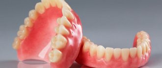
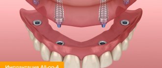
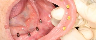
All this suggests that with proper daily care and timely treatment using modern technologies, the teeth of each of us have every chance of being preserved into old age. And, unlike our ancestors who lived centuries and millennia ago, people of the current generation have every opportunity to come to the VivaDent clinic and use implantation and prosthetic technologies if any of the teeth could not be saved.
First lower molar
Average age of teething: 6 years
Average age of root formation: 9-10 years
Average length: 21.0mm
The mandibular first molar erupts earlier than other permanent teeth and most often requires endodontic treatment. It usually has two roots, but sometimes three roots are found, with two canals in the mesial root and one or two canals in the distal root.
The distal root is easily accessible for endodontic cavity preparation and mechanical treatment.
The doctor can directly see the opening(s) of the canal. The distal root canals are wider than those of the mesial root. Sometimes the mouth is wider in the buccolingual direction. This indicates the presence of two channels or a slit-like channel with a complex network-like configuration that can complicate cleaning and shaping.
The mesial roots are usually curved, with the greatest curve in the mesiobuccal canal. The orifices of the canals at the bottom of the pulp chamber are usually clearly separated from each other and located buccally and lingually relative to the upper tubercles.
The tooth often undergoes extensive filling. It almost always experiences a strong chewing load, so the coronal pulp cavity can be obliterated. It is easiest to identify the mouths of the distal canals.
Then the mouths of the mesial canals are found, which will be located in the above locations in the same horizontal plane.
Since the mouths of the mesial canals lie under the mesial tubercles, they may not be detected during normal cavity preparation. In this case, to determine their location, it is necessary to remove the hard tissue of the tubercle or filling. During access preparation, overhanging molar cusps need to be ground down [15]. Remember that this tooth, like all other lateral teeth, after endodontic treatment requires complete restoration of the entire occlusal surface area. Therefore, to identify anatomical landmarks and orifices, it is better to make a wider access cavity than to skip one or more channels for the sake of “sparing” preparation, which may cause failure.
Skidmore and Bjoradal [11] found that approximately one third of the mandibular first molars examined had four root canals. If there are two canals, “they either remain separate with separate apical foramina, or unite and form a common apical foramen, or communicate with one another by transverse anastomoses partially or completely... If the tooth, instead of the usual triangular shape, had a more rectangular shape, this would allow better vision and explore a possible fourth canal in the distal root.”
In the area of bifurcation of the roots of the lower molars there are several orifices of additional canals [9]. They are usually impossible to clean and shape, and are rarely visible except incidentally on radiographs when they are filled with root cement or heated gutta-percha during treatment. It would be correct to assume that if irrigation solutions tend to clean the canal from protein decomposition products, then the area of root bifurcation in the pulp chamber must be thoroughly cleaned (remove denticles, etc.) so that the solutions can reach the small mouths of the canals.
All infected dentin, leaking fillings, and pulp denticles must be removed before endodontic treatment begins. It is recommended to completely cover the cusps with a restorative structure after endodontic treatment.
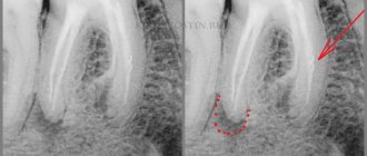
Tooth roots
The processes of bone tissue - the roots of the tooth, hold it in the jaw - and in this they are very similar to the roots of plants. But, in addition, in each root process there is a void inside - this is the root canal. The same nerve fibers and capillaries that go higher into the pulp, and from below are brought out into the jaw through a tiny hole at the tip of the tooth root, are laid in it, like wires in a protective cable. This hole is called apical.
Depending on the type and size of the tooth, there may be one root, or there may be two.
Second lower molar
Average age of teething: 11-13 years
Average age of root formation: 14-15 years
Average length: 19.8 mm
The crown is somewhat smaller in size than the first molar, and the more symmetrical second lower molar is characterized by a close arrangement of roots. The roots gradually converge distally and have closely spaced apices.
Exposure is made at the mesial portion of the crown with access extending only slightly distal to the central sulcus. After trephination of the cavity with a fissure bur with a cutting apex, an elongated ball-shaped bur is used to expand it until free access is achieved. The distal root angles often allow for a smaller opening than the lower first molar.
Particular attention should be paid to the shape of the mouth of the distal canal. A narrow oval orifice indicates the presence of a slit-like lumen in the distal canal, which requires more careful treatment.
All carious tissue, leaking fillings and denticles should be removed and replaced with a suitable temporary filling prior to endodontic treatment.
The lower second molar is most susceptible to vertical fractures. After preparing the access, but before starting endodontic treatment, the clinician should examine the floor of the pulp cavity using fiber optics. For mesial-distal crown or root fractures, the prognosis is extremely unfavorable.
To avoid vertical fractures after endodontic treatment, it is necessary to completely cover the cusps with a restorative structure.