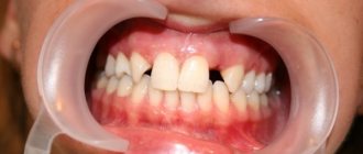
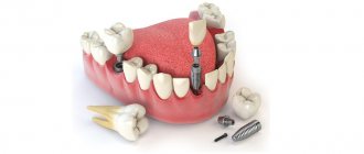
Dental modeling is a set of manipulations to prepare for the installation of orthopedic structures. Used to visually demonstrate the potential appearance of an artificial smile model and check the comfort of wearing a denture. At this stage, the doctor reproduces with wax the external aesthetic shape of the complete dentition and individual units.
The second name for modeling is the Wax-up technique. It is carried out during dentures and implantation by taking a personal impression of the oral cavity.
The wax sample is used as a base for the production of a permanent orthopedic product. To obtain an ideal impression, carious and destroyed units are reconstructed with liquid wax. The technician begins further manipulations after the composition has completely hardened. The specialist determines the accuracy of the connection between the created model and the tooth and determines the correct distribution of the chewing load.
There should be no pain during fitting. After receiving data on the anatomical structure of the visitor’s dental system, the physician determines the tactics of further treatment and begins restoration.
Features of the procedure
Wax-up for aesthetic restoration is popular among orthopedists. Wax is a malleable compound that can be molded. With its help, an accurate impression of the tooth is created, taking into account the anatomical features.
The accuracy of manipulation affects the comfort of the artificial prosthesis. Following the technology will relieve the patient from pain when talking and eating.
The Wax-up technique is safe and does not cause discomfort. This is due to the absence of violation of the integrity of the oral mucosa. There is a risk of cross-reaction in those who suffer from allergic reactions to bee products. In this case, replace the composition.
Simple instructions for modeling teeth using wax
U. Gafurov, dental technician, founder of international dental courses dentin.uz (Uzbekistan)
The profession of a dental technician begins with the morphology of teeth and the correct approach to this aspect allows you to achieve a high professional level. Anyone who missed this stage suffers for many years from a lack of knowledge and skills in creating the anatomical shape of teeth.
We recommend working on creating a symmetrical shape first. You don't have to start with wax, it's better to start with painting. You can find free activities to develop your ability to create symmetry by following the link.
After the symmetry stage, you need to master the skills of working with an electric spatula and wax. (Fig. 1).
Rice. 1.
You need to feel how the wax behaves at high and low temperatures. With the correct movement, you can get a larger (Fig. 2) or smaller portion of wax (Fig. 3).
Rice. 2.
Rice. 3.
After these important steps - creating symmetry and managing the wax, you can begin to model the anatomical shape of the teeth using wax. If we know how to spell the word “sky” in Russian, then we can easily cope with this task. But if we are asked to write this word in Chinese, difficulty arises (Fig. 4).
Rice. 4.
If we don't know the anatomy of each tooth, before wax modeling, we will feel the same as when writing a Chinese character. The book “Textbook of Dental Prosthetic Technology, Part 1” (A. Homann, W. Hielscher) is suitable for solving this problem. This book describes every detail, every part of the tooth in as much detail as possible.
The purpose of our material is to share with colleagues simple instructions for modeling teeth. But without the steps described above, the instructions will not be of much use.
The first step is to mark a center line with a pencil, perpendicular to the horizon (Fig. 5).
Rice. 5.
The second step is to apply one layer of wax at high temperature (Fig. 6).
Rice. 6.
The third stage is the creation of interdental contact surfaces (Fig. 7).
Rice. 7.
The fourth stage is the creation of a cutting edge, taking into account the anatomical data of the teeth (Fig. 8).
Rice. 8.
In order to get the most suitable tooth shape at once, we recommend forming a cutting edge from 33 to 43 teeth for the lower teeth, and from 12 to 22 teeth for the upper teeth. If you model each one separately, without creating a cutting edge according to our recommendation, at some point you may notice a gross error. The height of the above teeth from the first and second, or third and fourth quadrants may turn out to be different, this is one of the most unpleasant moments for a dental technician.
The fifth stage is to model the vestibular surface in parts: the first part is medial, then the equator, then the distal, after which you can fill in the central part of the tooth (Fig. 9 - 12). Also, other teeth in parts (Fig. 13), without delving into small details (textures, ridges, grooves).
Rice. 9.
Rice. 10.
Rice. eleven.
Rice. 12.
Rice. 13.
As soon as we have the preliminary shape of the above teeth, we can begin to form the parts (Fig. 14). This process is also best done in parts: the medial part, the equator and the distal part.
Rice. 14.
Detailed video instructions can be found at the link (
).
About the author
Gafurov Ulugbek, dental technician, founder of international dental courses dentin.uz, Uzbekistan, Tashkent
Gafurov U., dental technician, founder of international dental courses dentin.uz, Uzbekistan, Tashkent
+998946679055
Simple instructions for modeling teeth in wax
Annotation. The profession of a dental technician begins with the morphology of teeth and the correct approach to this aspect allows you to achieve a high professional level. The purpose of our material is to share with colleagues simple instructions for modeling teeth.
Annotation. Profession of a dental technician begins with the morphology of the teeth and the proper approach to this aspect allows to achieve a high professional level. The purpose of our material to share with colleagues a simple manual simulation of the teeth.
Key words: Tooth modeling, wax modeling, tooth morphology.
Keywords: Modeling teeth, modeling wax, morphology of teeth.
Purpose of application
The need for Wax-up is due to the following factors:
- Reproduction of dental units from wax is the starting stage of restoration work. It is required for planning future work of dentists. Wax impressions help to track the individual characteristics of the structure of teeth and avoid errors during implantation.
- Calculation of the cost of upcoming dental procedures.
- Temporary prosthetics. The specialist takes samples for the production of intermediate prosthetic restorations made of plastic. The technology is used when additional time is needed to create fixed prostheses.
Types of wax modeling
The most important part of prosthetic practice in dentistry is diagnostic wax-up. Its essence lies in the fact that the dentist and dental technician create a model of future work from wax. Wax is the most effective material for this purpose, since it is incredibly plastic and, when hardened, quickly takes the desired shape. Thanks to this, the technician manages to create the desired form, like a sculpture - his own artistic creation. Such wax modeling must be practiced before all types of work in a dental laboratory in order to work out all its details in advance.
The main practice of any orthopedic dentist involves the use of functional wax modeling. When preparing for orthopedic treatment, it is necessary to model not only the general shape of the teeth, but also the position of each individual tooth. The finished prosthesis or crown should be strong and comfortable for the patient, and the temporomandibular joint should not be under tension.
Groups of teeth and their functions
It should be noted that each group of teeth and individual teeth perform different functions, and this is important to consider when working with an orthopedist and technician. It is customary to distinguish the following functional areas of the dentition:
- Anterior group of teeth. This includes the incisors on both jaws. For them, the main function is the guiding function when moving the lower jaw forward. In addition, this group of teeth is important for recognizing food and determining the force of chewing, and also affects a person’s speech and the position of his lips. It should also be remembered that the incisors determine the aesthetics of the face and the entire dental system.
- A lateral group of teeth that includes the molars (6th, 7th and 8th teeth). Their main function is related to chewing and supporting properties.
- Premolars (4th and 5th teeth). They are involved in chewing and lateral movements.
- Fangs. These teeth perform the most important function of “all-round protection” of the teeth. When the lower jaw moves forward, as well as during lateral movements, all teeth become separated. Thanks to the fangs, the dental system is normalized and returns to its normal position.
The success of any treatment depends on many factors that the dentist must consider. The dental system of each person is completely unique and is influenced even by such features as the nutritional system or character traits.
Types of wax modeling
The following types of wax modeling are used in dentistry today:
- Modeling for planning the upcoming restoration: in case of severely damaged teeth, creating an accurate model helps to determine the volume of required preparation of dental tissues and agree with the technician on the creation of a future restoration (if we are talking about a ceramic filling, veneer or crown).
- Modeling for the creation of prosthetic structures, including in order to agree with the patient on the type of dentition after orthopedic treatment.
- Diagnostic modeling to determine the condition of the bite and options for its correction. Wax models are also made during the process of correcting the bite to accurately assess its condition and changes occurring.
- Modeling for planning the placement of implants or combined removable-fixed prostheses.
In recent years, with the development of technology, new electronic diagnostic devices have appeared. The accuracy of measurement increases, which means treatment becomes more effective. A modern device for wax modeling is called an articulator. It is programmed individually for each patient, for which the necessary parameters are measured. These measurements are carried out using condylography during preparation for treatment.
It remains to add that in order to conduct wax modeling, both the orthopedic dentist and the dental laboratory specialist must have the highest qualifications.
The result of this joint work is the patient’s individual occlusion project. It is carried out by installing temporary crowns, which are fixed by an orthopedist, taking into account the preservation of functionality and aesthetic properties.
If you have a problem similar to that described in this article, be sure to contact our specialists. Don't diagnose yourself!
Why you should call us now:
- We will answer all your questions in 3 minutes
- Free consultation
- The average work experience of doctors is 12 years
- Convenient location of clinics
Single contact phone number: +7
Make an appointment
Indications and contraindications
The orthodontist prescribes dental diagnostics after a clinical examination if there are indications:
- Abrasion of units due to malocclusion and other pathologies.
- Destruction of the supragingival area due to advanced forms of caries.
- Distinctive color.
- All that remains of the units is the root base.
- Edentia.
- Multiple pathologies.
Contraindications:
- dental pathologies in acute form;
- recovery after radiation therapy;
- drug addiction;
- diseases of the jaw tissue;
- inflammation of various etiologies.
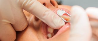
Modeling methods
Dentists use two methods of diagnostic modeling.
- Indirect
This technique is based on modeling a plaster model. Does not require prolonged presence of the visitor in the clinic. The orthodontist takes an impression of the oral cavity and passes it on to the technician to form the structure in the laboratory.
Precise finishing of the sample edges is carried out for user comfort. This method is chosen by the dentist to work with teeth located in hard-to-reach places.
- Straight
It is a combination of two technologies - Wax-up and Mock-up. Modeling is carried out directly in the patient’s mouth. Suitable for working on single structures.
The specialist fills the tooth cavity with liquid wax or a plastic mass. Reproduction and pinning are then performed, after which the resulting model is retrieved. Based on the resulting layout, a composite sample is created.
A person walks with the installed temporary system for a short time to evaluate its convenience and correct manufacture. The method is suitable for people who want to improve their bite or suffer from increased enamel wear.
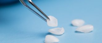
Material characteristics
When carrying out the procedure, two types of beeswax are used:
- 1 type Found application in direct modeling.
- Type 2 Used with indirect technology. The wax is of lower quality, but the result of the work is at a decent level.
Recommendations must be followed when selecting, storing and using the material:
- the use of colored waxes allows you to achieve clear contrast;
- the frozen forming composition is rigid and does not crumble;
- absence of crumbs and impurities in the heated mass;
- absence of chips and cracks when scraping;
- compliance with storage periods;
- competent selection of the type of material depending on the technique;
- selection of soft types of material.
Preparatory stage
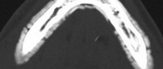
Before starting to reproduce the dentition, the following recommendations are followed:
- inspection is carried out;
- CT and X-ray diagnostics of the jaw are performed;
- treatment of dental diseases;
- compliance with hygiene rules, sanitation of the oral cavity.
The manipulation is carried out after a full examination and dental therapy.
Step-by-step algorithm
Stages of the procedure:
- taking a plaster (silicone) impression of the patient’s jaw at the site of future placement and on the opposite side;
- preventing jaw closure using silicone bite pads;
- determining the relationship between the upper jaw and the skull using a facebow;
- transferring samples onto a gypsum base;
- fixing the model on the articulator;
- modeling a wax cast on plaster models;
- Demonstration of the layout to the client.
The Mock-up technique involves transferring the mock-up to the mouth. Algorithm of the doctor’s actions:
- obtaining silicone keys;
- gentle removal of deposits;
- fixing the layout with special glue;
- placing composite or polyurethane into impressions, fixation;
- impression taking, grinding and polishing;
- assessment of work quality.
The duration of making a model depends on the specific structure and development of the jaw and location.
Pros and cons
Making wax models is a painstaking and technologically complex process. The popularity of the procedure among dental technicians and orthodontists is due to its advantages:
- adjustment of nuances;
- no need to get used to wearing permanent orthopedic structures;
- absence of discomfort, pain;
- gentle preparation process;
- patient participation in the process of adjusting the model using the indirect method.
The disadvantages include reduced accuracy with the indirect method. The volume of the material changes as it hardens.
Text of the book “Fundamentals of clinical dental morphology: a textbook”
4.1.4. Mandibular lateral incisor
The mandibular lateral incisor occupies the second position in the lower dental arch. It is larger than the lower medial incisor, with a well-defined crown angle feature.
The child erupts at 8-10 months. The change to a permanent lower lateral incisor occurs at 7–8 years.
In vestibular and lingual norms
(Fig. 68) the shape of the crown is close to the shape of an irregular quadrangle. The cutting edge line is relatively straight. The mesial angle of the crown is less than the distal one. The distal corner of the crown is rounded.
Rice. 68. Lateral incisor of the lower jaw, right.
a – vestibular norm; b – language norm.
The approximal contours noticeably converge towards the enamel-cementum junction. The medial contour is longer than the distal one.
The curvature of the enamel-cementum border in the direction of the root is more pronounced on the lingual side.
The transition of the approximal contours of the crown to the corresponding contours of the root is better visible from the distal side.
The root is cone-shaped, deviated distally (a sign of root position). The approximal contours of the root are somewhat convex in the middle third, smooth or concave in the apical and cervical thirds. The “waist” of the tooth is more pronounced along the distal contour. The apex of the root of the formed tooth, as a rule, is located distal to the USV.
On the vestibular surface
On the crown there is a vertical median ridge, which is separated from the lateral vertical ridges by weakly expressed depressions. The median and lateral vertical ridges in the cervical third merge with a clearly visible tooth belt.
The transition of the contact contours of the crown to the corresponding contours of the root is more pronounced on the distal side.
Lingual surface
more prominent than that of the medial incisor of the lower jaw. The marginal scallops, of which the distal one is the most developed, are delimited from the median scallop by pits. The distal fossa is deeper than the medial one. The median and marginal ridges in the cervical third of the crown merge with the lingual tubercle and the belt of the tooth.
The lingual surface of the root is already vestibular. The mesial and distal root surfaces converge lingually, so that in the lingual norm both approximal root surfaces are visible.
In medial and distal norms
(Fig. 69) the crown is shaped like a triangle, the most acute angle of which is located at the cutting edge.
The vestibular contour is convex at the tooth belt and relatively straight to the cutting edge. The lingual contour is more extended than the vestibular one, with a convexity at the level of the lingual tubercle and a concavity further to the incisal edge.
In the distal norm
The lingual contour of the crown is formed by the distal marginal ridge and has a concavity above the lingual cusp.
The line of the enamel-cementum border is convex in the direction of the cutting edge, and with a greater amplitude of curvature in the medial norm than in the distal one.
Rice. 69. Lateral incisor of the lower jaw, right.
a – medial norm; b – distal norm.
The vestibular and lingual contours of the crown, passing into the corresponding contours of the root, form the “waist” of the tooth. The transition of the contours of the crown to the root on the lingual side is smoother than on the vestibular side.
The contours of the root can be either convex or concave and reflect the uneven topography of the surface. The root apex is located near the USV (usually on the vestibular side of it). On the cone-shaped root, a vertical groove is visible on the distal side, which can serve as an additional sign of lateralization of the tooth.
In occlusal norm
(Fig. 70, a) the mesial-distal size of the crown somewhat prevails over the vestibular-lingual one.
Rice. 70. Lateral incisor of the lower jaw, right.
a – occlusal norm and section at the level of the base of the crown; b – tooth cavity.
The vestibular and lingual contours of the crown are convex. The degree of curvature of the lingual contour is greater than that of the vestibular one. The point of greatest convexity of the vestibular contour is slightly shifted to the medial side, and the point of greatest convexity of the lingual contour is distally.
Along the vestibular contour, a medial-distal slope is noticeable (a sign of crown curvature). The approximal contours are approximately equal in length. The cutting edge is located closer to the vestibular contour.
On horizontal sections, the root has the appearance of a laterally compressed oval, on the distal contour of which a concavity is visible.
Tooth cavity
(Fig. 70, b) corresponds to the external shape of the tooth. The cavity of the crown is narrowed in the vestibular-lingual direction and imperceptibly passes into the root canal. In the cavity of the crown there are small depressions that continue to the corners of the crown.
The lumen of the unformed root canal is relatively wide. A tooth with a resorbing root has a narrow canal. The diameter of the hole at the root apex corresponds to the width of the canal.
Anatomical options.
In
the vestibular and lingual norms,
the shape of the crown resembles an irregular quadrangle, usually trapezoidal. There are options for a rectangular or oval crown.
On the cutting edge, tubercles are more common in unformed teeth. The distal angle of the crown is usually larger than the medial one and is often rounded. The ridges and tubercle on the lingual surface vary slightly in size. There are sometimes vertical grooves on the lateral surfaces of the root.
In medial and distal norms
The crown is close to the shape of a triangle. The cutting edge is located along the USV or is slightly shifted to the lingual side.
The height of the tooth can be from 14.4 to 16.5 mm, while the height of the crown is 5.2–7.3 mm, the height of the root is 9.2–10.6 mm. The mesial-distal size of the crown ranges from 4.1 to 5.5 mm, the neck - from 3 to 3.7 mm. The size of the crown in the vestibular-lingual direction is from 4 to 4.8 mm, in the cervical area - from 3.2 to 4.5 mm.
4.2. Group of fangs
Milk fangs –
single-rooted teeth with a crown pointed on all surfaces, which are located in the middle part of each half of the dental arch distal to the incisors (third position). Primary canines erupt at 16–22 months and are replaced by permanent canines at 12–13 years.
The child has 4 primary canines (two in each dental arch):
– canines of the upper jaw
(right and left);
– fangs of the lower jaw
(right and left).
What is common in the anatomy of primary canines, as well as permanent ones, is the presence of a crown pointed on all surfaces and the longest root.
Primary canines differ from permanent canines in their smaller size and more symmetrical location of the main tubercle in relation to the approximal surfaces of the crown.
The upper milk canine is larger than the lower one.
The main signs of lateralization are uninformative.
To determine whether a canine belongs to the right or left half of the dental arch, the combination of structural features of the crown and root is taken into account.
4.2.1.
The canine of the upper jaw The canine of the upper jaw occupies the third position in the upper dental arch, just like the permanent tooth of the same name, it has a crown pointed on all surfaces and the longest root. The primary canine is less variable than the permanent canine.
The upper primary canines erupt at 16–20 months, and are replaced by permanent upper canines at 11–13 years.
In vestibular and lingual norms
(Fig. 71) the crown is close in shape to a pentagon.
Rice. 71. Maxillary canine, right.
a – vestibular norm; b – language norm.
The occlusal contour consists of two segments, which at the junction (the position of the tip of the “tearing cusp”) form an angle close to a right angle. The mesial segment of the occlusal contour is located more vertically and is somewhat longer in length than the distal one. The apex of the main tubercle coincides with the USV. The angle formed by the IVS and the mesial segment (slope) of the occlusal contour is smaller (sharper) than the angle between the IVS and the distal segment of the occlusal contour.
The approximal contours are relatively short, with the distal contour being longer than the mesial contour.
The line of the enamel-cementum border is slightly curved towards the root on both the vestibular and lingual sides.
The transition of the approximal contours of the crown and root is clearly visible and more pronounced on the mesial side. The approximal contours of the cone-shaped root are relatively smooth. The root apex is located near the USV.
On the vestibular surface
The crown has a vertical median ridge located along the length from the “tearing tubercle” to the tooth girdle. On both sides of the median ridge there are less protruding ridges, separated from it by depressions (mesial and distal).
On the lingual surface
The marginal ridges are well defined, which are delimited from the median ridge by triangular pits. The median ridge runs from the top of the main cusp to the base of the crown, where it merges with the marginal ridges, the lingual cusp and the tooth belt. The zygomatic tubercle is located approximately midway between the approximal contours of the crown. The lingual surface of the root is narrower than the vestibular one, therefore there is a convergence of the approximal contours of the root towards the lingual side.
In mesial and distal norms
(Fig. 72) the shape of the crown resembles a triangle, the base of which faces the neck of the tooth.
Rice. 72. Maxillary canine, right.
a – mesial norm; b – distal norm.
The line of the occlusal contour is connected to the vestibular and lingual contours on the vestibular side of the USV. The vestibular contour is convex with the greatest curvature near the neck (in place of the belt). The lingual contour is convex in the cervical third at the location of the lingual tubercle and relatively smooth throughout the rest of the length.
The line of the enamel-cementum border is slightly curved towards the occlusal contour. On the distal side, the enamel-cementum border is less convex than on the mesial side.
The transition of the contours of the crown to the corresponding contours of the root is well defined. The angle formed at the junction of the vestibular contours of the crown and root is greater than the angle formed at the junction of the lingual contours of the crown and root.
The contours of the cone-shaped root are uneven. The vestibular contour is often convex throughout its entire length, the line of the lingual contour is flattened. The root apex is rounded and located near the USV.
In the mesial norm
due to the contours of the vertical mesial ridge of the crown, the contour of the vertical median ridge of the vestibular surface protrudes. A mesial depression is noticeable between these ridges.
In the mesial norm, the mesial slope of the “tearing tubercle” is visible, which runs from the top of the main tubercle to the most protruding point of the mesial surface. The outline of the median ridge of the lingual surface protrudes from the contours of the mesial marginal ridge.
In the distal norm
the vestibular surface of the crown is visible with distal and median vertical ridges and a recess separating them.
From the apex of the main tubercle to the most protruding point of the distal surface, the distal slope of the “tearing tubercle” is visible, which is shorter than the mesial slope. From the distal side, the outline of the distal marginal ridge and the outline of the median ridge on the lingual surface are visible.
In occlusal norm
(Fig. 73, a) the contours of the crown resemble a rhombus in shape. The mesial-distal size of the crown prevails over the vestibular-lingual one.
The vestibular and lingual contours are convex. The degree of curvature of the lingual contour is greater than that of the vestibular one. The points of greatest convexity of the vestibular and lingual contours are located quite symmetrically with respect to the approximal surfaces.
The mesial contour of the crown is wider than the distal one, which can be used as an additional sign of tooth lateralization.
Rice. 73. Maxillary canine, right.
a – occlusal norm and section at the level of the base of the crown; b – tooth cavity.
In the occlusal norm, a convex vestibular surface with vertical ridges and a belt is clearly visible. Between the vertical enamel ridges there are depressions, of which the mesial one is more pronounced. On the lingual surface, the median and marginal ridges are clearly visible, converging towards the tooth girdle and clearly visible in the occlusal norm. The median and marginal scallops are separated by pits. The distal fossa is slightly deeper than the mesial one.
Tooth cavity
(Fig. 73, b) to a certain extent repeats its external shape. In the cavity of the crown, in the area of the tubercles and corners of the crown, recesses for the pulp horns are noticeable. The transition of the crown cavity to the root canal is smooth, without noticeable boundaries. The dimensions of the crown cavity and root canal depend on the degree of tooth formation. A tooth with a developing root has a larger cavity than a tooth with a resorbing root.
Anatomical options.
The primary canine is less variable than the permanent canine.
In the vestibular norm
The size of the angle formed by the slopes of the “tearing tubercle” varies less in the primary canine than in the permanent canine.
An additional tubercle is often found on the distal slope, which is less pronounced than that of the permanent canine.
Relief of the lingual surface
determined by the severity of the lingual tubercle and ridges. The zygomatic tubercle can “split” into two parts, which in the middle or occlusal third merge into one and pass into the median ridge.
In mesial and distal norms
the line of the enamel-cementum border can be straight or convex to varying degrees towards the occlusal contour. The bend of the enamel-cementum border on the mesial surface is slightly greater than on the distal surface.
The height of the tooth ranges from 18.2 to 20.2 mm, while the height of the crown is 6.4–7.6 mm, and the height of the root is 11.8–13.5 mm.
The mesial-distal size of the crown ranges from 6.4 to 7.4 mm, the neck - from 4.2 to 5.3 mm. The size of the crown in the vestibular-lingual direction can be from 5.4 to 7 mm, in the cervical area - from 4 to 5.5 mm. 4.2.2.
Canine of the lower jaw The canine of the lower jaw occupies the third position in the lower dental arch, somewhat inferior in size to the upper canine. The sign of crown curvature is weakly expressed.
Erupts at 16–18 months. The change to a permanent lower canine occurs at 11–13 years of age.
In vestibular and lingual norms
(Fig. 74) the contours of the crown form a pentagon, the most acute angle of which corresponds to the apex of the main tubercle.
Rice. 74. Canine of the lower jaw, right.
a – vestibular norm; b – language norm.
The occlusal contour is formed by the slopes of the “tearing tubercle”, converging to its apex at an acute angle. The mesial slope is longer than the distal one. The position of the apex of the main tubercle approximately coincides with the USV.
The occlusal contour line smoothly transitions into the contact contours of the crown, which converge towards its base.
The enamel-cementum border is slightly curved towards the root on the vestibular side and slightly more on the lingual side.
The apex of the root of the formed tooth practically coincides with the USV. The contours of the crown without sharp boundaries transform into the corresponding approximal, relatively smooth and symmetrical contours of the root.
The surface reliefs of the crowns of the lower and upper canines are similar. On the vestibular surface
From the top of the “tearing tubercle” in the direction of the neck of the tooth, an enamel ridge runs in the shape of a triangle, the base of which is the belt. On both sides of the enamel ridge there are depressions, of which the mesial one is better expressed than the distal one.
Lingual surface
less prominent than that of the antagonist of the same name. The marginal ridges at the base of the crown merge with the lingual tubercle. A weakly defined median ridge runs from the top of the “tearing tubercle” to the lingual tubercle. On both sides of the median ridge there are depressions, of which the distal one is larger.
In mesial and distal norms
(Fig. 75) the contours of the crown resemble a triangle, the base of which is located at the neck of the tooth.
Rice. 75. Lower jaw canine, right.
a – mesial norm; b – distal norm.
The apex of the main tubercle is projected, as a rule, on the vestibular side of the USV.
The vestibular contour of the crown is unevenly convex. Its greatest curvature is noted in the cervical third; throughout the rest of its length, its convexity is insignificant.
The line of the lingual contour is concave from the “tearing tubercle” to the lingual tubercle, at the level of which (the cervical third of the crown) the line of the lingual contour is usually convex.
The enamel-cementum border is convex towards the crown and differs little in both norms.
The contours of the crown in the area of the enamel-cementum border have a noticeable transition into the corresponding contours of the root.
The contour lines of the cone-shaped root are often vaguely curved. Often the vestibular contour of the root is somewhat convex in the cervical third and concave throughout the rest of the length, and the lingual contour is, on the contrary, convex in the middle third and concave in the cervical and apical thirds. The root apex is located along the USV, often rounded.
Mesial surface
The root is convex with a smoothed relief. A vertical groove runs along the distal surface of the root.
In occlusal norm
(Fig. 76, a) the contours of the crown are shaped like an isosceles triangle with rounded corners at the base, which is the mesial contour.
Rice. 76. Canine of the lower jaw, right.
a – occlusal norm and section at the level of the base of the crown; b – tooth cavity.
The vestibular contour has a slope in the mesial-distal direction (a sign of crown curvature). The point of greatest convexity of the lingual contour corresponds to the lingual tubercle, which is displaced mesially from the USV. The vestibular and lingual contours of the crown converge towards the distal contour, which can be used as an additional sign of lateralization of the tooth.
The vestibular-lingual size of the crown is not much inferior to the mesial-distal one. On horizontal sections of the root, the vestibular-lingual size significantly prevails over the mesial-distal one.
Tooth cavity
(Fig. 76, b), corresponds to its external outlines. The cavity has small depressions in the direction of the protruding areas of the crown (corners of the crown and the tip of the “tearing tubercle”) and bends in the canal area, repeating the curvature of the root.
The volume of the cavity relative to the external dimensions of the tooth depends on the stage of its development. In a tooth with an unformed root, the relative volume of the cavity is significant. The root canal is large in diameter with the apical opening corresponding in size. A tooth that is in the stage of root resorption has a relatively small cavity volume. The root canal of such a tooth is narrow and opens at the apex with a pinpoint-sized hole.
Anatomical options.
In
the vestibular norm,
the slopes of the main tubercle are often somewhat rounded and converge at an angle close to a straight line. As a rule, the mesial slope of the “tearing tubercle” is longer than the distal one, although there are options when both slopes are equal in size or the distal one is slightly larger than the mesial one.
By changing the relief of the vestibular surface. As a rule, on the vestibular surface there is a large enamel ridge running from the tooth belt to the top of the main tubercle. Often the median enamel ridge is displaced mesially and divides the crown into two unequal parts. The lateral enamel ridges can be expressed differently and are separated from the median ridge by deltoid depressions. More often, the distal depression is less pronounced than the mesial one. With weak development of the lateral enamel ridges, the entire vestibular surface becomes uniformly convex.
The anatomical formations on the lingual surface are variable in degree of development. The severity of the ridges and lingual tubercle is very diverse and determines the features of the relief of the lingual surface. Sometimes the lingual tubercle is absent. In the mesial and distal norms, the line of the enamel-cementum border can be convex towards the crown or almost straight.
The root is slightly flattened in the mesial-distal direction and in cross sections has an oval or close to triangular shape. Root bends are variable.
The height of the tooth ranges from 16.2 to 18.7 mm, with the height of the crown being 6–8.2 mm and the height of the root being 10.6–11.7 mm. The mesial-distal size of the crown ranges from 5 to 6.1 mm, the neck - from 3.6 to 4.2 mm. The size of the crown in the vestibular-lingual direction can be from 4.8 to 5.7 mm, in the cervical area - from 3.8 to 5 mm.
4.3. Molar group
Primary molars –
teeth with a multi-tubercular chewing surface and several roots. Molars are located in the distal parts of the dental arch and occupy the fourth and fifth positions.
Primary molars erupt from the 14th to the 30th month and are replaced by permanent small molars (premolars) from 8 to 13 years.
The child has eight primary molars:
– first and second molars
upper jaw (right and left);
– first and second molars
lower jaw (right and left).
Primary molars are the largest teeth in the primary dentition. Second primary molars, unlike permanent teeth, are much larger than first molars.
Primary molars of the upper jaw have three roots - two vestibular and lingual, the lower jaw - two roots - mesial and distal.
A characteristic morphological feature of primary molars is the predominance of the mesial-distal diameter over the height of the crown. In maxillary molars, the vestibular-lingual size of the crown is larger than the mesial-distal size. In mandibular molars, the mesial-distal diameter of the crown is larger than the vestibular-lingual diameter.
The deciduous molars have a well-defined belt, and in the first molars it is most developed.
The roots of primary molars are pincer-shaped. The “waist” of the tooth is well defined. Of the main signs of lateralization, all primary molars are characterized by the sign of crown curvature.