Indications and contraindications Pros Cons Implantation process Alternatives
Teeth transplantation from your own dentition is a procedure that is used as an alternative to implantation. Every person has teeth that are not involved in the chewing process, do not affect the bite and do not perform any important functions. These are wisdom teeth. They are the ones who most often become donor teeth. But not always.
There are teeth that grow outside the arch, spoil the smile, and provoke inflammation. You can get rid of the problem through orthodontic treatment: installing splints, plates, braces. There are times when the only option is deletion. Then the tooth has a chance to benefit its owner. It can be transplanted to a place where a tooth is lost or planned to be pulled out.
Another source of donors for transplantation is supernumerary teeth. Some people grow double teeth. Such units are direct candidates for removal. At the same time, they can be beneficial by replacing a sick or lost brother.
Replacing an artificial root
Sometimes it happens that the patient’s body rejects the implant. Most often this happens due to the fact that the body perceives the implant material as a foreign body. In this case, the dentist has no choice but to remove the implant. Fortunately, nowadays it is possible to reuse an implant of a different design. Reimplantation is necessary in the following cases:
- implant rejection;
- dentist mistakes;
- use of a defective implant;
- the patient’s failure to comply with the dentist’s recommendations;
- deterioration of general health;
- expired implant.
Separately, it is worth mentioning jaw injuries. Sometimes even a minor injury causes the artificial structure to begin to wobble. In this case, you have to resort to reimplantation.
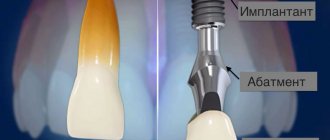
At the same time, a worn-out crown can be replaced by installing it on the previous artificial root. The same applies to replacing the abutment - the connecting link between the crown and the implant.
How much does a tooth transplant cost?
Each dental clinic sets its own prices for autotransplantation. And the price depends on the status of the center, the qualifications of the doctor, the complexity of the operation: whether the removal will take place during the operation or whether the work will take place in an already empty place, the anterior or chewing units will be replaced. But on average, transplanting your own tooth will cost from 25,000 to 45,000 rubles.
It seems that transplanting your own organ to yourself is much cheaper. Since there are no costs for expensive materials: implant, abutment and crown, or the work of a dental technician. “Everything is yours” as they say. But the price is not much lower than implantation.
How is the old implant removed?
First of all, it is worth saying that if any problems arise with the implants, then you should only contact the clinic where they were installed. This is important, including for financial reasons. The savings are explained by the fact that a new design calculation is not required.
Most implant manufacturers usually guarantee their impeccable service for 5-7 years. Reputable manufacturers provide such a guarantee for decades and even for life. Of course, the cost of such implants is very high, if not unaffordable for most of our fellow citizens. In addition, reimplantation is not considered the fault of the manufacturer, and therefore has to be paid for again. Only some clinics do it for free in order to raise their authority.
This dental operation is performed in the following sequence:
- disinfection of the oral cavity;
- crown removal;
- removal of the implant from the jawbone.
After this, the patient will have to undergo examinations to determine the cause of implant rejection. After doctors determine the cause, the treatment stage begins.
Delayed replantation of injured teeth
Complete tooth dislocation (also known as tooth avulsion) is characterized by complete displacement of the tooth from the socket and occurs in 0.5-3% of cases among all injuries of the dentofacial apparatus. The frequency of such lesions increases sharply in children aged 7 to 9 years, which is associated with incomplete development of the roots, as well as the fact that the alveolar bone and periodontal ligament at this age are the least resistant to the effects of extrusive forces during teething. The etiology of complete dislocation varies depending on the type of bite. Luxation of primary teeth usually occurs as a result of being struck by a hard object, while permanent teeth are more often injured as a result of falls, fights, sports injuries, car accidents and child abuse. In both permanent and temporary dentition, avulsions are more common in the upper jaw with a predominant lesion of the central incisors. Over-overlapping of teeth and underdevelopment of the lips are potential etiological factors predisposing to injury. Although, as a rule, complete dislocation of only one tooth occurs, multiple avulsions are also known with parallel involvement of the supporting soft tissues as well as the lips.
The main goal of treating avulsed teeth is to preserve and treat the adjacent supporting tissues and replant the problematic teeth. The success of the last manipulation depends on the general health of the patient, the degree of root formation, the time that has passed since the injury, as well as the storage environment of the dislocated tooth. The time that has passed since the complete traumatic removal of the tooth from the socket, as well as the environment in which it is stored outside the oral cavity, have a key impact on the condition of the periodontal ligament cells. The purpose of this study is to present two clinical cases of delayed replantation of avulsed central incisors after a long extra-alveolar period of dry storage.
Clinical cases
Clinical case 1
An 8-year-old boy was referred to a pediatric dental clinic after a fall that resulted in a tooth injury. The incident occurred 27 hours before the report while the child was playing on the school playground. The child was subsequently examined by emergency medical personnel at a local hospital, who did not reveal any neurological damage or other general medical complications. The child's parents kept the avulsed tooth dry in a piece of paper and brought it with them to the clinic. They denied the child had any underlying medical conditions. During an intraoral examination, it was diagnosed that the central left maxillary incisor (tooth 21) was completely dislocated (photo 1). In the 11th tooth, an uncomplicated crown fracture with dentin damage, dislocation and excessive mobility, as well as lacerations of the mucous membrane on the palatal side were observed. During vital testing, the tooth reacted positively. The patient had a mixed dentition; also diagnosed with severe carious lesions due to poor hygiene.
Photo 1. Complete dislocation of the left upper incisor.
Periapical and panoramic radiographs showed no evidence of fracture of the alveolar bone wall or adjacent bone tissue. An examination of a dislocated tooth revealed a fracture of the coronal enamel, an open root apex, and remains of periodontal tissue on the root surface.
After informing the patient's parents of the possible risk, the dental socket was carefully rinsed with saline under local anesthesia (Maxicaine, Vem Drugs, Istanbul, Turkey). The tooth root was thoroughly cleaned of necrotic and dry remains of periodontal tissue.
Endodontic treatment prior to the replantation stage was performed outside the mouth by filling the root canals with mineral trioxide aggregate (MTA) (BioAggregate, DiaDent, Burnaby, BC, Canada). The tooth was then slowly placed back into the socket with slight pressure.
A wet cotton ball and glass ionomer cement (Ketac Molar, 3M/ESPE Dental Products, St. Paul, MN, USA) were used to temporarily restore the access cavity. The position of the transplanted tooth was checked using clinical and radiological methods. The tooth was splinted with a flexible ligature (0.195-inch round twist-flex arch wires, 3M Unitek, Monrovia, CA, USA) using a composite (Clearfil Majesty Esthetic, Kuraray, Tokyo, Japan (Figure 2, Figure 3).
Photo 2. Splinting a dislocated tooth using an orthodontic ligature and composite.
Photo 3. Periapical radiograph after replantation of a traumatized tooth.
In addition, the patient was given recommendations regarding hygiene and the need to use chlorhexidine (Klorhex, Drogsan, Ankara, Turkey) for rinsing during the stabilization period, and a soft diet was prescribed.
A prophylactic course of antibiotic therapy with amoxicillin trihydrate/potassium clavulanate (Beecham Laboratories, Bristol, TN, USA) at a dose of 625 mg/day. was prescribed for a period of one week.
The patient was also referred for tetanus vaccination.
Parents were informed of the importance of regular clinical and radiological follow-up visits.
The patient was reexamined two weeks later, but no clinical or radiographic pathological changes were identified.
Four weeks later, at the next visit, the splinting structure was removed, and the restoration of the destroyed tooth crowns was completed with a composite (Clearfil Majesty Esthetic, Kuraray Tokyo, Japan).
At the third month of observation, percussion testing of the reimplanted tooth revealed a change in percussion sound due to tissue ankylosis.
12 months later the right central incisor was found to have lost vitality; Calcium hydroxide (Sultan Chemists Inc., Englewood, NJ, USA) was used to complete the process of apexogenesis.
At the follow-up visit 18 months later, the transplanted tooth was in a stable and functional position, but showed signs of initial replacement resorption, ankylosis and infraocclusion of approximately 0.5 mm (Figures 4 and 5).
Photo 4. Frontal view 18 months after injury, slight infraposition of the problematic tooth.
Photo 5. Evaluation of the replanted tooth after 18 months.
The patient will be monitored until the growth period is completed and, if necessary, complete and appropriate treatment will be provided. Cone beam computed tomography was performed to evaluate the relationship between the roots of the lateral incisor and the permanent canine.
Clinical case 2
A 10-year-old boy was referred to a pediatric dental clinic after falling from a bicycle, which resulted in dental injury. The avulsed tooth was not placed in any special container or environment, but was brought to the clinic in a dry state 7 hours after the accident. The patient's parents denied any underlying systemic illnesses, and there was no history of loss of consciousness or vomiting. Examination revealed no additional extraoral injuries. During an intraoral examination, complete dislocation of the maxillary right permanent central incisor (tooth 11) was diagnosed (photo 6). Cracks and damage to the enamel were found in the left central incisor (21 teeth). The patient was diagnosed with a permanent dentition with mild crowding and deep incisal overjet. The level of oral hygiene was excellent and no carious lesions were found.
Photo 6. Complete dislocation of the right upper incisor.
On periapical and panoramic radiographs, no traces of an alveolar bone fracture were found, and upon examination of a dislocated tooth, a fracture of the coronal enamel and a closed form of the root apex were diagnosed.
After examination, an algorithm was applied for the treatment of avulsed permanent teeth with a closed root apex and a long extraoral time.
Root canal treatment was performed outside the mouth by filling them with MTA. A wet cotton ball and glass ionomer cement (Ketac Molar, 3M/ESPE Dental Products, St. Paul, MN, USA) were used to temporarily restore the access cavity. Necrotic and dry periodontal tissue debris was also carefully removed from the root surface.
After local anesthesia, the empty tooth socket was thoroughly washed with sterile saline solution. The tooth was placed in place under light pressure after the clot was removed from the socket. The correctness of replantation and tooth position were determined on periapical radiographs (Figure 7).
Photo 7. Splinting a dislocated tooth using an orthodontic ligature and composite.
The dentition was splinted from canine to canine with a flexible ligature (0.195-inch round twist-flex arch wires) (Figure 8).
Photo 8. Periapical radiograph after replantation of a traumatized tooth.
The instructions given to the patient's family were similar to those described in Case 1 (diet and hygiene recommendations). In addition, a prophylactic course of antibiotic therapy with amoxicillin trihydrate/potassium clavulanate at a dose of 1000 mg/day. was prescribed for a period of one week. Parents were informed of the importance of maintaining good oral hygiene and regular clinical and radiological follow-up.
Two weeks after replantation, the patient was examined, but no clinical or radiological signs of pathological changes were found. The splint was removed four weeks after replantation at a follow-up visit. Restoration of damaged tooth crowns was performed using a composite.
Three months later, signs of ankylosis of the transplanted tooth were discovered during percussion.
Clinical and radiographic monitoring were also carried out at 6 and 12 months.
At the 12-month visit, clinical and radiological examinations demonstrated satisfactory functional and aesthetic results, as well as signs of initial resorption and ankylosis without any evidence of infraocclusion (Figure 9 and Figure 10). The patient will be monitored until the growth period is completed and, if necessary, complete and appropriate treatment will be provided.
Photo 9. Frontal view 12 months after injury.
Photo 10. X-ray studies after 12 months: no signs of pathology and resorption.
Discussion
Treatment protocols for completely avulsed permanent teeth vary, but the bottom line is that immediate replantation is the ideal choice. However, this procedure cannot always be performed immediately. The decision regarding the treatment of completely avulsed teeth is related to the degree of formation of the root apex (open or closed) and the condition of the periodontal ligament cells. The condition of the ligament cells depends on the storage environment and the time that has passed since the injury. The duration of the extraoral period significantly affects the result and directly correlates with the level of vitality of periodontal cells. Clinical studies have shown that teeth transplanted within the first 5 minutes after luxation have a better prognosis for treatment. All periodontal ligament cells lose their viability after 60 minutes of storing the tooth in dry conditions. The environment for storing and transporting the tooth in extraoral conditions is also vital. In patients with a prolonged extraoral period, teeth should be preserved in a suitable carrier or medium, such as Hank's balanced salt solution, saline, milk, saliva, until they are replanted by the dentist.
In these cases, the teeth were kept dry in paper, and their extraoral residence time was more than 60 minutes (27 hours and 7 hours in clinical cases 1 and 2, respectively). Treatment in the presented cases was carried out in accordance with the accepted replantation protocol described by the International Association of Dental Traumatology. This protocol suggests that if a tooth has been kept in dry conditions for more than 60 minutes prior to replantation, endodontic root canal treatment should be performed prior to ex-oral root canal treatment. Given the lack of any chance of pulp revascularization, as well as the necrotic nature of the changes in the periodontal ligament, it is advisable to perform root canal treatment outside the oral cavity.
According to the protocol and literature on delayed replantation, periodontal ligament cells lose their viability when replantation is delayed, resulting in a poor long-term prognosis. Most complete dislocations occur before the patient's facial apparatus becomes fully formed. Preventing resorption of surrounding bone tissue and supporting the tooth in the dentition are critical until the end of facial growth. Replantation helps restore aesthetic appearance, functional occlusion, and also prevent physiological trauma due to the loss of the front tooth. If the avulsed incisors in these clinical cases had not been replanted, treatment might have consisted of prosthetic restoration of the defect, orthodontic gap closure, or autotransplantation of another tooth into the defect space.
The outcome of replantation must be carefully monitored using clinical and radiological methods. Ankylosis in children and adolescents is often associated with infraposition of replanted teeth, and in both cases reported, signs of ankylosis were present. Although no infraposition of the tooth was found in the second case, slight infraocclusion was diagnosed in the first case when comparing the problem tooth with the adjacent central incisor. In the future, when the degree of infraocclusion increases by more than 1 mm, a decoronation procedure may be necessary.
Conclusion
Despite the long period of extra-alveolar exposure of teeth in dry conditions, teeth after delayed replantation can remain stable and functional units of the dentition. In patients who continue to grow, the use of replantation as an alternative treatment is indicated to maintain the surrounding bone for the next few years until conditions for dental implantation are achieved.
Authors: Selcuk Savas, Ebru Kucukyilmaz, Merve Akcay, and Serhat Koseoglu
Oral hygiene after implant removal
For several hours after removal of the implant, you should avoid eating altogether. You should absolutely not drink alcohol or smoke for two days. This can cause inflammation not only in the gums, but also in the jawbone. During the first day after surgery, you should not brush your teeth or even rinse your mouth with elixir solutions. Then you can brush your teeth only with a soft toothbrush. In this case, irrigators and flosses should not be used at the site of implant removal.
Do I need to insert a tooth myself?
In case of incomplete dislocation, the crown that protrudes strongly from the gums at the scene of the incident can be carefully inserted into place without applying significant effort. This will prevent complete tooth loss, the likelihood of which is quite high. But you shouldn’t touch a knocked-out baby tooth; it’s better to quickly go to the dentist to identify possible damage to soft tissue and bone.
If a piece of a tooth breaks off, you should rinse your mouth with clean water, try to find the fragment and rush to see a qualified doctor.
Sequence of re-implantation
The first stage is examination and treatment.
At the second stage, the surgical operation is performed directly. At this time, the supporting part of the implant is installed in the bone bed, which in most cases is first increased in size using a special cutter.
The third stage of re-implantation begins one to two weeks after root installation. At this time, an incision is made on the mucous membrane to expose the upper part of the implant. This is necessary to install a gum former or an abutment made of titanium. The dentist begins the fourth stage only after three to six months. At this time, a dental crown is installed.
How does natural tooth transplantation work?
The operation is carefully prepared, the patient must undergo examination, diagnosis and at the time of the intervention not suffer from acute diseases.
A tooth can be transplanted either into the socket of a newly extracted tooth, or a place for it can be created artificially using dental instruments. It is important that as little time as possible passes between the removal of the donor and its transplantation. Otherwise, the tissues that normally hold the tooth in the socket - the periodontal ligaments - will die. The most common procedure is wisdom tooth transplantation. Removing it can be a problem in itself. The roots of such teeth are complex, often intertwined. Carrying out an operation without damage requires great skill.
- Preparation
. Sanitation of the oral cavity and professional cleaning are necessary to eliminate the possibility of infection. - Diagnostics
. A computed tomography scan and measurements are required. - Making a 3D model
. This stage is carried out only in clinics with appropriate equipment. In dentistry it is simpler; the tooth is adjusted by eye during transplantation. - Preparing the bed
. If the tooth has been lost previously, the bed is formed artificially. If removal occurs during replantation surgery, then the hole formed in the gum is used. - Trying on a 3D model
. Based on the scan, an exact copy of the donor is made. It is used during the surgical stage to form a bed or adjust it. - Removing a donor
. It is performed under anesthesia. Requires special care, because you need to preserve all the tissue. - Installation
of the donor, its
fixation
with a splint. - Bite removal
. The chewing tubercles are polished so that the tooth does not come into contact with the antagonist and does not participate in chewing (temporary measure). - Recovery period
. It lasts differently for everyone, it takes about 2-3 weeks. - Depulpation
.
Removal of the nerve (pulp). During transplantation, the pulp receives damage that leads to death. If it is not removed, decay products can cause inflammation and the work will be in vain, the tooth will not heal. After removal, calcium preparations are introduced into the cavity and a temporary filling is placed. another 2 weeks,
the canals are filled. - Bite restoration
. The surface relief, previously polished, is restored with composite materials. - Prosthetics
. An inlay or crown is made to correct any discrepancies and protect the tooth.
Indications and contraindications for natural tooth reimplantation
This type of operation is the exception rather than common practice. It is impossible when there is atrophy of a section of the jaw bone, as well as when adjacent crowns are closed.
When is reimplantation indicated:
- loss due to crown injury;
- dislocation when removing molars;
- jaw bone fracture;
- root perforation;
- canal obstruction;
- injury when removing adjacent crowns.
Contraindications for reimplantation:
- extensive caries;
- the presence of diseases of the jaw bone tissue in acute form;
- gum disease;
- severe curvature of the roots;
- cardiovascular diseases;
- mental illness;
- acute infectious disease;
- oncological neoplasms;
- acute radiation sickness.
Indications for use
Autologous tooth transplantation may be necessary in the following cases:
- tooth loss due to injury;
- the need for removal due to widespread caries (deep type);
- congenital absence of a tooth when there was no germ;
- removal is done for orthodontic purposes, since the tooth affects the malocclusion.
You can find cases of transplanting your own tooth in childhood or adolescence. This is due to the fact that implantation is contraindicated before the age of 18, since the jaw bones are growing and are not yet fully formed. And the survival rate of your own tooth is almost the same as that of an implant, and with a good outcome of the operation, it will grow and develop along with its owner. The work of the orthopedist will move the teeth so that the empty space of the taken donor will not be noticeable.
Which tooth can be reimplanted?
A lost tooth can be implanted only if it meets certain conditions. These are:
- integrity of the tooth crown;
- straight tooth roots;
- proper storage of a lost tooth (up to two days in a sodium chloride solution at a temperature of 37 - 40 degrees).
The success of this operation greatly depends on the professionalism of the dentist. This is especially true when the tooth is not lost, but is severely loosened due to injury. Such a tooth sometimes has to be carefully removed and then put back in place.
Kinds
Traditional orthopedic classification distinguishes two treatment formats:
- Vital restoration - used in situations where the crown and pulp remain unharmed, is not accompanied by canal filling, and involves splinting the row using wire or styracrylic tape.
- Devital restoration is a more complex procedure that combines extraction of the problematic tooth, removal of root processes and filling of the canals with composite material. The element is reintegrated into the row after processing is completed.
The choice in favor of one of these methods depends on the results of the examination conducted at the preliminary stage.
Advantages of reimplantation
Perhaps it’s worth repeating and saying that reimplantation can only be successful if you consult a doctor in a timely manner. As for the advantages, these include:
- preservation of natural teeth;
- the operation is performed once;
- the possibility of implanting a lost tooth after two days;
- quick implantation;
- long service life (5 - 20 years);
- the cost of the operation is low.
It is also worth considering as an advantage the fact that when a natural tooth is implanted, the overall architecture of the dentition is not disturbed. Inflammation during implantation is practically not observed. Neighboring teeth do not move, and degeneration of the jaw bone area does not occur.
Preparatory measures
First, you should visit your dentist. To decide whether dental surgery (replantation) can be performed in this case, the doctor carefully examines the condition of the socket, the oral cavity and the prolapsed unit. For a complete clinical picture, an x-ray will be required. If no contraindications are identified, the oral cavity is prepared for the upcoming manipulation:
- Healing caries, if there is one in the mouth.
- Professional teeth cleaning.
- Tartar removal.
- Thorough antiseptic treatment.
Sequence of the operation
Reimplantation of a natural tooth is always done under local anesthesia and in several stages. Their order is as follows:
- anesthesia;
- careful extraction of the tooth while maintaining the size of the socket;
- keeping the extracted tooth in a sodium chloride solution;
- treatment of the extracted tooth, if necessary, and removal of plaque;
- cleaning the socket and gum pocket;
- disinfection of the socket and gum pocket;
- placing the tooth in its original place.
Complete implantation of the tooth usually occurs on the 20th day after surgery.
General overview
Technically, replantation is a set of medical procedures that result in the return of a damaged tooth to the alveolar socket. This technique is implemented quite rarely, which is due to the need for a number of mandatory conditions to coincide. The basis for choosing this method of dental treatment is the diagnosis of dislocation, chronic periodontitis, which negatively affects the condition of bone tissue, as well as the occurrence of problems with single-rooted frontal incisors that form the smile area.
Reimplantation of a natural tooth with splinting
In some cases, the installed tooth must be strengthened. This technique is called splinting. Its essence is to install a special thin plate or aramid thread on the inner surface of the tooth crown and on two adjacent teeth. In addition, splinting can be done using caps. In the latter case, the patient will have to wear them for a month, or even a month and a half.
Splinting in most cases is used for dislocated teeth, as well as single-rooted ones. Multi-rooted teeth are installed without splinting. The splinted tooth is excluded from the bite by slightly grinding down the tubercles on its upper part.
How it is carried out: stages of treatment
Autotransplantation requires a tooth with healthy characteristics. For these purposes, a rudiment with a dental follicle, which contains cells ready for growth and development of the root system, works very well. Wisdom teeth are taken to replace 6 and 7; if the front tooth is injured, then any suitable 4.5 can be taken. Often, for upper losses, the donor is the lower teeth, because the lower tissues are restored well.
- Diagnostics. X-rays and CT scans are used to evaluate the condition of the jaw bone and tooth roots;
- Creation of a computer model of the donor tooth. It is printed on a 3D printer based on CT data;
- Extraction of an infected or decayed tooth;
- Treatment of the hole with an antiseptic and antibiotic. If the place has been empty for a long time, the hole is formed surgically;
- Trying on a 3D model of the tooth so that the shape of the hole for the roots fits perfectly;
- Removal of a healthy tooth or germ. One of the important points is to preserve the ligamentous apparatus. The success of donor integration depends on its viability;
- Transplantation of the removed tooth to a prepared site. The time between extraction and installation should be no more than 25 minutes for the donor tissue to function normally;
- Splinting and suturing.
After a few days, the sutures are removed, and after 25 days, the splints are removed. Then the orthopedic correction begins.
Postoperative period
During the first two hours after tooth reimplantation, you should under no circumstances eat or drink. Then for 24 hours you should stop cleaning and rinsing your mouth, spitting, smoking and drinking alcohol. In addition, physical activity should be avoided during this time. Under no circumstances should you eat solid food for three to four days after surgery. It is advisable not to eat anything other than thin first courses. During this time, some patients may experience tissue swelling. In such a situation, you have to apply compresses and take antibiotics and sometimes painkillers.
A prerequisite is to visit the dentist two days after the operation.
Preparation for replantation
Before the operation, the doctor examines the patient’s oral cavity and selects the appropriate technique.
Mandatory training includes such manipulations as:
- treatment of identified dental diseases;
- cleansing tooth enamel from stones and pathogenic plaque;
- cutting off the upper part of the root;
- cleaning and filling carious cavities (if any).
After preparation and elimination of defects, the patient is sent for an x-ray. Using X-rays, the doctor assesses the quality of the filling and then begins replantation.
Important! Immediately before the procedure, you should not eat, smoke, drink alcohol or exercise, and your mouth should be clean.
What could be the consequences?
If the implantation process proceeds normally, it is usually completed 4 to 6 weeks after surgery. The process of tooth implantation itself can proceed in different ways. It depends on what preliminary measures the dentist had to apply
During periodontal preparation, the periosteum of the alveoli and the remains of periodontal tissue are completely preserved.
With periodontal fibrotic preparation, the alveolus and tooth roots are only partially preserved. During osteoid preparation, the periosteum of the alveoli and periodontium is completely removed.
The fastest way to implant a natural tooth is with the periodontal preparation method. On the contrary, this process occurs most slowly with the osteoid method.
After reimplantation, intact and completely healthy teeth last the longest. If the operation is not performed accurately, it may happen that the tooth roots begin to dissolve, causing the tooth to become very loose and have to be removed.
Advantages and disadvantages
As for the advantages, the following points can be highlighted:
- Possibility of preserving the original tooth.
- High percentage of engraftment, even when compared with dental implantation.
- A relatively painless process.
- Quick results.
- A replanted unit will last up to 15 years if properly maintained.
Among the disadvantages, a large number of contraindications can be noted. In addition, dental services are not performed if the crown part of the tooth has at least some defects. Some are stopped by too long a recovery period, which entails a special diet and careful attitude to the oral cavity. Another disadvantage is the unpredictability of the situation. The body can perceive this manipulation in different ways, and situations when a tooth is rejected, despite the fact that it is native and stands in a physiological place, cannot be ruled out.
In fact, this manipulation is very complex and requires a professional approach. Even the slightest mistake on the part of the doctor can lead to an unfavorable outcome. Contact only trusted dentists so as not to be disappointed with the final result.