Most people associate dentistry only with dental diseases, forgetting that the soft tissues of the oral cavity can also have pathologies, causing discomfort and pain. Moreover, some gum inflammations can be dangerous to a person's overall health. Often, bumps appear on the gum tissue - this is what purulent neoplasms or fistulas on the gums are commonly called. A lump on the gum is a symptom of a serious complication that occurs for many reasons, which only a dentist can identify.
A lump appeared on the gum: what could be the reason?
A lump on the gum, by and large, is a compaction that is caused by damage to periodontal tissue in a certain area. The cause of the lesion is another point on which the treatment plan and possible complications in the absence of timely medical care depend. First of all, the doctor determines the nature of the appearance of the lump on the gum, which comes in two types.
- Infectious nature.
Infection caused by pathogenic bacteria is the most common cause of lumps and lumps in the gum area. - Non-infectious nature.
This includes injuries, mechanical, chemical damage, as well as other factors not related to the activity of microorganisms.
A lump on the gum above a tooth or under a tooth (in the case of the lower jaw) appears much more often as a result of infections, however, the treatment regimen is individual for each case, depends on the specific symptoms and is drawn up after collecting an anamnesis. Next, it makes sense to describe in more detail the diseases and pathologies that provoke the appearance of seals on the gums.
Infectious diseases that can cause a lump on the gum
- Periostitis (flux).
Periostitis, or flux, usually occurs against the background of complications of caries, pulpitis and periodontitis, when infection and pus extend beyond the periodontal tissues and penetrate into the soft tissue area. You can notice with the naked eye that there is a swollen lump on the gum. - Complications of periodontitis (granuloma, cyst).
When infections occur at the root apex, a periodontal abscess occurs. Gradually, pus penetrates into the periosteum area, and then directly under the mucous membrane. Thanks to its elasticity, the patient often does not feel any unpleasant symptoms from the lump that appears, so he is in no hurry to see a doctor. Quite dangerous are cases when the pathology takes the form of a fistula, which looks like a small white lump near the tooth. The fistulous tract allows pus to come out, but this does not mean that the inflammatory process will disappear on its own; - Gingivitis and periodontitis.
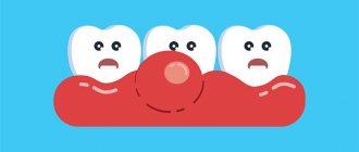
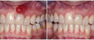
With periodontal inflammation, swelling and hardening of the gums are almost always observed. Gingivitis is considered the initial stage of the disease, therefore it is characterized by the so-called red ball (gum tumor), while with periodontitis, purulent discharge is observed, and the tumor acquires a beige or grayish tint. In some particularly advanced cases, against the background of periodontal problems, gumboil may appear, which visually looks much more noticeable.
Prevention of gum disease
All preventive measures to prevent gum disease come down to simple rules of oral hygiene, as well as timely and regular visits to the dentist. Since caries is treated in the early stages, professional dental care prevents all kinds of inflammatory diseases of the soft tissues of the oral cavity. In addition, you need to take into account that proper nutrition and the absence of bad habits are also the key to a healthy and beautiful smile. And it is also important to remember that a painkiller pill simply relieves a pain symptom, but does not cure. In this case, the disease passes into a more acute or chronic stage and develops into serious illnesses. Therefore, at the slightest manifestation of an inflammatory process on the gums, you need to consult a doctor and not self-medicate.
Non-infectious diseases that can cause a lump on the gum
- Epulis.
A small growth that has some kind of stalk, which occurs mainly due to injuries, malocclusions, improper prosthetics, during teething (in children), and also due to hormonal disorders (less often). In appearance, epulis may resemble a tumor due to gingivitis, so diagnosing the disease may require x-rays and histological examination. - Ecostosis.
This pathology is a bone growth that is formed due to a jaw anomaly. The main cause of ecostosis is considered to be a genetic predisposition, but pathology can develop after injuries and traumatic tooth extraction. In this case, the bone extends beyond its usual boundaries, resulting in the formation of a noticeable lump. The disease is not accompanied by an inflammatory process, but it can occur due to mechanical damage to the growth. - Hematoma.
It is formed as a result of a strong blow and may well go away on its own or with the use of special means. If the tissue structure is disrupted as a result of injury, the help of a surgeon is needed.
It hurts/doesn’t hurt: in what cases does pain occur?
Periostitis is considered the most painful. With this complication, the main inflammatory process is localized in the area of the bone and periosteum, which causes severe pain. Seals caused by the inflammatory process in the periodontium (gingivitis, periodontitis) are disturbing when pressed, thermal and mechanical influences are applied. Epulis and ecostosis, as a rule, are not accompanied by pain, but when damaged and inflamed, they also disturb the patient.
A lump near a tooth with periodontitis may not cause noticeable pain, especially if it has a fistulous tract. It is important to understand that the absence of pain does not mean that treatment can not be carried out. In most situations, the tooth can be saved, but some patients are very negligent about their health and consult a doctor in extreme cases. A lump above (or under) a tooth is quite an alarming signal, which often indicates the development of quite serious diseases.
Preventive measures
Preventing gum bumps is quite simple. The main methods will be daily thorough oral hygiene and timely visits to the dentist.
To prevent the appearance of cones, rinse with aloe juice diluted with water in a ratio of 1:4. For the same purpose, use an infusion of fresh sorrel leaves: to complete the procedure, 2 tbsp. of crushed raw materials, brew 200 ml of boiling water and leave for at least 1 hour.
For patients who have a lump on their gum, early diagnosis and appropriate treatment are of great importance. Only a qualified specialist will help you safely get rid of gum formation, minimizing the risk of developing unwanted complications.
This article is for informational purposes only, please consult your doctor for details!
Lump after crown installation
Improper denture fitting is a very common cause of lumps on the gums. Most often, errors occur at the tooth preparation stage, when the depulpation procedure is carried out. Poor canal filling provokes the development of infection, which, in turn, leads to periodontitis. Another common reason is a poorly made prosthesis or defects during its installation. In this case, the prosthesis injures the mucous membrane and causes swelling, abscesses and growths. Treatment almost always involves removal of the prosthesis and subsequent replacement of the prosthesis (if possible). The sooner a complication is detected, the higher the chance that it can be successfully treated. A well-installed prosthesis should not cause pain, discomfort or injure the mucous membrane. The tooth under the crown should also not hurt, since the dental nerve is killed before permanent prosthetics.
Diagnosis and treatment of gum formations
A gingival lump can be easily identified during a visual examination and instrumental examination. To clarify the diagnosis, an x-ray or biopsy may be indicated.
Treatment methods are determined by the root cause of the formation on the gums. First of all, you will need to limit the further development of the infection and eliminate pain.
Traditional treatment is as follows:
- fistula
– elimination of pus by rinsing (using Chlorhexidine, Miramistin, decoctions of medicinal herbs, saline solution);
- epulis
– surgical treatment (removal with a scalpel, cryodestruction or diathermocoagulation);
- periodontitis
– unsealing and cleaning of root canals, removal of accumulated pus, mouth baths made of soda solution or herbal decoctions;
- periostitis
– opening the tooth and placing special medications under a temporary filling (if there is no positive result, the diseased tooth must be removed);
- gingivitis
– cleaning of periodontal pockets, antibacterial and antiseptic treatment, removal of pathological formations.
Traditional medicine offers treatment with freshly squeezed juice of Kalanchoe leaves, used in the form of lotions. Another option is to chew the leaves, having previously cleared them of the film. A popular remedy for bumps in the mouth is rinsing with regular vodka.
Treatment of lumps on the gum
The treatment plan depends entirely on the results of the diagnosis, which is designed to determine the root cause of the lump or ball on the gum, which is why the patient should not self-medicate, but go to the doctor as soon as possible. If the cause of the lump is an infection, then you need to eliminate the cause of its appearance. For complications of pulpitis and periodontitis, treatment can take a long time. The lump is opened, a drainage is installed to pump out the pus, after which the doctor prescribes conservative or surgical treatment to the patient, depending on the severity of the disease. In case of periodontal inflammation, professional cleaning and sanitation of the oral cavity are carried out, special ointments and antiseptics are prescribed. In more complex cases, curettage, physiotherapy, and so on are used. With epulis and ecostosis, pain and inflammation are often absent, so the decision about intervention is made together with the patient. Most often, the pathology will be removed after surgery: even a small bump on the gum (especially in the smile area) spoils the aesthetics, so it is understandable that a person wants to get rid of it.
Clinical picture
Features of the manifestation of the clinical picture will depend on the root cause of the pathology and the age of the patient.
In an adult
In adults, the cause of lump growth is most often infectious diseases, which are characterized by the following forms of manifestation:
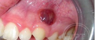
Fistula
Formed as a result of inflammation of dental or periodontal tissues. A fistula is a formation that rises above the level of healthy gums, with a diameter of 3 to 5 mm .
In the center of the elevation there is a white dot , which defines a channel for the release of pus from the source of inflammation. The gums in the area of elevation are hyperemic and painful.
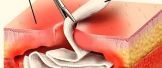
As a rule, the formation of a fistula signals the growth of an infectious process. To ensure the outflow of exudate, you need to visit a dentist, who will open the lump and put drainage into its cavity.
After the outflow of all contents, treatment is carried out to relieve inflammation and the source of infection. Even with regular release of pus without concomitant therapy, the inflammatory process is not eliminated and can take a chronic form.
More details about the fistula in the following video:
What are the types of non-carious lesions on human teeth?
Let's discuss topical anesthesia in dentistry here.
The price of a filling for a front tooth https://www.vash-dentist.ru/lechenie/zubyi/plombyi/iz-chego-skladyivayutsya-tsenyi.html is announced at the link.
Periodontitis
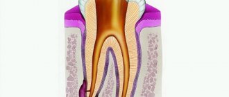
It is characterized by the appearance of a dense, voluminous lump about 1 cm in diameter . The formation is characterized by slight pain that occurs upon palpation of the gums. The cause of periodontitis is teeth affected by caries with inflamed canals and pulpitis, which contribute to the accumulation of bacteria at the base of the root apex.
Gradually, a granuloma forms at the site of accumulation , and without subsequent treatment it degenerates into a cyst . Treatment options for the pathology will depend on the degree of neglect of the process.
In case of minor inflammation, carious tissue is removed, the canals are cleaned, and a medicinal preparation is placed in the tooth cavity, which is covered with a temporary filling. After the inflammation is removed, the tooth is once again cleaned of pathological tissues and filled, completely restoring the coronal part.
Periostitis
It is a complication of periodontitis, in which the infection affects the bone tissue of the jaw. Periostitis is manifested by the growth of lumps on the gums, swelling and redness of the periodontium. In the area of inflammation, constant acute pain occurs, which intensifies as the formation grows .
At the same time, a person experiences an increase in lymph nodes and body temperature. By the deterioration of the general condition one can judge the severity of the pathology.
Treatment of pathology begins with cleaning the canals into which the medicine is placed. After the inflammation is relieved, the medicine is removed, and a new medicine is placed in its place and covered with a temporary filling.
Treatment lasts about 2 months. Only after the source of infection and the consequences of inflammation have been eliminated, the tooth is filled with a permanent composite.
After tooth extraction
The cause of the development of a lump in this case may be a hematoma or inflammation . A hematoma is formed due to damage to blood vessels, which is not uncommon during the process of tooth extraction.
During the normal course of the process, the hematoma does not cause discomfort and resolves after some time. Its soreness and change in color may signal the onset of inflammation.
Also, inflammation can occur without a hematoma, as a result of a blood clot breaking off and bacteria entering the hole. Pain that increases every day after tooth extraction is an indicator of developing inflammation.
If the pain is accompanied by redness of the gums, severe swelling and bleeding, then you need to quickly contact a dentist who will carry out the necessary therapeutic procedures.
Cyst
The reason for its development is advanced periostitis. In this case, a voluminous lump with a diameter of 1 cm appears on the gum , which has a dense structure . The lump can cover both the area of the affected and adjacent tooth.
Soreness and hyperemia are not observed, but there is a constant putrid odor from the mouth. If a cyst is detected in a multi-rooted tooth, partial root resection is performed while preserving other roots and neurovascular bundles.
If a cyst is diagnosed in a single-rooted tooth, the only treatment option is its removal.
Fibropapilloma
It is a small, slowly growing formation on the gum , benign in nature. It occurs due to the proliferation of periodontal tissue cells.
The tumor is painless on palpation and acts only as an aesthetic defect. To eliminate this problem, surgical intervention is used.
How dangerous is a dental x-ray in the first weeks of pregnancy for a child?
Read the link https://www.vash-dentist.ru/lechenie/zubyi/infiltratsionnaya-anesteziya-v-stomatologii-chto-eto-i-kak-provoditsya.html about the technique of infiltration anesthesia in dentistry.
What is dental physiotherapy here is a description
The child has
White bumps on the gums in childhood can appear during the eruption of temporary or permanent teeth, and can also act as complications of dental diseases. In each case, they have different causes and a specific clinical picture:
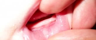
hard lump
Occurs during the period of eruption of units at the site of the future tooth. In this case, there is slight swelling of the gums and pain , which prevents the child from chewing food normally.
As a rule, teething occurs within a few days and the accompanying symptoms disappear. In isolated cases, an additional hematoma may form at the site of the bump.
To reduce pain and relieve inflammation of the gums, it is recommended to treat them with special gels or ointments for children. For example, Dentinox or Kalgel.
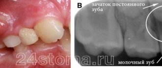
If the lump has formed next to the milk unit, then this indicates the growth of a permanent tooth. If the lump increases significantly and the tooth does not fall out for a long time, you should consult a dentist.
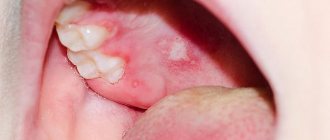
Purulent lump
The appearance of a lump of this nature requires immediate attention to the dentist, as it indicates the development of a serious inflammatory process , which not only affects the tissues of the oral cavity and jaw, but can spread to other organs. The growth of a tumor near a baby tooth will lead to its removal.
An abscess on a permanent tooth is treated by preparing the tooth and canals from tissues affected by caries. The medicine is placed into the cleaned cavity and covered with a temporary composite. After the inflammation has been eliminated, the tooth is restored with permanent filling material.