Lipoma and skin atheroma are two common types of benign neoplasms. They require exceptionally attentive treatment, since in some cases (though, fortunately, not often) they can degenerate into malignant tumors. The appearance of atheroma may not cause suspicion - at first it usually does not cause much inconvenience. However, even if the tumor is not painful, you should still see a doctor. Often a lump (lipoma) on the neck or scalp gradually increases in size, in this case you need to visit a doctor urgently - the new growth will need to be examined to determine whether there is a risk of developing cancer.
Lipoma and atheroma are often similar in appearance, and patients often do not distinguish them from each other, defining them under the general name “wen.” Let's try to figure out what the difference is between a lipoma and an atheroma, and also what to do if you have one of these formations.
Services and prices
Removal of lipoma more than 3 cm
7000 ₽
Sign up
Removal of lipoma up to 3 cm
4000 ₽
Sign up
Lipoma is the most common soft tissue tumor and consists of fat cells surrounded by a thin fibrous capsule. Popularly, such a neoplasm is called a wen.
Often, a person who is attentive to his body may stumble upon such a subcutaneous formation. Such a finding can cause, at a minimum, caution, and sometimes fear of the oncological process. The unknown is always scary. If you have any concerns, you should consult your doctor. In most cases, subcutaneous neoplasms in soft tissues are benign and do not pose a threat to the owner of the wen. However, lipomas still require the supervision of a specialist who must recognize a malignant process, if one occurs.
Peculiar red flags, signs that make you wary, are rapid tumor growth, pain, and the presence of two or more similar formations on the body.
By external signs, lipoma is little distinguishable from liposarcoma, hygroma, subcutaneous cyst, hematoma, parasitic invasion, inflammation or consequences of injury. Therefore, it is important that any neoplasms be examined by a doctor.
First of all, the question of the malignancy of the tumor is decided at the appointment. Liposarcoma occurs more often in middle-aged and elderly people. This is an aggressive and fast-growing tumor that can put pressure on surrounding organs and tissues and cause pain. Situations when an existing lipoma degenerates into a malignant tumor rarely occur. However, the oncological process requires a radically different approach to diagnosis and treatment, so it is important to diagnose it as early as possible.
Lipomas may have a hereditary predisposition. This fact, combined with the spread of wen to other parts of the body, makes the doctor suspect lipomatosis. Lipomatosis accompanies a number of hereditary syndromes such as Madelung's disease and Dercum's syndrome. Diseases with a family history require a special approach to therapy.
What is atheroma?
Atheroma is a cyst of the sebaceous gland that occurs when its duct is blocked. Sebum cannot come out and accumulates inside, it stretches the gland, and it turns into a cystic cavity. This can happen for a variety of reasons, often due to trauma to the skin.
Atheromas most often occur where there is a lot of hair and sebaceous glands: atheromas on the scalp, face, neck, back, genitals.
In its manifestations, atheroma resembles a lipoma: it is also a “bump” that is located under the skin, does not cause pain and easily moves when pressed. But there is one characteristic sign: usually there is a hole in the skin in the area of atheroma through which contents with an unpleasant odor are periodically released. Inside the atheroma there is a substance that resembles cottage cheese or paste in consistency.
Atheroma is a benign formation. It's not cancer. The only thing that is dangerous about it is the possibility of suppuration. An infected and inflamed “bump” swells, turns red, and becomes painful. Sometimes it opens and pus comes out along with fat.
Atheromas can degenerate into malignant tumors, but this happens very rarely.
Reasons for appearance
There are many factors that influence the formation of lipomas. Among the reasons are hereditary predisposition, impaired metabolism of fatty acids in the body, liver disease, pancreatic disease, non-compliance or violation of hygiene rules.
For a long time it was believed that soft tissue injury predisposes to the development of lipomas, but this fact was subsequently refuted in research. Thus, doctors agree that one reason that would explain all the processes has not yet been found. However, predisposition to gastrointestinal lipomas has a proven connection with a gene mutation on chromosome 12. In other cases, the reasons remain unknown.
By structure
Lipomas according to these characteristics are divided into:
- Classic (there is only fatty tissue inside);
- Angiolipomas (have vessels inside);
- Hibernomas (in the fiber there are formations similar to the formations of hibernating animals);
- Myelolipomas (hematopoietic and adipose tissue are located together);
- Myxolipomas (contain mucous tissue elements inside);
- Myolipomas (muscle fibers are found together with fatty tissue);
- Fibrolipomas (there is connective and fatty tissue inside).
Symptoms and classification
Lipomas are distinguished by anatomical location into lipomas of the head, face and neck, lipomas of the trunk, extremities, chest (mediastinum), mammary gland, gastrointestinal tract, internal organs, retroperitoneal tissue, spermatic cord. There are also rare localizations in the myocardium, lungs, and meninges.
Another classification of adipose tissue tumors involves a clinical division:
- Lipoma surrounding nerve structures is called perineural . Due to compression of the nerves it can cause severe pain. Removal of perineural lipomas differs from subcutaneous lipomas and requires a highly qualified surgeon;
- A tumor growing in the spinal canal (usually in the lumbar region) is called lumbosacral lipoma; Mostly occurs in children and is combined with underdevelopment of spinal structures;
- Lipoma of the joint and its structures (synovium, vagina, tendons);
- Intermuscular lipomas are formed from areas of adipose tissue between muscle fibers;
- Angiomyolipoma is a tumor of fatty and muscle tissue, which in most cases grows in the kidneys and pancreas. Middle-aged and mature men are more predisposed to its formation;
- Subcutaneous lipoma is a formation of varying sizes in the subcutaneous fat tissue. In everyday life it is usually called wen.
Lipomas usually occur alone. However, some patients discover several tumors at once. Such cases are most often associated with hereditary diseases and require careful study by specialists. The most common places for lipomas to form are the neck, back and limbs.
A subcutaneous lipoma is a mobile, elastic seal in the form of a lump or ball, which does not cause pain when pressed. A neoplasm can cause pain if it grows beyond its capsule into healthy tissue or due to compression of adjacent nerves.
For example, a wen located on the head can cause headaches, and the same formation on the neck can cause hoarseness and difficulty swallowing.
Gastrointestinal lipomas differ from their subcutaneous counterparts. Small formations in the intestine do not cause symptoms and are often discovered incidentally during an instrumental examination of the gastrointestinal tract. However, if it increases in size, this happens when the tumor reaches 2 or more centimeters in diameter, the lipoma can block part of the intestinal lumen and cause intestinal obstruction, intussusception, stool problems, abdominal pain and even bleeding.
Features of pathogenesis
There is no consensus among specialists about the neoplasm. Some sources consider it to be hypertrophied fatty tissue, others - a true tumor. If the process occurs in a place where there is no fatty tissue, then it is associated with a change in the structure of connective cells.
One theory states that the accumulation of adipocytes in a certain zone is due to chromosomal gene abnormalities responsible for the production of TAG lipase. The enzyme is responsible for providing energy through the breakdown of fats.
Some experts are confident that the appearance of subcutaneous wen is explained by the active deposition of lipids in adipose tissue. The prerequisites for the formation of a lipoma appear during embryonic development, during its formation. In adult patients, wen can form anywhere. In rare cases, massive development of benign neoplasms is observed.
Treatment methods
The treatment method is selected based on the location of the tumor, size and medical history of the patient. First of all, the doctor must make sure that the patient’s neoplasm is benign. To do this, the doctor carefully examines the patient, collects anamnesis and, if necessary, refers him to the necessary tests. For the differential diagnosis of lipomas, ultrasound examination of soft tissues, computed tomography, magnetic resonance imaging are used and, if a malignant tumor is suspected, a tumor biopsy is taken.
The most common type of tumor from adipose tissue is a subcutaneous lipoma, which does not cause dysfunction of organs, systems and does not threaten the patient’s life, and removal is performed only for cosmetic purposes.
Also, indications for surgical treatment of lipoma are large tumor sizes, 5 cm or more, and the presence of symptoms caused by the tumor.
There is no effective conservative treatment for lipomas. It is also worth noting that traditional methods such as heating and applying ice do not affect tumors from adipose tissue. However, they can cause serious complications if the neoplasm is of a different nature. For example, when atheroma is heated (atheroma is an accumulation of sebaceous secretion that clogs the duct of the sebaceous gland and causes inflammation), inflammation may spread and infect healthy tissues that are nearby.
Among the surgical methods for treating lipomas are:
- lipoma excision
- liposuction
- laser removal
- endoscopic method (for gastrointestinal lipomas)
Diagnosis of lipomas
Since the development of a neoplasm in the body practically does not cause symptoms, the patient may not be aware of its presence for a long time. Therefore, in most cases it is diagnosed accidentally during a preventive examination or treatment of other pathologies.
If the localization of the formation allows the doctor to palpate, the specialist can determine the lipoma even during a standard physical examination. However, the placement of a node does not always allow it to be detected by superficial palpation. At the same time, quite a few dangerous diseases have symptoms similar to lipoma. Therefore, diagnostics are carried out not only to identify a neoplasm, but also to exclude other diseases and clarify the benign quality of the process.
A comprehensive examination includes a number of laboratory and instrumental examinations, the need for which is determined individually by the attending physician in each medical case:
- Blood tests. The most accessible method of primary assessment of the body’s condition. The results of the study allow us to identify the presence of pathological changes, inflammation, viruses or bacteria.
- X-ray examination. Depending on the location of the compaction, an X-ray of the chest, abdominal cavity, and extremities is prescribed. Allows you to diagnose a formation, identify its exact location, and also analyze the condition of bone tissue and structures.
- Ultrasonography. Scanning soft tissues and organs allows you to determine the size of the node, identify the clarity of its contours, and analyze the contents. Not the most informative examination method for suspected lipoma, since even in the presence of a capsule, the compaction is often difficult to visualize using ultrasound waves.
- CT scan. Allows you to establish an accurate diagnosis and distinguish lipoma from malignant neoplasms if there is suspicion.
- Magnetic resonance imaging. It is prescribed, if necessary, to evaluate the signs of compaction and distinguish it from malignant liposarcoma. Using this method, the diagnosis is established with maximum accuracy.
- Biopsy. Tissue collection from the compaction and their further cytological and histological analysis. Allows you to exclude the possible oncological nature of the pathology.
The examination also reveals the reasons that caused the formation of a compaction in the body. If other chronic diseases are a prerequisite for the development of pathology, additional diagnostics are also carried out for an accurate diagnosis and further effective treatment.
Advantages and disadvantages of various methods
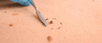
- Lipoma excision.
Excision of a lipoma is the simplest and most affordable way to remove a wen. The operation is performed under local anesthesia. The surgeon injects the lipoma with an anesthetic and removes the tumor along with the capsule through an incision in the skin. Removal of the fibrous capsule is an undeniable advantage of this method. This prevents the re-formation of lipoma in the old place, which means it reduces the risk of relapse to a minimum. This method also allows one to examine the histological structure of the tumor. In general, the operation lasts no more than half an hour.
- Liposuction.
Liposuction allows the tumor to be removed through a small hole. A special device is inserted into the cavity of the wen, which destroys the lipoma. Many doctors and patients love this method for its minimal invasiveness and good cosmetic results. However, the disadvantage is the inability to remove the fibrous capsule of the wen, which creates the possibility of future relapse of the tumor.
- Laser removal.
Laser techniques are used to eliminate tumors larger than 3 cm. The method is gentle, carries minimal risks of bleeding and infection, and does not leave wounds or scars.
How are atheromas and lipomas treated?
Atheromas are treated surgically; the type of operation will depend on the size of the formation. Small sebaceous cysts can be removed with a laser. Most often, atheromas are large in size and are removed with a scalpel. The “bump” on the skin is surrounded by two incisions, then the cyst is peeled out and removed along with a small piece of skin. Stitches are placed on the wound.
Atheroma cannot be cured by “sucking out” the contents with a needle and syringe. It is imperative to remove the walls of the stretched sebaceous gland - if they remain, they will begin to produce sebum again, and the cyst will grow again.
If atheroma suppurates, surgical treatment is performed, and the doctor may prescribe a course of antibiotics. Lipomas do not need to be treated. The operation is performed if:
- education is large and growing rapidly;
- the lipoma is in an inconvenient place and is constantly in the way;
- bothered by soreness;
- The patient himself insists on removing the lipoma.
Wen is removed in the classic way, using a scalpel. Relapses are possible, but extremely rare. Typically, atheromas and lipomas are removed on an outpatient basis; hospitalization is not necessary. Anesthesia is also not needed - local anesthesia is sufficient. The operation lasts on average 15–20 minutes.
Make an appointment by phone +7 (495) 120-08-07.
Preparation for the procedure
In case of subcutaneous lipoma removal, no special preparation is required. The mini-surgery is performed on an outpatient basis, meaning it does not require hospitalization. The surgeon performs all necessary manipulations under local anesthesia. Thus, the procedure is painless for the patient.
Giant subcutaneous lipomas, as well as neoplasms of the intestines, internal organs, and peritoneum require more serious and thorough preparation. Operations of this type are carried out with hospitalization of the patient. Before the intervention, samples are taken and, if necessary, additional studies are done. Operations performed under general anesthesia require restriction of water and food on the eve of the operation.
Treatment of lipomas with pharmaceutical products
Those who prefer to use pharmaceutical drugs should purchase Vishnevsky ointment. This product should be applied to an adhesive plaster and attached to the affected area, after 2 days the plaster should be replaced with a new one.
You can also use hydrogen peroxide. This drug should be regularly lubricated. After a few days, the skin on the affected area will burst and the contents of the wen will leak out.
lipoma treatment
How a doctor can help you remove a wen
It is highly advisable that lipoma removal be performed by an experienced specialist. After a thorough examination, a dermatologist may prescribe the following procedures to remove wen:
- Mechanical cleaning. This method is considered the simplest. The procedure is carried out by piercing the wen with a needle, after which all its contents are removed. Sometimes the procedure is performed under local anesthesia.
- Removal of lipoma with laser . Laser therapy is considered the most progressive method for removing fatty tissue. The procedure is very quick and does not leave scars. In addition, the possibility of the wen reappearing in the same place is excluded.