General information
A malignant neoplasm in the tongue comes from the mucosal epithelium.
Despite the fact that tongue tumors are uncommon, they have an aggressive course, so early detection is important. Distinctive features of this disease are early metastasis (including distant), rapid growth, resistance to treatment, high recurrence rate and mortality. At the time of diagnosis, half of the patients had affected regional lymph nodes. Recurrence is observed in 30-50% of patients. If we consider the localization, then most often a tumor of the tongue occurs on the lateral surface, much less often there is cancer of the root of the tongue, the back and the tip. A neoplasm of the root of the tongue is classified as oropharyngeal cancer. The asymptomatic course of the disease makes early diagnosis difficult. Tumors are either not visible in the early stages or initial changes are regarded as a consequence of trauma to the tongue. Due to the lack of alertness of patients and doctors regarding malignant neoplasms, most cases are diagnosed with a full clinical picture or already in an advanced stage.
The highest incidence is observed after 50 years of age, but this disease also occurs in people under 30 years of age. In men, this pathology is diagnosed 5 times more often. In 95% of cases, tongue tumors are squamous cell carcinoma. Squamous cell carcinoma of the tongue is an aggressive tumor and insensitive to treatment ( chemotherapy ), but radiation treatment .
Possibilities of ultrasound diagnostics in identifying initial tumors of the tongue
Ultrasound scanner HS60
Professional diagnostic tools.
Assessment of tissue elasticity, advanced 3D/4D/5D scanning capabilities, BI-RADS classifier, options for expert cardiological studies.
Introduction
In socially and industrially developed cities, the incidence of malignant neoplasms (MNT) is still growing, which is directly related to environmental pollution, the spread of infectious diseases and addictions. Also, despite the increased quality of training of medical personnel in oncology and the development of medical equipment for diagnostics, mortality from tumor changes is not decreasing. The average annual growth rate of newly diagnosed oral cancers in Russia is 2.33% (standardized rate per 100,000 population in the period from 2008–2018) per year, the average mortality rate in 2022 was 6.84% [1].
The main treatment method for early stages of tongue cancer is surgery [2], and in more rare cases, radiation and/or chemotherapy. To plan radical surgical treatment of malignant tumor pathology of a given localization, even with small sizes, it is important to accurately assess the size of the tumor (especially its thickness) and local extent (involvement of anatomically important structures - muscles, vessels) and transition through the midline to the contralateral side. Assessing the thickness of the tumor is important because it can help in determining the strategy for postoperative management of the patient, since there is a relationship between the thickness of the tumor of the tongue mucosa and secondary damage to the regional lymph nodes of the neck [3–7].
Ultrasound examination (ultrasound) in the primary and clarifying diagnosis of malignant tumor changes in the oral cavity (in particular, tumors of the tongue) is currently used quite rarely. This is explained, first of all, by the accessibility of the oral cavity tumor for visual inspection and palpation. However, ultrasound performed by a trained specialist allows for an objective assessment of the local extent of the tongue tumor with high accuracy. Despite the high information content, low cost and availability of ultrasound, the method is still not in full demand in practice and in the world literature. The number of publications over the past 5 years is insignificant [8].
Based on this, the main purpose of ultrasound of the tongue with an intracavitary sensor is the diagnosis of small-sized tumors of the tongue (T1), assessment of local prevalence (including the depth of invasion) in patients with “initial” tumor changes or suspicions of them.
Material and methods
From 2016 to 2022 at the Moscow Oncology Research Institute named after. P.A. 29 patients came to Herzen regarding the appearance of superficial, flat formations on the mucous membrane of the tongue. At the time of treatment there was no established diagnosis. The changes were localized in the mobile part of the tongue (p/3 – 4 patients), base (sr/3 – 13 patients) and in the area of the tongue root (mainly near the border with sr/3 – 12 patients). Of the 29 patients, 13 (44.8%) were women and 16 (55.2%) were men. In 2 patients, the age was 25 and 27 years, in the remaining patients the age ranged from 42 to 65 years.
To study changes in the mucous membrane of the tongue, an expert-class ultrasound device with a laparoscopic sensor with a scanning frequency of 10 MHz was used. An expert-class ultrasonic device with a microconvex intracavitary sensor operating in the range of 3–10 MHz and a linear sensor with a frequency of 5–12 MHz was also used.
During the study, the patient was in a sitting or lying position with the oral cavity as open as possible. The patient fixed the tip of the tongue with his hand with a gauze pad and moved it in the opposite direction from the area of interest, thereby creating maximum space for maneuvering the ultrasound sensor. The scanning surface of the sensor was installed as perpendicular to the area of interest as possible. Whenever possible, the study was carried out in mutually perpendicular planes in gray scale mode (B-mode) and using color Doppler mapping (CDC).
The study assessed: 1) precise localization; 2) tumor size: thickness (depth of invasion), length of the tumor and its width; 3) structure of changes; 4) the presence of vessels and features of blood flow in the zone of changes; 5) involvement of anatomically important structures (involvement of the lingual artery, spread to the midline and to the contralateral part of the tongue, spread to the floor of the mouth) [9].
The mucous membrane of the tongue is represented sonographically as three linear structures. The superficially located structure (immediately adjacent to the sensor) has increased echogenicity and is quite homogeneous, the second is of reduced echogenicity and is quite homogeneous, the third structure (adjacent to the muscle tissue) is slightly increased echogenicity and has a homogeneous structure. The muscles of the tongue are echographically displayed as layers of linear structures of moderately reduced echogenicity (the echogenicity of the mucous membrane is somewhat lower than the echogenicity of the muscle tissue); the muscle bundles are separated from each other by thin layers of slightly increased echogenicity, like a “layer pie”. Each individual layer is homogeneous. In general, the muscle bundle (within the tongue) with clear and even boundaries has a connective tissue capsule, which on ultrasound looks like a hyperechoic layer with clear and even contours (Fig. 1). The normal thickness of the tongue mucosa on ultrasound ranges from 1 mm (the lower surface of the underwater part of the tongue) to 2.5 mm (the area of the root of the tongue), depending on the part of the tongue.
Rice. 1.
Echogram of unchanged mucous membrane of the tongue.
All patients included in the study group underwent MRI and verification as a result of cytological or histological examination. All patients selected for the study group had confirmed squamous cell carcinoma of the tongue.
results
Ultrasound of the tongue mucosa showed exophytic (papilloma-like) changes in 8 (27.6%) patients, and mixed growth types in 21 (72.4%) patients. All the formations we identified had a significantly reduced echogenicity (compared to the surrounding tissue), in the central parts of an almost homogeneous structure, but in the peripheral parts of these changes the structure was somewhat heterogeneous, the contours of all identified formations were uneven and unclear (Fig. 2).
Rice. 2.
An echogram of tumor changes in the tongue mucosa obtained using a linear sensor. Crosses indicate tumor changes.
During CDC, blood flow was not determined in 10 (34.5%) patients, and moderately expressed blood flow in the area of interest was determined in 19 (65.5%) patients (Fig. 3) [10]. In 19 (65.5%) patients with blood flow determined by CD, the thickness of the changes approached or exceeded 5 mm.
Rice. 3.
Echogram of tumor changes in the mucous membrane of the tongue using color circulation.
According to ultrasound data in this sample, the minimum visualized thickness of tumor changes was 2.2 mm (±0.2) (Fig. 4).
Rice. 4.
Echogram of tumor changes in the mucous membrane of the tongue, obtained using an intracavitary sensor. Crosses indicate tumor changes.
To plan and carry out further treatment, it is extremely important to clarify the degree of local prevalence. Based on the fact that tumor changes, unchanged mucosa and tongue muscles have different echographic patterns, we can differentiate them from each other and evaluate their relationship.
Visualized changes, ranging in thickness from 2.2 mm to 4 mm (in 9 patients - 31%), had a “layer” of unchanged tissue between them and the muscle tissue. Thus, according to the ultrasound picture, the tumor did not spread to the muscles of the tongue, which was confirmed by the results of a histological examination of the postoperative material.
In the remaining cases (20 patients - 69%) there were signs of involvement or intimate adherence to the muscles of the tongue. We determined invasion into muscle tissue by the following criteria: 1) the border between tumor changes in the mucous membrane of the tongue and muscle tissue was not visualized or was visualized in fragments; 2) the border between the mucous membrane and the muscle of the tongue is deformed; 3) there was no unchanged part of the mucosa between the tumor changes and muscle tissue (intimate adhesion) (see Fig. 2).
Taking into account the size of tumor changes and their location in patients included in the study, the described changes did not extend to the midline of the tongue, the lingual artery, or the floor of the mouth.
Theoretically, tumor changes in the mucous membrane of the tongue of “small size” can spread to the mucous membrane of the floor of the mouth, to the midline and to the contralateral parts of the tongue with appropriate localization - the lower, inferolateral surface of the tongue (the area of adjacent mucosa of the tongue to the mucous membrane of the floor of the mouth) and on the back of the tongue near midline. In our sample there was 1 (3.4%) patient with an exophytic papilloma-like formation located in the tip of the tongue and in the midline and without spreading beyond the mucous membrane.
All patients participating in the study underwent MRI, according to which 2 (6.9%) patients had no evidence of a tumor process (patients with tumors 2–3 mm thick). In 3 (10.3%) patients, according to MRI data, there was a suspicion of the presence of tumor changes in the mucous membrane of the tongue. In the remaining patients (24 - 82.8%), the MRI picture corresponded to tumor changes.
All patients underwent surgical treatment. According to the histological report, these changes in the mucous membrane of the tongue corresponded to malignant tumor changes. Also, according to the conclusion of a routine histological examination, the involvement of muscle tissue was established in 22 (75.9%) patients; according to ultrasound data, invasion of muscle tissue was determined in 20 patients.
The sizes of tumor changes obtained by ultrasound and MRI coincided within the error of the method (±1–2 mm).
The main difficulty in studying pathologies of the tongue mucosa is the insufficient opening of the oral cavity as a result of pathological processes that cause dysfunction of the temporomandibular joint (trismus, inflammatory changes and their consequences, post-radiation fibrosis, etc.).
Conclusion
Despite the device- and operator-dependence of ultrasound diagnostics, it allows one to visualize tumor changes in the oral cavity and, in particular, tumor changes in the mucous membrane of the tongue. Based on the results of the study, ultrasound diagnostics, on a par with MRI, makes it possible to visualize “initial” tumor changes in the mucous membrane of the tongue and assess the local prevalence, and in some cases surpasses it.
Literature
- Kaprin A.D., Stalinsky V.V., Petrova G.V. Malignant neoplasms in Russia in 2022. M., 2022: 31.
- Omura K. Current status of oral cancer treatment strategies: surgical treatments for oral squamous cell carcinoma // Internat J Clin Oncol. 2014; 19: 423–430.
- Liu B., Amaratunga R., Veness M. et al. Tumor depth of invasion versus tumor thickness in guiding regional nodal treatment in early oral tongue squamous cell carcinoma // Oral Surg Oral Med Oral Pathol Oral Radiol. 2020; 129(1):45–50.
- Loganathan P., Sayan A., Hsu DWK et al. Squamous cell carcinoma of the anterior tongue: is tumor thickness an indicator for cervical metastasis? // Clin Paper Head Neck Oncol. 2017; 46:407–412.
- Sohail Ahmed Khan, Sadaf Zia, Syeda Uzma Naqvi et al. Relationship of Oral Tumor Thickness with the rate of lymph node metastasis in Neck based on CT Scan // Pak J Med Sci. 2017; 33(2):353–357.
- Tarabichi O., Bulbul MG, Kanumuri VV et al. Utility of intraoral ultrasound in managing oral tongue squamous cell carcinoma: Systematic review // Laryngoscope. 2019; 129(3):662–670.
- Thomas JW, Klein Nulent, Rob Noorlag et al. Intraoral ultrasonography to measure tumor thickness of oral cancer: A systematic review and meta-analysis // Oral Oncol. 2018; 77:29–36.
- Farhoud Faraji, Stephanie F. Coquia, Meghan B. et al. Evaluating oropharyngeal carcinoma with transcervical ultrasound, CT, and MRI // Oral Oncol. 2018; 78: 177–185.
- Solovyov V.A., Reshetov I.V., Mitina L.A., Stepanov S.O., Kazakevich V.I., Shcherbina V.G., Borodina N.B. Ultrasound examination for cancer of the tongue and floor of the mouth // Medical visualization. 2015; 1:26–31.
- Chika Yamamoto, Kenji Yuasa, Kazuhiko Okamura et al. Vascularity as assessed by Doppler intraoral ultrasound around the invasion front of tongue cancer is a predictor of pathological grade of malignancy and cervical lymph node metastasis // Dento maxillo facial Radiol. 2016; 45(3):20150372.
Ultrasound scanner HS60
Professional diagnostic tools.
Assessment of tissue elasticity, advanced 3D/4D/5D scanning capabilities, BI-RADS classifier, options for expert cardiological studies.
Classification
Depending on the location, cancer is distinguished:
- Bodies. These include tumors of the tip, dorsum and lateral surface (most often the middle of the lateral surface). This localization occurs in 70% of cases.
- Root (accounts for 20% of all cases).
- Lower surface (sublingual area).
By growth pattern:
- Exophytic form (this includes papillary and ulcerative forms of tumors growing outward).
- Endophytic (growing inside the tissue of the tongue - infiltrative and ulcerative).
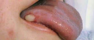
Papillary form a is a dense papillary growth of a mushroom shape. There may also be raised plaque-like growths that have clear boundaries.
Ulcerative (occurs in 50% of cases). Characterized by a superficial ulcer surrounded by a ridge. The ulcer is constantly increasing in size. At first, the ulcer does not bother the patient, but as it grows, pain and bleeding appear. The ulcer can become infected and inflamed, making diagnosis difficult.
Infiltrative form . The tumor grows into the thickness of the tissues, and they become denser. With diffuse growth, the compaction spreads to the entire tongue, which impairs its mobility.
The infiltrative-ulcerative form is characterized by thickening of the tongue with the presence of deep ulcers.
According to histological composition:
- Adenocarcinoma.
- Squamous cell carcinoma.
Classification according to the TNM system.
- Tis "cancer in place."
- T1 Tumor no more than 1 cm. Not accompanied by complaints, discovered by chance during examination.
- T2 ≤2 cm. There are ulcers and areas of compaction.
- T3 >4 cm. The tumor is equal in size to half the tongue. Metastases to the occipital lymph nodes, postauricular.
- T4 Locally advanced cancer that occupies the entire tongue and invades the tissues of the mouth and face. Metastases to internal organs (brain, liver, heart) and bones.
Symbol N - the presence of metastases in the lymph nodes:
- N0 - No lymph node involvement.
- N1 One node on the side of the tumor is affected.
- N2 Metastasis: one metastasis no more than 6 cm, several up to 6 cm on one side or both sides up to 6 cm.
- N3 Metastases larger than 6 cm.
With T1 tumors, lymph nodes are affected in 40% of cases, with T4 stage in 85%. A reliable factor for metastasis is the depth of invasion - 4 mm is considered a critical value. Most often, metastases are found in the submental, submandibular and cervical nodes (upper third).
Histopathological differentiation:
- G1 High degree.
- G2 Medium.
- G3 Low.
Causes
- Bad habits. Among which, smoking is of particular importance. The influence of carcinogenic substances in tobacco in the development of cancer has long been proven. The risk increases with duration and intensity of smoking. The risk increases further with alcohol consumption.
- Occupational hazards (contact with radiation, work in the oil industry).
- Constant traumatization of the mucous membrane (poor-quality dentures and damaged teeth, poor treatment of fillings, and constant biting of the tongue are important).
- Chewing mixtures (betel nut, non-smoking tobacco), consuming spices that have a carcinogenic effect.
- Oral infection.
- Pre-tumor conditions ( leukoplakia , Bowen's disease , ulcerative-erosive form of lupus erythematosus , papillomatosis , erythroplakia , leukokeratosis ).
- Action of oncogenic viruses. The connection of this pathology with chronic human papillomavirus infection and HIV . The oncogenic effect of viruses is associated with blocking or the influence of tumor suppressor genes.
- Long-term use of immunosuppressive drugs.
Symptoms of tongue cancer
During the course of the disease, the initial stage, developed and advanced, is distinguished. It is important to know and identify the symptoms of the initial stage. Tongue cancer initially has an asymptomatic course. What does the neoplasm look like?
It may manifest itself:
- whitish spots similar to plaque;
- compaction and slight redness;
- painless nodules;
- superficial ulcers or cracks.
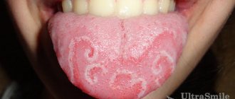
Most often, such changes are located on the lateral surface and are mistaken for glossitis (inflammation of the tongue). It is important that conservative treatment (treatment with antiseptics, sea buckthorn oil) does not help. It is rare, but it still happens that already at an early stage discomfort (burning or tingling) appears when eating and pain.
Photo - initial stage of cancer of the lateral surface of the tongue
Signs of tongue cancer at an advanced stage are characterized by pain, which is observed in 100% of cases. It has different intensities, can be local and diffuse, and also radiate to the temple, various areas of the mouth and ear. Patients are bothered by a feeling of numbness. When an infection occurs, bad breath appears. If the tumor begins to disintegrate, necrosis products irritate the mucous membrane and cause salivation. Bleeding of the tongue may also occur.
The advanced stage is characterized by an aggressive course and rapid invasive growth. Signs in an advanced stage are massive destruction of the tumor and surrounding tissues, including bone structures, and metastasis to local lymph nodes (occipital, submandibular, cervical and mental). With this disease, distant metastases occur in the lungs, bones, brain and liver. As the tumor disintegrates, the smell from the mouth increases, difficulty swallowing, difficulty speaking, numbness of the tongue and bleeding are observed. Common symptoms include weakness, weight loss and exhaustion.
Symptoms of tongue root cancer
This localization, which is observed in 20% of cases, is characterized by a rapid and aggressive course. There is a sore throat and discomfort when swallowing as the tumor reaches a large size. It often spreads to the auditory nerve. Patients have impaired tongue mobility.
Stages
According to the generally accepted classification, tongue oncology has four stages, differing in the degree of spread of pathological tissue.
- The size of the affected area does not exceed 2 centimeters, the process has not spread to the lymph nodes and other organs.
- The tumor grows and grows into muscle tissue, but does not exceed 4 centimeters in size. Lymph nodes and other organs are not affected.
- The tumor spreads within the tongue and exceeds 4 centimeters in size, or can be of any size with metastasis to one regional lymph node.
- The malignant process spreads to most of the tissues of the oral cavity, metastases penetrate into distant lymph nodes and organs - liver, lungs, brain, bone tissue.
Tests and diagnostics
To clarify the diagnosis, patients are prescribed:
- Biopsy of a tumor formation (or scraping and impression smears from the surface of erosions).
- CT and MRI of the affected area with intravenous contrast. This study makes it possible to assess the extent of the tumor and the depth of invasion into the tongue tissue.
- CT scan of the facial bones with contrast if there is a suspicion of tumor spread to the bones of the jaw and skull.
- Ultrasound of the lymph nodes of the neck.
- Puncture of the affected cervical nodes with cytological examination.
- Ultrasound of the abdominal cavity to exclude distant metastatic process.
- Chest X-ray to exclude metastases.
- Osteoscintigraphy for metastatic lesions of skeletal bones.
Questions and answers
Is there a cure for tongue cancer?
Treatment of a tumor will be most successful if it is detected at an early stage of the disease. As the cancer process spreads to other organs, the effectiveness of treatment decreases, however, even in the most advanced cases, oncologists strive to prolong the patient’s life as much as possible.
How to determine tongue cancer?
It is impossible to independently determine that a spot or growth that appears on the tongue is a cancerous tumor. A histological analysis of the tissue is necessary to confirm or refute the suspicion of tongue cancer. If there are any suspicious signs, you must contact a qualified oncologist and undergo a special examination.
How long do people live with tongue cancer?
According to medical statistics, with timely detection and treatment of tongue cancer, patients live from 5 to 15 years. In the absence of medical care, the patient’s life expectancy does not exceed one and a half years.
Attention! You can cure this disease for free and receive medical care at JSC "Medicine" (clinic of Academician Roitberg) under the State Guarantees program of Compulsory Medical Insurance (Compulsory Medical Insurance) and High-Tech Medical Care. To find out more, please call +7(495) 775-73-60, or on the VMP page for compulsory medical insurance
Diet
Diet for tongue cancer
- Efficacy: therapeutic effect for a month
- Timing: constantly
- Cost of products: 1300-1500 rubles per week
Difficulties with swallowing in the patient before and after surgery force the installation of a nasoesophageal tube. Through it, broth with meat and eggs beaten in a blender, sour cream, cream and other high-calorie liquid products are introduced. A nutritional mixture must be prescribed in addition to the main diet . This is a liquid high-calorie and high-protein mixture enriched with micronutrients. This can be Nutrizon Advance , Nutrizon Protein Advance , Nutrizon Energy , Nutrizon Protein Intense , Supportan Mixture for enteral nutrition, Fresubin original , Fresubin Energy , Nutricomp . With the transition to natural nutrition, a diet table with liquid dishes is organized - puree soup, boiled fish and meat (veal), poultry (turkey, chicken), whipped in a blender, meat and fish soufflé, liquid omelet, yoghurt, milk, cauliflower, broccoli in the form of mashed potatoes, mashed potatoes.
Diagnosis and treatment of tongue cancer in Moscow
If you find external signs of tongue cancer, contact the Medicina clinic for prompt and high-quality diagnosis. Once the diagnosis is confirmed, qualified oncologists provide treatment in accordance with the protocols of the world's leading clinics. The examination is carried out using the most modern diagnostic equipment available at the moment. For treatment and the subsequent rehabilitation period, we offer comfortable wards in the inpatient department, where patients receive professional care and high-quality medical service.
Prevention
- Quitting bad habits (smoking, alcohol).
- Elimination of tongue injury (high-quality treatment of fillings after their installation, correct selection and correct installation of dentures, timely treatment of dental chips).
- Regular oral hygiene, including professional.
- Timely detection of precancerous diseases of the tongue ( leukoplakia , dyskeratosis , papillomatosis ).
- Diet rich in vitamins . It has been proven that vitamin A and β-carotene are important in the prevention of neoplasms. With a deficiency of carotenoids, there is a risk of squamous cell carcinoma .
Forecast
With early diagnosis and radical treatment, the prognosis for recovery from tongue cancer is more favorable. It is believed that complete recovery from cancer is impossible and the goal of treatment is to achieve long-term remission of 5-8 years. Thus, with this pathology, the five-year survival rate ranges from 65 to 85%. Complete remission within 5 years after surgery and radiation therapy with T1 is achieved in 80% of patients, with T2 in 60% of patients, and with T3-4 does not exceed 35%. Metastases in the lymph nodes are an important prognostic factor and, if present, survival rate is halved.
How long do people live with stage 4 tongue cancer or with relapse after chemotherapy? Such patients have a poor prognosis: they live no more than 3.5 months while on maintenance treatment. The addition of cetuximab to chemotherapy increases survival by 2 times.
Treatment
The treatment strategy depends on the stage of tumor development and the spread of the cancer process to other tissues. The most commonly used combination of several basic methods.
- Surgery. For a tumor whose size has not reached 4 centimeters, as a rule, it is limited to the removal of malignant tissue of the tongue and affected lymph nodes, if any. If the malignant process has spread to surrounding organs, surgical treatment is combined with other methods.
- Radiation therapy. Dosed hard radiation is used as the main method for some types of tumors that have not reached a large size. When malignant tissue grows, radiation precedes surgery to reduce the size of the tumor. After the intervention, radiation therapy is prescribed to destroy the remaining cancer cells and avoid relapse. In case of inoperable cancer of the tongue with metastases to distant organs and large-scale damage to surrounding tissues, radiation can prolong the patient’s life.
- Chemotherapy. Drugs that destroy cancer cells or inhibit their growth are usually used in combination with radiation therapy as a palliative treatment for inoperable patients, as well as before and after surgery. Polychemotherapy is most effective for the treatment of poorly differentiated tumors and metastases to distant organs.
- Targeted therapy. Targeted drugs do not destroy the tumor. By blocking a certain type of protein, they prevent cell division and the further development of the malignant process. They are used only after molecular genetic analysis confirms the presence of the desired type of proteins.
List of sources
- Alieva. S.B., Alymov Yu.V., Kropotov M.A., Mudunov A.M., Podvyaznikov S.O. Cancer of the oral mucosa. Oncology. Clinical recommendations / Ed. M. I. Davydova. – M.: Publishing group RONC, 2015, pp. 27-37.
- Romanov, I.S. Features of regional metastasis of squamous cell carcinoma of the oral cavity, detected during preventive lymph node dissections. / Romanov, I.S., Yakovleva L.P., Udintsov D.B., Dzhumaev M.G., Tsiklauri V.T. // Dentistry. – 2012. – T.91, No. 4. — P. 28-31.
- Romanov I. S., Yakovleva L. P. Issues in the treatment of oral cancer. Farmateka 2013; (8):59–63.
- Paches A.I. Tumors of the head and neck. 5th ed., supplemented and revised M.: Practical Medicine, 2013. P. 119‒146.
- Romanov I. S. Prospects for the use of cetuximab in the treatment of squamous cell carcinoma of the head and neck / Oncology. Hematology. Chemotherapy. - 2015. - No. 17/1.