Most dental procedures are performed under local anesthesia. It allows for comfort for both the patient and the doctor.
Mandibular anesthesia is a conductive anesthesia that is performed for a mobile jaw and allows you to reduce the pain threshold of the lingual and inferior alveolar nerves. The technique is quite complex, and to obtain a high-quality result, a clear understanding of the anatomical structure of the dental system is necessary.
Using this anesthesia, the sensitivity of the following areas is blocked:
- half of the units of the dentition (from the insertion side);
- alveolar bone tissue;
- gum mucosa;
- half lip;
- the area under the tongue and the tip of the tongue;
- chin skin
What is mandibular anesthesia
In pediatric dentistry, when treating primary teeth, they most often resort to mandibular anesthesia - anesthesia of the lower alveolar and lingual nerves. The inferior alveolar nerve arises from the mandibular nerve, passes through the mandibular foramen into the canal and branches to each tooth.
The action of the anesthetic solution is directed to the mandibular opening. Until the age of six, it is at the same level as the chewing surface of the primary molars, and then rises by 6 mm at the age of 9 and another 3 mm at the age of 12. Therefore, such exposure is not advisable until the age of six.
Since the inferior alveolar nerve is covered by a bony visor, it can only be reached in the groove of the mandibular nerve. The solution falls down under the influence of gravity. Before manipulation, it is recommended to palpate the edges of the mandibular ramus in order to determine the target point as accurately as possible. The needle is placed slightly above the mandibular foramen.
Palpation introduction
Before insertion, the injection site is palpated, which makes it possible to determine the location of the opening of the lower jaw. This is the area where the mandibular nerve exits.
The effect occurs after 10-15 minutes and lasts for 2-3 hours.
After administration, the patient feels numbness, coldness and tingling in the half into which the drug was injected.
Advantages and disadvantages
The advantages include:
- reducing the risk of complications by obtaining anatomical landmarks using the palpation method;
- elimination of sensitivity occurs even in complex clinical cases;
- long-lasting action of the anesthetic;
- complete blocking of half the jaw, which allows you to treat several areas during one appointment.
Disadvantages of the tactile method:
- increased morbidity if the technique is not followed;
- a feeling of discomfort for the patient due to complete numbness of the anesthetized half of the jaw;
- the likelihood of biting the mucous membranes until the effect of the substance stops.
Apodactyl insertion
To implement this method, it is necessary to accurately take into account anatomical features. The localization of the pterygoid fold of the movable jaw, passing behind the molars, serves as a guide to clearly determine the location of the needle entry.
The duration of anesthesia is exactly the same as with the tactile method.
Procedure:
- Injection is administered at the medial end of the fold at the point where the upper and middle thirds border.
- The direction of the needle is perpendicular to the bone.
- The cylinder of the injection mechanism is localized on the opposite side of the premolars.
- The distance the needle advances to the bone is one and a half to two centimeters. This is where the first dose of pain medication is administered.
- Next, the syringe is transferred to the incisors, and the needle is advanced another two centimeters.
- Carrying out an aspiration test. If the test result is negative, the remaining anesthetic is administered.
The area of sensitivity blocking and complications are the same as with the tactile method.
Advantages and disadvantages of the apodactyl method
The pros and cons of the considered method are practically no different from the previous one. The difference lies in the increased likelihood of complications, which is associated with a discrepancy between the exit site of the nerve ending and the anatomical structure of the pterygoid fold of the mandible.
There is another variation of performing apodoctylic mandibular anesthesia (using the Verlotsky technique). The procedure is carried out by analogy with the classical method, with the exception of the injection site. Introduction according to Verlotsky involves inserting the needle centrally between the molars of the upper and lower jaw.
Technique of mandibular anesthesia
In pediatric dentistry, this type of anesthesia is performed intraorally. There are various methods, but this one has proven to be the best.
- The dentist determines the point at the intersection of the two lines. The horizontal line is a line running along the chewing surface of the lower molars or 3-5 mm above. The vertical line follows the direction of the pterygomaxillary fold.
- The injection is made at the intersection of these lines.
- After the injection, the needle is gradually advanced, releasing the solution, along the bone by 1.1-1.2 cm. During this period, up to 1 ml of medication must be injected.
- The correct position is this: the bevel of the needle is directed towards the bone, the syringe is deflected in the opposite direction.
- After the injection, the soft tissue in the injection area is pressed against the bone for 1-2 minutes.
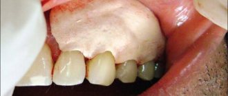
Possible complications after tooth extraction
After simple extraction of any tooth, complications occur quite rarely, provided that the doctor’s recommendations are followed.
The main thing is that the patient needs to remember that if the blood clot remains in the socket until healing, then infection and suppuration of the wound will not occur, and within two weeks a healthy gum mucosa will appear at the site of the clot. In case of loss of a blood clot, the unprotected wound surface of the socket is exposed to pathogenic microflora of the oral cavity; the accumulation of pathogenic bacteria provokes the development of an inflammatory process - alveolitis.
Infection of the alveolar socket is the most common complication after tooth extraction. In this case, the infection penetrates and affects the soft tissues surrounding the tooth. If an infectious infection of the hole is accompanied by the presence of throbbing pain and suppuration, this signals the development of an abscess. When alveolitis is advanced, there is a danger of the inflammatory process spreading to the jaw bone - the development of osteomyelitis.
Complex (traumatic) tooth extraction has more prerequisites for the development of inflammatory complications. If after removal there remains a piece of tooth infected with caries, curettage of the hole and extraction of the tooth fragment will be required.
Also, after a complex removal, there is a possibility of delayed bleeding, several hours after the operation. The causes of such bleeding can be high blood pressure, severe stress, the body's reaction to certain medications, excessive rinsing and irritation of the wound with hot food.
In rare cases, the nerve trunks of the lower jaw may be damaged during surgery. Simple or complex tooth extraction is an unpleasant operation, but there is no point in postponing a visit to the doctor if the tooth causes discomfort and pain, most likely its removal is only a matter of time. Modern ultrasonic removal of teeth on the upper or lower jaw will save you from unwanted consequences and many troubles associated with the onset of complications after surgery.
Incorrect position of the wisdom tooth in the row
When a wisdom tooth takes an incorrect position during growth or is partially erupted, pericoronitis (inflammation of the tissues around the tooth) often develops, this is due to the fact that part of the tooth remains in the gum, and the formed periodontal pocket covering the tooth is difficult to clean from food debris , which accumulate and create an environment favorable for the development of pathogenic microflora.
A common disease of wisdom teeth is caries, since food particles remain in the interdental space with the adjacent tooth due to difficult access for cleaning, and plaque forms on the teeth. In this case, carious destruction spreads to the adjacent second molar.
Doctors recommend removing wisdom teeth if they have partially erupted or are incorrectly positioned in the dentition as early as possible, before complications occur and before their roots are fully formed.
Removal of impacted wisdom tooth
Defects in the development of third molars are caused by injury and destruction of the wisdom tooth itself and, if it puts pressure on the adjacent tooth, it damages the roots and surrounding tissues. This situation is observed due to improper growth, damage by caries, often inaccessible to treatment, surfaces of the wisdom tooth with the formation of a focus of infectious inflammation. These complications diagnosed by a doctor are absolute indications for tooth extraction.
This includes the removal of an impacted (unerupted) figure eight, when the tooth failed to erupt and remained embedded in the gum or bone.
Immersion may be tissue if the tooth has passed through the bone but is unable to penetrate the gum tissue, or bone if the tooth remains completely in the jawbone.
Follicular cyst of wisdom tooth
The formation of a follicular cyst can be asymptomatic, but the appearance of pain and high temperature already signals its active development and the presence of an inflammatory process. An increase in the size of a follicular cyst is dangerous due to the thinning of the jawbone tissue due to its replacement with a new growth.
Also, impacted and semi-impacted (partially erupted) figure eights are indicated for removal, since remaining covered by a large layer of dense mucous membrane, they put pressure on the surrounding soft tissues.
Prolonged injury to the mucous membrane from the inside contributes to the formation of pathogenic microflora. The appearance of redness, pain, and swelling indicates the occurrence of an inflammatory process, pericoronitis.
Simple tooth extraction
Simple tooth extraction, including wisdom teeth, is in most cases permissible in the upper jaw, which has a looser and softer structure and allows the tooth to be successfully grasped with forceps. If the tooth has fused roots and can be rocked, then its removal will not be difficult.
Simple removal of a wisdom tooth in the upper jaw can be done with little resistance.
Complex tooth extraction
The anatomy of the lower jaw itself prevents not only the eruption of third molars without complications, but also complicates the process of wisdom tooth extraction.
Difficult tooth extraction from below is due to the fact that the mandibular bone has a denser and stronger structure, and the roots of such teeth in the lower jaw are often curved and may have several processes, which complicates their smooth removal.
Bent or damaged roots of the eighth tooth below or above and its incorrect location, including complete or partial immersion in bone tissue, create a serious obstacle to its extraction. This situation is non-standard and requires labor-intensive efforts to remove wisdom teeth in difficult conditions. To provide quick access to the area where the tooth is located, the surgeon cuts the gum and periosteum.
During such an operation, it is possible to drill out a certain amount of bone covering the tooth or extract the problematic tooth in parts.
Ultrasound removal of wisdom teeth
At the Apex-D Dental Implantation Clinic, wisdom teeth are removed using an ultrasonic instrument.
Ultrasound surgery allows the removal of wisdom teeth, especially in complex cases, in the most atraumatic way.
The use of ultrasound in dental surgery makes it possible to perform surgery in extremely hard-to-reach places, minimize trauma to the soft and bone tissues surrounding the tooth, reduce the likelihood of complications after surgery and speed up the healing of the wound surface of the socket after tooth extraction.
Questions and answers on this topic: Tooth extraction, wisdom teeth removal
You can make an appointment at Apex-D Dentistry by calling the administrator at +7 and +7, or filling out an electronic form (the administrator will contact you at the specified phone number and agree on the date and time of the appointment).
What happens after the introduction of conduction anesthesia?
2-3 minutes after the administration of the anesthetic, the patient may feel numbness, tingling and other unpleasant sensations in the lower lip and tongue. The closer the solution was injected to the inferior alveolar nerve, the faster the reaction will occur.
First, pain relief is noticeable at the end of the tongue, then spreads to the lower lip and the corner of the mouth. As soon as the patient’s entire tongue and lower lip are completely numb, this indicates that the anesthesia has taken effect and therapeutic manipulations can begin.
Scope of application in dentistry
Mandibular anesthesia is used before any manipulation that is accompanied by pain:
- elimination of carious cavities;
- root canal treatment;
- extraction of teeth, including impacted ones;
- opening of abscess foci to extract pus;
- removal of sequesters;
- excision of the mucosal hood during teething;
- removal of cystic formations and tumors in the mobile jaw area;
- splinting for jaw fractures.
Material and methods
A topographic-anatomical study of the pterygo-maxillary space was carried out on cadaveric material from the Department of Topographic Anatomy and Operative Surgery of Tver State Medical University. The work was carried out on 12 preparations taken from fixed corpses of people of different sexes and ages using the methods of macro- and micropreparation, morphometry, photography and sketching. Data obtained during the study were entered into the protocol manually. A clinical study of the use of the universal apodactylic method of mandibular anesthesia was carried out on the basis of the dental clinic of the Tver State Medical University. The study involved 20 patients (10 men and 10 women) aged from 20 to 52 years, who gave informed consent to participate in the study.
Recommendations after the event
Any type of anesthesia is stress for the body, which must be minimized. Immediately after applying anesthesia, you must rise from the dental chair carefully to avoid dizziness. After pain relief, you must avoid drinking hot and alcoholic drinks. Do not massage the injection area or eat hot food. It is necessary to rinse your mouth with soda-salt or any other solution as prescribed by your doctor several times a day. It is better to sleep on the side opposite to the injection.