Stomatitis is a group of inflammatory processes of various origins that affect the oral mucosa. The main symptoms of stomatitis are the appearance of areas of redness and swelling of the mucous membrane, as well as blisters and erosions. These signs are accompanied by pain and burning while eating. The specification of the clinical picture depends on the type of stomatitis. The cause is determined after taking a smear from the affected area. Treatment is complex and includes symptomatic, early cleansing and antiulcer therapy. In mild forms of pathology, recovery is sufficient to maintain hygiene and sanitize the affected areas. Frequent relapses and a complicated course indicate the presence of a systemic pathology, the symptom of which is stomatitis.
Causes of stomatitis in adults
According to the etiology, stomatitis in adults occurs:
- traumatic;
- aphthous;
- catarrhal;
- ulcerative;
- candida;
- vesicular;
- bacterial (staphylococcal, streptococcal);
- angular;
- allergic.
Traumatic stomatitis develops as a result of injuries to the oral cavity caused by dentures, chips on tooth enamel, and food. After eliminating the traumatic element, the mucous membrane restores itself. Another reason for this type of pathology is the choice of hot or spicy foods. The result of such eating habits is a burn of the mucous surfaces of the mouth. Its development is provoked by:
- carious cavities with a sharp edge;
- fragments of teeth;
- burns;
- poorly installed crowns, dentures, braces;
- bad habit of biting lips, cheeks, tongue;
- malocclusion, pathological shape of teeth;
- addiction to excessively hot, cold or spicy foods;
- smoking;
- frequent consumption of foods that injure the mucous membrane (nuts, seeds).
The causes of the chronic form of aphthous stomatitis are not fully established. Presumably, the pathology is caused by staphylococci, adenoviruses, autoimmune reactions, and immune disorders.
Specific signs of aphthous stomatitis:
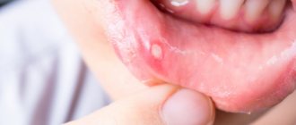
- the oral cavity is strewn with aphthae: round or oval ulcers;
- the center of the ulcer is covered with a gray or yellow coating, the edges are outlined by a narrow area of hyperemia.
Clinical picture of catarrhal stomatitis:
- swelling, hyperemia and painful discomfort of the oral mucosa;
- increased salivation;
- yellowish-white plaques of plaque on the mucous membrane;
- less often - slightly bleeding erosions, foul-smelling breath.
Ulcerative stomatitis occurs as a result of severe catarrhal stomatitis or independently. Sometimes considered a complication of catarrhal disease.
Candidal stomatitis (“thrush”) is a mycotic pathology caused by the hyperactivity of fungi of the genus Candida. Factors conducive to development include:
- immaturity of immunity in infancy;
- deterioration of the body's protective functions due to hematopoietic pathologies, infections, cancer, AIDS;
- old age (there is a natural decline of immune forces);
- hormonal changes during pregnancy;
- neglect of the rules of oral hygiene;
- use of dental prosthetic structures;
- drying out of the mucous membrane;
- pathologies of glucose absorption;
- taking glucocorticoid sprays;
- long-term antibiotic therapy.
Vesicular stomatitis develops as a result of contact with viruses of the genus Vesiculorus, which parasitize the body of cattle. Contamination occurs through the process of mosquito bites. Most often, this type of stomatitis affects veterinarians and farm workers.
Bacterial stomatitis occurs with a pathological increase in the population of bacteria of opportunistic microflora, usually against the background of weakened immunity.
Activation of staphylococcal and streptococcal stomatitis occurs against the background of:
- carious inflammations;
- oral injuries;
- purulent processes in periodontal pockets;
- reduction of local immunity of the oral cavity;
- non-compliance with antiseptic and aseptic rules during surgical and dental treatment.
A type of bacterial stomatitis is angular stomatitis. The cause of the pathology is not only streptococci, but also yeast-like fungi that are part of the symbiotic microflora of the human body.
Allergic stomatitis is a type of pathological autoimmune reaction. Clinical manifestations are polymorphic and include a group of dermatostomatitis, multimorphic exudative erythema and stomatitis of various etiologies (usually catarrhal or aphthous).
Types of angular stomatitis
The symptoms of the disease are largely determined by the form of the bacterial infection, which allows us to distinguish two main types of the disease:
- Streptococcal angular stomatitis. It is more common in children, develops due to a streptococcal infection entering the body and appears quite suddenly. One or more bubbles form near the oral cavity, in which clear liquid accumulates. At a certain point, they open, and erosions appear in these places, which cause pain, itching and burning.
- Candidal angular stomatitis. It is caused by exposure to yeast-like pathogenic microflora. This type of disease is, as a rule, sluggish in development, and bubbles do not form in this case. There is red erosion surrounded by a whitish fringe. Next, a white cheesy coating forms on the affected area - it can be safely removed, but after a few hours it appears again. This is a waste product of fungi. Candidal angular stomatitis develops with reduced immunity, which can be facilitated by frequent stressful situations, hypothermia or a common cold.
Types of disease
Based on the nature of the inflammatory elements formed on the mucosa, the following classification is accepted in dentistry:
- Aphthous stomatitis. Areas of inflammation look like oval and round ulcers - aphthae. This clinical form of pathology is divided into types:
- Necrotizing stomatitis. It occurs against the background of severe systemic pathologies. Characterized by the death of cells on the mucous surface in the mouth. Foci of inflammation are painful, on the 3rd-5th day of development of the pathological process they grow, and ulcers appear in their place. Aphthae heal within a month.
- Fibrinous stomatitis. The surface of aphthous ulcers is covered with a gray coating. From the moment of ulceration until the integrity of the mucous membrane is restored, 7-21 days pass. According to statistics, once every 1-3 years there is a relapse of fibrinous stomatitis.
- Scarring. Severe form of stomatitis. It begins with the formation of aphthous inflammations, growing day by day. Soon deep ulcers up to one and a half centimeters in diameter form in their place. Healing takes up to 12 weeks, and scars remain at the site of the ulcers.
- Glandular stomatitis. It affects the salivary glands, which are located throughout the oral cavity. Aphthae form at the exit of the salivary ducts. Painful when pressed.
- Deforming. The most severe variant of the pathology in its manifestations. Ulcers affect large areas of the mucous membrane and heal poorly. After the inflammatory stage, the ulcers heal with the formation of scars and scars that tighten the tissue, causing the oral cavity to become deformed.
- Catarrhal stomatitis is a process of pathology of the upper layers of the oral mucosa. Accompanied by bad breath, swelling and hyperemia of inflammation. The affected mucosal fragments are covered with plaques with a white coating, and tooth imprints remain on the gums and soft tissues of the tongue. There are severe pain, increased salivation, a slight increase in body temperature and weakness.
- Ulcerative gangrenous stomatitis. Severe form: the mucosa is affected by areas of ulceration and necrosis. The inflammatory process can destroy the entire thickness of soft tissue, down to the bone. Accompanied by severe weakness.
Cheilitis - symptoms and treatment
The lip consists of three sections: cutaneous, transitional (red border of the lips) and mucous. Deep in the lips is the orbicularis oris muscle, which is responsible for their movements.
The cutaneous section is similar to normal skin: it contains hair, sweat and sebaceous glands, and keratinizing epithelium. In the keratinizing epithelium, scales are formed that protect the underlying layers of the skin. Over time, the scales fall off and are replaced by new ones.
The transitional region is different from the skin: there is no hair and sweat glands, the sebaceous glands remain only in the corners of the mouth. They perform a protective function so that open areas of the lips do not dry out. The keratinizing epithelium gradually turns into non-keratinizing epithelium, i.e., protective scales do not form on it.
In the mucous membrane, the sebaceous glands completely disappear, the epithelium becomes non-keratinized and small salivary glands appear.
Cheilitis can be accompanied by various manifestations: scales, crusts, cracks, erythema, blisters and erosion.
Scales in cheilitis are silvery plates that form after the death and rejection of a layer of surface tissue. The keratinizing epithelium can only be seen under magnification, but the scales are immediately noticeable. They can also appear on non-keratinizing epithelium if its cells have died.
The next pathological element is the crust. It is similar to a scale, but is formed if the death of surface cells is complicated by inflammation. Fluid containing immune cells emerges from the blood vessels, and the scales swell and turn into crusts. Due to the fact that there is more fluid between the cells, the adjacent tissues increase. They begin to compress pain receptors, and pain occurs.
Cracks are painful defects in the form of straight lines. They occur when lips become dry and lose their elasticity. They can appear due to bad habits such as smoking and lip biting, as well as burns, physical and chemical trauma.
Cracks can heal and reappear after some time, often in the same place. This cycle can be seasonal, for example worsening in winter due to adverse weather conditions or weakened immunity.
Erythema is a red spot without clear boundaries, most often due to allergies. Usually painless, but sometimes its sensitivity increases slightly.
A vesicle is a small formation above the surface of the skin or mucous membrane. Inside it there is a liquid - blood plasma with living and dead immune cells and microorganisms; in more severe cases it is called exudate, or pus. The appearance of fluid is often preceded by an infection, to which the body's immune cells react.
Erosion is a pink or red formation that appears at the site of a large burst bubble. It is painful when touched. It differs from erythema by clearly defined boundaries and location below the level of the skin or mucous membrane, forming a small “pit”.
Symptoms of stomatitis in adults
Aphthous stomatitis. At the beginning, signs of general intoxication of the body and malaise, hyperthermia, soreness of the mucous membrane, which soon becomes covered with aphthous ulcers, are recorded. During the healing process, canker sores become scarred and leave scars.
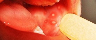
Herpetic stomatitis. When the oral cavity is infected with herpetic virus type 1-2, there is a high risk of developing acute herpetic stomatitis. Typically, the rapid beginning of the formation of the clinical picture: hyperthermia, pronounced signs of general intoxication of the body. During examination of the oral cavity, hyperemic, edematous mucosa with areas of bleeding gums is visible. Increased saliva secretion and bad breath may occur. A few days after the manifestation of the pathology, the mucous membrane becomes abundantly covered with small pustules and vesicles, and necrotic changes in the epithelium occur.
Catarrhal and ulcerative stomatitis. The symptom complex of these two types of pathology is similar at the beginning of the disease, but after a few days the ulcerative form is identified by specific signs: severe hyperthermia, fatigue, headache and swelling of the lymph nodes closest to the source of inflammation. Ulcerative stomatitis affects the area of ulcer formation throughout the entire depth of the tissue, catarrhal lesions are limited to the upper layer of the mucous membrane. The pain intensifies while eating, sometimes so severe that it forces you to refuse food. Symptoms develop progressively and are most noticeable in people with weakened immune systems.
Necrotizing ulcerative stomatitis, or Vincent's stomatitis, occurs as a result of the activity of symbiotic microorganisms: spindle-shaped bacteria and a type of spirochete. The main reason for the pathological activity of these microorganisms is weakened immunity. Clinical manifestations of necrotizing ulcerative stomatitis:
- scattering of ulcers and erosions in the mouth;
- low-grade fever;
- pain and bleeding when pressing on the gums;
- bad breath with a hint of rot;
- the pathological process begins from the gingival margin and spreads further.
Angular stomatitis is widely known as “jams”. This form of stomatitis is characterized by painful cracks in the corners of the mouth. A feature of angular stomatitis is that in other forms of the disease the inner surface of the oral mucosa is affected, and when jammed, the integrity of the outer mucous layer is disrupted. In addition to the seasonal decrease in immunity, among the provoking factors recorded:
- gastrointestinal diseases
- metabolic pathologies
- disturbances in the functioning of the endocrine system
- oral health problems
- long-term use of artificial hormones
- pathological growth of the streptococcus population;
- unbalanced diet;
- smoking and alcoholism;
- incorrect bite.
These factors create conditions for permanent irritation of the mucous membrane, which makes it vulnerable to erosion, attachment and the development of infections.
Allergic stomatitis, with all the polymorphism of manifestations, begins with dryness of the mucous membrane, itching sensations in the mouth and along the inner edge of the lips, the process of eating causes discomfort. The oral mucosa swells, becomes hyperemic, and in the absence of timely treatment, atrophy of the lingual papillae occurs.
Treatment of angular stomatitis or cheilitis
Angular stomatitis (mostly known as angular cheilitis) is a condition in which cracks, cuts, and lesions appear at the corners of the mouth. The irritation can spread to the lips, the inside of the mouth, and even lead to infection. Angular stomatitis refers to a wide range of associated infections and lesions, mainly associated with cuts that occur at the corners of the mouth.
Although there is no single cure for angular cheilitis, there are some things you can do to improve your condition. Below are several procedures.
Increase your vitamin intake
Often, outbreaks of angular stomatitis are the result of a weakened immune system. To improve the situation, it is necessary to increase the intake of vitamins, especially group B. You should also increase the intake of iron, since its lack can also be one of the reasons. Vitamins and iron are found in a variety of foods and also in dietary supplements.
Clean your dentures
Often outbreaks are associated with poorly cleaned dentures. If dentures are poorly fitted, they become a breeding ground for bacteria. Make it a rule to wash your dentures under water until they are squeaky clean.
Chapped lips
Often, angular cheilitis is accompanied by chapped lips. This occurs mainly because a person with chapped lips tends to lick them frequently. The problem is that saliva does not help in moisturizing the skin on the affected area. Angular cheilitis is common in children.
Infections
Angular cheilitis often appears as cracks or cuts in the corners of the mouth. This often leads to an infection that can spread throughout the mouth. Some doctors prescribe an antibiotic in such cases. Others recommend using 1% hydrocortisone cream, which is used to treat candida infections.
Home Remedies
There are also some home remedies that can help treat angular cheilitis, which are often surprisingly effective.
Just make sure you give it the attention it deserves. Angular stomatitis and cheilitis can be cured. Just take the first step.
It is always better to consult a good dentist for treatment of stomatitis or any other oral disease. If you can’t visit the dentist right now, then you should take all possible measures yourself. The main thing is to do this wisely, so that the effect of the actions taken does not turn out to be the opposite. Well, as soon as the opportunity arises, run to the dental clinic!
Make an appointment with a dentist by phone or through our website - the online booking form is in the upper right corner of this page.
Treatment of stomatitis in adults
It is important to start etiotropic treatment in a timely manner to prevent chronicization of the pathology. Therapy for stomatitis includes a set of measures:
- the symptomatic part of therapy consists of taking antipyretic drugs;
- in case of severe pain, painkillers are prescribed;
- the key stage of treatment is to find and eliminate the root cause of stomatitis: the causative agent or the underlying disease; which helps prevent relapses of the painful condition;
- local treatment involves the use of local agents with a healing and antibacterial effect;
- An important component of the treatment plan is the selection of a diet and the exclusion of products containing aggressive and irritating substances (menthol, sodium lauryl sulfate) from the regimen of regular hygienic treatment of the oral cavity.
A gentle diet includes adding soft fermented milk products, neutral vegetables and fruits, cereal porridges, lean boiled meat and low-fat broths to the diet. Tea and coffee should be avoided; it is recommended to replace them with decoctions, fruit drinks and water. Spicy, sour, hot dishes or food, parts of which can physically injure tissues affected by stomatitis, are excluded. After eating, you should rinse your mouth with water or a medicinal solution.
In cases of recontamination of ulcerated areas of the mucosa, drugs containing chlorhexidine bigluconate are used. Long-term use of these drugs has an unpleasant side effect: dark spots appear on tooth enamel and fillings, which disappear after the end of treatment.
Questions and answers
— Which doctor treats stomatitis in adults?
— The dentist deals with the treatment and prevention of stomatitis. He is also competent to diagnose the type of pathology and select an adequate therapeutic regimen. Since stomatitis in adults often acts as a symptom, consultation with another specialist is possible: a gastroenterologist, an infectious disease specialist, an endocrinologist, an allergist.
— How long does stomatitis last?
— Depending on the type and severity of stomatitis, the inflammatory process should subside within 7-10 days, and two weeks after the onset of the disease, complete recovery occurs. If alarming symptoms do not go away within the specified period, you need to contact your dentist again to find out the reasons or adjust treatment measures.
— What medicine helps with stomatitis?
— Unfortunately, a universal remedy for stomatitis is still unknown to science. Stomatitis is a group of pathologies, the causes of origin and manifestations are diverse. The development of pathology is influenced by factors such as the decline of the body’s immune forces, concomitant systemic diseases, and stress. The decision to prescribe a particular drug is made individually, depending on the clinical picture and characteristics of the body. When two different people have the same diagnosis, therapeutic measures differ.
Sources
The following materials were used in preparing the article:
- Anisimova I.V., Nedoseko V.B., Lomiashvili L.M. Clinic, diagnosis and treatment of diseases of the mucous membrane of the mouth and lips. - M.: 2008. - 194 p.
- Borovsky E.V., Mashkilleyson A.L. Atlas of diseases of the mucous membrane of the oral cavity and lips // Medicine - M.: 2001. - 703 p.
- Langle R. P., Miller K. S. Atlas of oral diseases: Atlas / Translation from English, ed. L. A. Dmitrieva. - M.: GEOTAR-Media, 2008. - 224 p.
- Danilenko S. M. The most common diseases of the oral mucosa // Consilium-Provisorium. - 2001. No. 6. - With. 6.