About two years ago, a filling with a pin was placed on the 7th lower tooth. The filling recently broke off, I went to the hospital, they put a new filling on the same pin, and when they aligned the filling with the bite, the pin became visible. Either it’s translucent, or it’s already polished along with the filling, but it’s visible on the cutting edge, although I can’t feel it.
Today I went and told the doctor that the pin was visible, she told me that this happens. Is this normal?
No, this is not normal, if only because in your case, if the coronal part is damaged by more than 30%, it is necessary to install not a filling with a pin, but a stump inlay and a crown.
In this case, you run the risk of chipping the tooth wall and, if the outcome is negative, you may lose the tooth altogether if the wall chip is below the gum.
I recommend that you go to paid dentistry and get quality service.
Contact phone number
Comments and reviews 10
After installing the fiberglass pin, the tooth was closed with a temporary filling. Part of this filling fell out. Installation of a temporary filling is planned in 4 days. Do I need to see a doctor soon or can I wait for a scheduled appointment? Answer
It is better to inform your doctor about the current situation. Answer
General dentist
Today I had a filling placed on a fiberglass pin on my front lower incisor (4, I think). On the inner surface of the tooth, on the filling, at home I saw a small white spot, is this the edge of the pin? Photos are not high quality, unfortunately. If it’s a pin, then what does this mean for me? Answer
The lower incisor or even the fourth tooth are not large in size, so it is quite possible that the edge of the fiberglass post is showing through or even reaching the surface. Ideally, it should be completely immersed in the composite structure, but if the translucency occurs on the inner surface, then there is nothing to worry about, because apart from aesthetic disturbances, which are important only on the front surface, there are no other negative consequences from the surface location of the fiberglass pin. Answer
Contact phone number
My wife got a filling on a pin today. I looked and was surprised that the golden pin was very visible from the tooth. My wife can't feel it with her tongue. Is it generally normal that it can be seen so clearly? Will it interfere later if the filling sags or wears off? The tooth is chewing! Answer
Of course this is not normal! The dentist selected a pin that was too large. Call for a follow-up appointment. Answer
Contact phone number
A month ago I got a light filling with a pin. For the whole month I have been feeling discomfort in the tooth (as if something was stuck), the pin is visible, the filling does not completely cover it, and when I touch the tooth with my tongue, I feel the border between the tooth and the filling! Should I redo it or is this the norm? Answer
Of course, it needs to be redone. This phenomenon has nothing to do with quality work. Answer
The pin from the filling is visible
We placed a filling on the 6th upper tooth, the golden pin is very visible. What should I do? Should I go to the doctor to have it redone? Or is this normal? What could be the consequences?
Hello. If the filling does not interfere with your height and there are no negative changes on the x-ray, this restoration does not have any negative consequences other than aesthetics and there is no point in redoing it. It is recommended to take an X-ray of this tooth to control the quality of treatment and with this image go to a face-to-face consultation with a dentist-therapist. Sincerely, dentist Andrey Aleksandrovich Syomkin.
Good afternoon. It needs to be redone.
There are no consequences, if you are not satisfied only with the aesthetics, then redo it.
A dentist I know told me that they haven’t used anchor pins on chewing teeth for 10 years. And they put composite posts or a stump inlay, and a crown on top. And that in any case it will be necessary to redo it later, since the anchor pin may crack the tooth root.
Yesterday I took x-rays, a dentist friend said that the pin was screwed in too large and was located too close to the edge of the root wall. I don't know what to do now? I'm very worried.
Good afternoon. There is no point in panicking. It is difficult to judge why the doctor did not install the pin deeper; perhaps the conditions were difficult, but in general there is nothing to worry about.
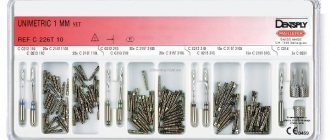
General classification of pins
Standard pins (pins theoretically designed to fit into any root). They are used for installation in slightly damaged teeth, if the level of destruction is above the gum. When installing a standard pin, it is necessary to adjust the tooth root to the pin using special tools. Standard pins can be metal or non-metal. Of the metal pins, today the best are titanium pins. They can also be made of steel, copper, brass and other metals. The pins can be plated with gold, but this does not give them any advantage. The pin set may be supplied with tools to install them. This option is much more convenient for the doctor, and is important for the patient because the installation accuracy is higher. You can ask your doctor how he plans to place the pin for you - using a set of tools or regular dental equipment. The first one will be preferable. Best choice: A titanium post made by a reputable manufacturer that includes the tools to install the post. In Family Dentistry, for example, standard pins from Maillefer (Switzerland) are used. Non-metallic pins are usually made by reputable companies and come with installation tools. They are installed less frequently, since the scope of application of these pins is insignificant. The advantage of non-metallic pins is their light transmission, which allows them to be used for highly artistic restorations, as well as as a basis for all-ceramic crowns (metal pins are completely lightproof). Family Dental uses non-metallic pins from the American company Bisco.
Individual pins or pin stump inlays. They are made taking into account the internal relief of the root of a particular tooth. Individual pins are more reliable than standard pins and are able to be held in complex roots. Their cost is more expensive, which is due to the complexity of manufacturing, in the process of which a dental technician is involved. Individual pins are made from the same metal from which the crown will be made, since the golden rule of prosthetics using metal is “The presence of one metal in the mouth.” Individual pins can be removable. This option is used on severely damaged chewing teeth, which will bear significant loads after restoration. A detailed explanation of their features will be discussed below, in examples of work done at Family Dental. Thus, we can summarize the features of individual pins (pin stump inlays):
- excellent reliability;
- the possibility of manufacturing from the same material as the crown material (gold-platinum alloy, titanium, cobalt-chromium alloy, etc.);
- Possibility of manufacturing from fiberglass for purely ceramic restorations (no metal);
- Possibility of use in severely damaged teeth with thin walls or with a level of destruction below the gum;
- the ability to make them collapsible if it is not possible to go deep into only one canal, and the tooth, after restoration, will bear a significant load.
Similar questions
Hello! I have a problem. A piece of either a tooth or a filling broke off, I went to the dentist. I was told that my tooth had already been treated as deep, possibly...
Hello! I am 23 years old! Even in childhood, the lower tooth 6 was healed, the tooth was very bad, all rotten, and when the doctor put a filling he said that there were guarantees on how long the tooth would last...
Hello! Please consult! There is no way to see a doctor in the near future. I need an approximate diagnosis. I took an x-ray of the healthy tooth and everything was fine. 3 from the edge...
Good afternoon, dear dentists! I ask for your help and advice. The fact is that in August I was given a large light-curing filling (I understand that this is it, because...
Types of pins and their main features
The pins are either fiberglass or metal. Fiberglass is used when installing tile crowns. Such an instrument does not show through the crown. It is quite easy to remove it if necessary. However, metal knitting needles are stronger, which is why they are used more often. Sometimes they are also called anchor.
Anchor pins are made from materials such as titanium, palladium or stainless steel. There are also pins made of brass and precious metals. They are used in the most difficult cases. Anchor pins are very popular because they make it possible to restore an almost completely destroyed tooth. It is only important that its root is intact.
The pins are fixed in the canal with phosphate cement. The use of ultrasound can help destroy it. This has to be done when repeated caries begins around the pin. Excessive strength of the material often causes displacement of the pin when chewing food, which can lead to damage to the prosthesis. In this case, the entire structure will need replacement or correction. Most often, the violation is detected already at the stage when it is already quite difficult to cure the tooth. In this case, you have to remove the pin along with the tooth. In some cases, the tooth can be saved. In this case, a small part of the dental tissue is removed around the pin using a drill, and then the pin is carefully unscrewed. After removal, re-treatment is carried out, the canals are properly processed and fillings are placed again.
Parapulpal pin wires are non-metallic. They are made from gold or special stainless steel, and then they are necessarily coated with polymer. Such knitting needles are used for retention of fillings and their reinforcement. A special feature of parapulp pins is that they are installed in hard tissue without touching the pulp. The absence of actions inside the tooth cavity ensures the complete elimination of the likelihood of contracting any infection or the development of inflammatory processes in the oral cavity. The disadvantage of such tooth strengthening means is the limited possibilities of use. The fact is that these products are located very close to the surface. They often break teeth and need to be reinstalled.
Dentists divide pin instruments into two groups: standard and special. The first ones are used to eliminate minor damage, and are available in the form of cylinders or cones. Special devices are made in a variety of shapes. Pin devices are also distinguished by fastening methods. Among screw materials, the main drawback is the danger of the tooth part breaking off during screwing.
Pin visible
Moderator: Lesya
Pin visible
Post by Alena310893 » Tue Jan 30, 2018 13:14
Re: Pin visible
Post by Lesya » Fri Feb 16, 2022 13:22
The tooth is very badly damaged, and it is not surprising that you were advised to remove it. It's easier, but not better. So you're lucky to have found a doctor willing to keep him. The fact that the doctor restored it unfortunately does not mean that this is all. With such destruction, a crown must be placed on the tooth. I'm sure the doctor told you about this. Therefore, if you plan to install a crown in the near future, then it’s not scary that you see the pin. If you don’t install a crown, then such a tooth will not serve you for long. The fact that there are no bumps on it, and it is clearly lower in bite, indicates that the doctor is going to put a crown.
And here is the main question about the channels and the old pin. I would still remove it, since it is not exactly along the channel. And I also treated the canals, because judging by the pictures, the filling in the canals is far from good. If the tooth has been standing like this for more than 10 years and has never hurt, then you can take a risk. But if this filling is a year or two old, the tooth bothers you not even much, but periodically, then it will be wrong to put a crown on it, without treating the canals. After all, if he gets sick under the crown, then it will have to be cut and thrown away, and the canals will still have to be re-treated. Conclusion: If the tooth has never bothered you and the filling (of the canals) is more than 7-10 years old, get a crown. If the tooth sometimes bothers you, the filling is 1-2 years old - you need to re-treat the canals, and only then put on a crown. But I would recommend a crown in any case. Without it, you will lose your tooth. After all, the doctor is not a magician. He molded a huge filling on the pin for you, and the walls of the tooth are almost completely gone. Such a tooth will not last long for chewing.
Installing a seal on a pin
A damaged tooth is restored using a pin in the case where the tooth's nerve has been removed, but there is still at least a wall that can hold the filling substance.
Using a post when installing a filling will not only create a more dazzling smile. With this procedure, you can restore the chewing function of the tooth and thereby significantly simplify the patient’s life.
A pin is essentially a screw made of a special material. The dentist inserts it into the tooth canal. The pin is the basis for the filling substance. It is on the screw that the seal is applied. The strength of this design allows the patient to wear it even with serious injuries. Let's take a closer look at when this method of treatment is used.
Who needs a filling on a pin?
- Dentists install a filling on the pin for patients whose enamel is severely damaged
- This procedure is also necessary for those who have had a tooth completely removed or knocked out.
- In addition, the pin allows you to achieve the necessary rigidity when installing prostheses.
To install a filling on a pin, you only need a couple of hours of free time and one trip to the dentist.
However, it is worth noting that such a serious procedure has contraindications. Everyone should familiarize themselves with them before going to the doctor.
The fact is that the procedure for installing a seal on a pin is considered a surgical intervention. That is why it cannot be performed if you have gum inflammation and periodontal disease, carious enamel damage and insufficient root thickness, as well as the formation of a jaw cyst. But the main contraindications are disorders of the central nervous system and cardiovascular diseases. In all these cases, the pin cannot be installed.
This method of dental treatment has a major advantage. Installing a filling on a metal pin will cost the patient significantly less than dental crowns. In addition, with this treatment option, the tooth will look much better from an aesthetic point of view. It is also important for patients that the pin allows them to preserve the root of the tooth and restore its chewing functions. Another advantage is that the filling on the pin lasts for quite a long time - about ten years, but only with proper use and timely oral hygiene. The procedure for the patient occurs without any discomfort and takes a couple of hours. After installing the filling substance, neither the fiberglass nor the titanium pin will cause any pain or discomfort.
Unfortunately, this method of treatment also has disadvantages. After a few years, the patient’s tooth walls may become thinner and even the enamel may wear off. In addition, there is a threat of caries development and corrosion of the metal pin.
No one excludes the possibility of individual intolerance to such intervention. In this case, the structure may not take root and then the tooth will have to be removed. It is impossible to predict this, but you have the power to eliminate all allergic reactions.
View full version : Pins visible
Olgi I will support the previous speakers, a crown is definitely needed. Concerning
2. Will this affect the faster destruction of the filling due to the heterogeneity of the chewing surface. What you call a filling is actually a tooth stump, that is, the doctor did everything correctly, preparing the tooth for prosthetics, removing it from the bite. And in this case, the goal is usually not to make a relief chewing surface, outlining all the anatomical features of the tooth. By refusing a crown in this case, you take full responsibility for this tooth, since without a crown, even if something breaks (as already said, thin walls), no one will most likely redo it for free (I wouldn’t) . Discuss this issue with your doctor so that it doesn’t come as a surprise to you later. And it will definitely break sooner or later.
I don’t agree about stamping crowns. If I were you (if the reasons are financial), having found the funds to restore a tooth with pins, I would rather save up and eventually make metal-ceramics.
Vector is very effective, in my opinion (more precisely, my opinion on the results after the treatment), the price justifies itself.
With the advent of the German Vector device, there is no longer any doubt about the effectiveness of ultrasonic methods for treating periodontitis, since thanks to its unique design, the main problem is eliminated - the randomness of the beating of the cleaning instrument. Ultrasonic vibrations are transmitted to the tool through a resonant circuit, as a result, ordered vibrations are imparted to the tool - it moves strictly along the tooth surface being cleaned. Lateral vibrations no longer irritate the gum tissue in the periodontal pockets and do not destroy the surface of the tooth roots, so Vector is even suitable for the treatment of periodontitis in patients with high sensitivity. Due to the orderliness of the vibrations, the depth of penetration of ultrasound also increases - up to 11 mm, which in many cases makes it possible to do without surgical treatment methods. During the treatment of a periodontal pocket, a mixture containing hydroxyapatite particles 10 nm in size is supplied to the working instrument. Vibrating in an orderly manner in an ultrasonic field, these particles completely remove subgingival deposits and endotoxins from the periodontal pocket and polish the tooth surface. In addition, hydroxyapatite promotes rapid gum recovery after the procedure. Patients respond very well to the treatment carried out with the Vector device, since during and after cleaning the periodontal pockets there is practically no discomfort. Therefore, in many cases it is possible to do without anesthesia. Thanks to the orderliness of the vibrations, the depth of penetration of ultrasound also increases - up to 11 mm, which in many cases makes it possible to do without surgical treatment methods.
Why is a pin inserted into a tooth?
A pin is a special rod-shaped structure made of metal (titanium, brass, steel, precious metal alloys) or non-metallic materials (fiberglass, ceramics, carbon fiber), which is inserted into the dental root canals and helps strengthen the damaged tooth. It can have different shapes: conical, cylindrical, screw, and can be installed in an active way (screwing into dentin) or passively (fixed with filling material when filling tooth canals).
The dentist selects pins depending on the condition of the dental tissues and root canals, as well as depending on the method of restoring the tooth on a pin. The load to which the restored dental unit will be subjected is also taken into account: it will be different for chewing and front teeth, and it also matters whether the tooth will be a support for a prosthetic structure or a free-standing unit. Very often, pin restoration precedes the installation of a metal-ceramic crown, either a separate crown or a supporting crown of a bridge.
If a tooth unit still has healthy roots or can be successfully treated, it is not necessary to remove it at all - installing pin structures will allow you to save the tooth root and “grow” a new crown onto it. But you need to know that if the thickness of the root canal wall is less than 2 mm, if there is a complete absence of crowns on the teeth in the “smile zone,” as well as with some serious diseases of the internal organs, the method of restoring a tooth on a pin cannot be used.
If you have a problem similar to that described in this article, be sure to contact our specialists. Don't diagnose yourself!
Why you should call us now:
- We will answer all your questions in 3 minutes
- Free consultation
- The average work experience of doctors is 12 years
- Convenient location of clinics
Single contact phone number: +7
Make an appointment
Filling on a tooth using a pin, restoration of a tooth on a pin
Petrov Oleg Viktorovich
Chief physician, director of the clinic, implant surgeon, candidate of medical sciences at the branch of the street. 8th Krasnoarmeyskaya, 3
He began studying maxillofacial surgery and dentistry at the department of the same name at the Military Medical Academy, and almost all subsequent postgraduate education was associated with training at this oldest educational institution. After graduating in 1992, he underwent advanced training at the faculty of retraining and advanced training in the “surgical dentistry” cycle. And in the period from 1996 to 1999, he studied at the faculty of medical management, specializing in “maxillofacial surgery and dentistry.” Candidate of Medical Sciences.
For six years he was the head of the dental department of the hospital. During this period, he repeatedly underwent advanced training in various areas of dentistry from “therapeutic dentistry” to “implantology”. Out of love for his specialty, two rationalization proposals were born. When treating patients, I prefer a systematic approach, using all knowledge to solve the patient’s problems.
Knowledge of implantology and improvement of implant treatment methods in complex clinical cases took place at the bases of well-known European universities: a one-year cycle in implantology at the Frankfurt University. Goethe; cycle on bone grafting at the Medical University of Vienna. Participated in international implantological congresses in Italy, England, Monaco. This made it possible to use the acquired knowledge and skills to solve practical problems of restoring patients’ missing teeth.
I have been working at the Astra Clinic since 2005. Managing a friendly team of doctors, I try to use all known modern methods of dental restoration to achieve aesthetic and functional results.
Nemchenko Dmitry Vladimirovich
Surgeon, surgeon-implantologist at the branch of Tikhoretsky Ave., 22/13
Graduated from the Military Medical Academy named after. Kirova S.M. in 1992. In the specialty of maxillofacial surgery, he specialized at the MAPO and continued to work as a maxillofacial surgeon at city hospital No. 15. Having gained rich clinical experience in the treatment of various diseases of the maxillofacial region, he noted that in Russia very little attention is paid to the rehabilitation of patients and, in particular, implantation. In the nineties, implantation in Russia was in its infancy, and to obtain practical skills and theoretical knowledge it was necessary to turn to foreign sources. For this purpose, he interned and assisted in operations at the maxillofacial department in the clinic in Luleå (Sweden) under the guidance of prof. Ulf Blomback. I acquired the basics of my knowledge of implantology under the guidance of the head. Member of MAPO Professor Vasiliev A.V. While continuing to work on implantation, he took part in international symposiums and congresses dedicated to this topic. In 2010-2011 he studied at the University. Goethe in Frankfurt am Main and, after successfully passing theoretical and practical exams, received a European certificate as an implantologist. In 2014, he completed training in Berlin on plastic surgery of soft tissues in the area of installed implants to create aesthetic restorations of the dentition. I have been working at the ASTRA-3 LLC clinic on Tikhorektsky Prospekt 22/13 since 2008. Every year I perform about 1000 successful operations to install implants. I pay great attention to the problem of bone tissue deficiency and ways to solve it. I am proficient in various methods of bone tissue augmentation: sinus lifting, harvesting and transplantation of bone blocks, 3D restoration of the bone crest using the Curie technique. Extensive knowledge, existing experience and modern technologies help me in the work that I consider my calling.
Beginning of the implantation operation with Nemchenko D.V.
Patient's review of the work of Zybina M.O. and Nemchenko D.V.
Review of a foreign patient about the work of Nemchenko D.V.
Elimination of defects in hard dental tissues can be carried out by using pin structures, such as:
- pin tooth;
- pin stump structures;
- restorations on pins.
A pin restoration is a microprosthesis designed to replace a defect and restore the coronal part of a tooth and is tightly fixed to the tissues using a special rod. In fact, such a filling with a pin is installed when there is doubt about the effectiveness that a tooth restoration without a pin (a simple filling) can demonstrate.
We speak of restoration on root pins when the shank of the rod is located inside the canal (filling root canals with pins). When restoring with parapulp pins, the rod is placed in the thickest areas of dentin. This design makes it possible to restore the coronal zone of chewing and anterior teeth without removing the pulp and installing artificial crowns.
How is tooth restoration performed on a pin?
Despite its apparent simplicity, restoring a tooth on a pin is a rather painstaking and time-consuming process that requires a lot of experience and a certain skill from the dentist. This procedure is carried out in several stages:
- Anesthesia, providing access to root canals (if necessary, if this has not been done before - depulpation).
- Cleaning root canals from pulp residues and infected tissues.
- Inserting the pin into the canal so that it enters the bone. Next, it is securely fixed with filling material.
- Restoration of the coronal part with filling material or an artificial crown.
- Checking the success and reliability of the restoration. It is usually carried out 1-2 days after restoration; if necessary, correction and polishing are carried out.
Such restoration is several times cheaper than full-fledged prosthetics, so dentists advise people who have partially or completely destroyed dental units in their mouths not to rush to remove them and first try restoration using pin structures.
Veronika Aleksandrovna Ivantsova, dentist at the 32 Dent clinic, says: “The success of restoring teeth on a pin depends not only on the doctor, but also on the patient. If the patient follows the diet prescribed by the doctor, that is, refuses solid fibrous food for a while, if he carries out all hygiene procedures on time and takes the medications prescribed by the doctor, the restoration will be successful and durable. And, of course, one should not forget to visit the dentist every six months for examinations and timely identification of possible problems with the dentition and gums.”
General indications and contraindications
Typically, pin filling methods are used when the nerve has been removed and at least one tooth wall has been preserved. A similar need may arise when a tooth is damaged due to untimely treatment of caries, pulpitis, periodontitis, etc. It is important to understand that in case of severe tooth decay, the absence of a crown part, this method is not applicable and requires consultation with a dentist regarding the installation of a pin tooth or an artificial dental crown with core pin insert.
Parapulpal pins (aka pins)
Parapulpal pin – a rod about 5 mm in length, with a radius of mainly 0.35 -0.4 mm, without/with thread, which is installed in dental hard tissues when it is necessary to hold fillings in cavities of the IV and II classes, severe destruction of the dental crown and preserved pulp (not used in the treatment of pulpless teeth).
The material of the rod can be titanium, steel, gold alloys .
The pin is usually inserted into the subgingival dental area, due to the properties of root dentin that are suitable for this purpose. The pin is located at a distance of 0.5 mm from the enamel/dentin border and from the pulp, 1-1.5 mm from the marginal area of the tooth.
In accordance with the decay of the tooth, the required number of pins is inserted. Thus, when intervening in the area of the front teeth, one rod is sufficient, while to restore chewing teeth it is necessary to use as many pins as there are no chewing drills.
Examples of work with pins performed at the Family Dental.
Application of a standard titanium anchor pin from Maillefer (Switzerland)
The patient complained of a cracked bridge with worn-out crowns. An examination was carried out, x-rays were taken and a diagnosis was made. The old bridge was removed. It was decided to strengthen the upper canine, which served as a support for the bridge, with a standard anchor pin.
The picture shows the root of the tooth, from the side of the chewing surface, prepared for a standard pin. The root is destroyed above the gum, the walls are thick - all this allows the use of a standard pin with the same reliability as an individual pin, and the cost of a standard pin is lower.
An analogue of a pin is inserted into the prepared root. If necessary, you can take an x-ray with an analogue to ensure that the pin is correctly positioned in the root.
The photo shows the pin next to the analogue.
The pin is inserted into the root, and the position of the pin relative to the opposing teeth is checked.
With the help of a rubber dam, the tooth root is isolated from the effects of saliva, which increases the reliability of operation. You can read more about the use of rubber dam here .
The cement secures the pin into the root of the tooth.
The so-called stump is molded from the restoration material on the pin - the basis for the future crown.
The final view of a tooth prepared for a crown. The tooth is ground with a shoulder, which makes it possible to make a higher-quality and durable metal-ceramic structure. You can read more about this in the section “ Metal ceramics ”. The dark rim around the tooth is a special retraction thread that is placed between the gum and tooth in order to move the gums back and prevent injury to the gums by the bur.
Stages of restoring a filling on pins
- cleaning the cavity and leveling its walls;
- formation of a pin channel;
- installing a pin into the channel and securing it;
- installation of a seal.
The parapulpal pin is an additional device, which is why, to ensure the quality, strength and duration of the result, other methods are used in parallel to ensure the retention of the filling. Professionalism in working with channels plays an important role.
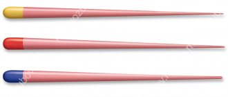
Sometimes the pin can be seen through the filling. To prevent such defects, special coatings are used, for example, gold plating (yellow color is more natural than steel), plastic.
Everything you wanted to know about dental pins in dentistry
The use of dental pins in Kirov dentistry is a very common phenomenon, since it is this technology that allows you to save a tooth in a number of cases. If the treatment involves depulpation (removal of the dental nerve), a pin must be placed in place of the cleaned canals - we dedicated our new article to these devices. From this material you will learn what a pin is, what types there are, how they are installed, and what are the advantages/disadvantages of the decision to use a similar approach in dentistry.
What is a pin?
A dental pin is a specialized structure that is installed in the internal cavity of the tooth root after removal of the nerve. This approach allows you to preserve the root itself and, as a result, the original structure of the jaw, which is very important for every patient.
It is also worth understanding that using a pin to save a tooth is a much more affordable option from a financial point of view when compared with prosthetics.
As a rule, a pinned tooth after treatment requires the installation of a crown (or bridge), and the very possibility of using pins is limited by a number of requirements:
- The thickness of the root walls must be at least 2 millimeters;
- The depth of unsealing the canal is at least 2/3 of its length;
- Possibility of complete (and high-quality obturation of the canal);
- Availability of conditions for giving a conical or cylindrical shape to the channel.
To put it in very simple terms, pins in dentistry are a kind of reinforcing reinforcement when restoring the shape of a tooth after removing nerves and cleaning the canals.
How are pins used in dentistry?
Today, there are five key areas for the use of pins in dentistry in Kirov. Let's consider and comment on each option:
Extension of the crown part of teeth on a pin is a key direction in the implementation of the technology;- Prosthetics with pins in cases where it is not possible to attach the prosthesis to adjacent teeth;
- Reinforcement of a tooth with a removed nerve using a pin insert;
- Treatment of periodontal diseases using combined structures for splinting teeth;
- Carrying out operations to replant your own teeth, where dental pins have also found their use.
Modern dentistry today is simply impossible to imagine without the use of pins.
This is the same indispensable tool for a dentist as, for example, a burr or filling material. Of course, in 8 cases out of 10, these pin structures are used to build up the crown of a tooth, however, many other procedures are also impossible without the use of such products.
Types of pins for teeth
There are several options for making pins for solving dental problems today – let’s look at the most popular and common ones:
- Anchor pins, which can be active or passive, depending on the fixation method. Active ones are screwed into the root of the tooth, which can cause complications. Passive pins are installed in a simple way and are used for reinforcement after tooth depulpation;
- Fiberglass pins. This modern material has a number of important advantages, including elasticity, hypoallergenic properties, ease of installation (in just one visit to the dentist) and the ability to quickly remove in case of dental retreatment;
Carbon fiber pins. In its texture, this material is as close as possible to the natural dentin layer - this is a guarantee of increased strength and reliability;- Parapulpal pins. They are a metal base with a polymer coating;
- Stump tab. It is used in case of serious damage to the crown, since this design is a microprosthesis. Production is carried out individually according to casts.
Pins, like filling compounds, are made from a wide variety of materials: each has its own characteristics and cost, as well as indications. That is why the choice depends on the recommendations of the dentist, who will select the best option based on the characteristics of the disease.
Installation of a pin in Kirov
The pin installation process depends on the type of structure and material used. However, as a rule, the universal treatment algorithm looks like this:
- Inspection and identification of the possibility of using pins for tooth treatment. Typically, this may require an x-ray or even a CT scan;
- Unsealing the canal (if there is a filling) or removing the nerve (in case of a depulpation procedure);
- The pin is installed in the channel, and its excess is cut off (in the case of using carbon or glass fiber);
- Fixation of the pin in the canal using composite compounds;
- Restoration of the crown part of the tooth, which, as a rule, occurs during a second visit to the dentist. The reason is the need for complete hardening of the composite in the channels.
After completion of treatment, such a tooth is closely monitored, analyzing possible pain and other complications.
If there is a feeling of discomfort or pain, especially after removal of the nerve, there is no need to worry, as this is a completely natural phenomenon. After a few days, the pain should subside, otherwise it is better to consult a doctor.
Disadvantages and contraindications
And this article should be completed by familiarizing yourself with the list of disadvantages and contraindications of using pinning technology. Among the negative points are:
- There is a risk of complications if the dentist makes mistakes;
- There is a possibility of allergic reactions;
- Limited service life of a “dead” tooth with pins.
As for contraindications, the key reason for the impossibility of implementing the technology may be the condition of the root walls (insufficient thickness, severe destruction, obstruction of canals, etc.).
In any case, the decision on the possibility of using dental pins is made by the dentist at the initial appointment.
Sign up for a consultation at Dr. Efremov’s Dentistry and get answers to all your questions! Information by phone: 8 (8332) 255-717 . Be healthy and take care of the beauty of your smile!
Indications for installation of anchor pins
- strengthening teeth without pulp before restoration with filling materials;
- strengthening teeth without pulp that are more than half destroyed before temporary restoration with composite materials (if orthopedic intervention is planned in the future) - the so-called. temporary filling of the root canals of teeth with pins;
- restoration of pulpless teeth to retain the filling in a situation where further crown coverage is expected;
- preventive strengthening of teeth in case of severe thinning of the dental crown due to endodontic intervention.
Posts are not used for the purpose of strengthening a tooth root that has been weakened due to endodontic manipulation . They only improve the fixation of the filling. It is also important to consider that the presence of a structure increases the risk of root fracture due to excessive forces.
The intracanal rod can be characterized by active fixation (with a thread for screwing in) or passive (fixation of the pin in the root canal is achieved by cementing).
The use of posts (anchor pins) is possible only with:
- high-quality tooth treatment endodontically;
- absence of pathologies in the peri-apical zone of the tooth root;
- complete removal of dentin affected by caries;
- the ratio of the coronal part of the rod and its part inside the canal = 1:2;
- careful filling of the canal fragment from the apex of the root to the end of the rod (from 0.5 cm);
- the ratio of the width of the walls of the tooth root canal and the thickness of the rod = 1:1 (the root wall is a millimeter thick!).
If the tooth has thin roots, then conical rods are usually installed, while thick ones are cylindrical. The selection of a specific design for a tooth is carried out on the basis of an x-ray.
Recovery and care after treatment
To prevent unwanted consequences and ensure full recovery, it is important to follow a number of rules:
- take medications prescribed by your doctor and follow dietary recommendations;
- exclude the consumption of rough foods that injure the mucous membranes;
- carry out thorough hygienic care without injuring the tooth;
- Get regular check-ups with your dentist.
Painful sensations after filling should gradually subside over several days. If the tooth continues to hurt, the canals are bothering you, the pain increases, if the filling with the pin falls out, you need to consult a doctor.
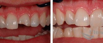
In a situation where the pin is visible from under the filling, you should also visit a specialist, since this phenomenon may be the result of errors in installing the structure on the tooth, a violation of the seal, the development of secondary caries around the filling, etc.