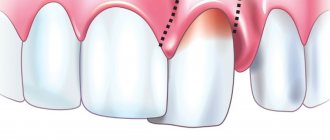
Description of the pathology
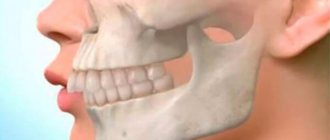
The normal shape of a row of permanent teeth resembles an elongated horseshoe. The lower jaw is naturally slightly narrower than the upper jaw - this is necessary for better closure of teeth and chewing food.
Narrowing of the jaw is called its reduction in diameter - the appearance of a “dent”. This happens more often with the top than with the bottom. It is formed by more porous bone and is more closely connected in the patterns of its development with the upper palate and other elements of the skull. But in fact, the process can affect both jaws, only the right or left side of one of them, or even both.
Another weak but obvious pattern is the increased frequency of curvature in the premolar area (4-5 teeth on each side), while “concave” anterior teeth are less common.
Classification
According to the clinical manifestations of narrowing of the jaw arch, the anomaly is divided into several forms, each of which has its own characteristics :
- Narrowing of the jaw along its entire length . It is characterized by an elongated anterior section towards the frontal region. In this case, the teeth have a fan-shaped position. The lateral incisors fit tightly to the central ones.
- Tight jaw. The main sign of the anomaly is a saddle-shaped shape and compression of the dentition in the area of premolars and molars.
The compression in this case is uneven and does not extend to other parts of the jaw.In addition to narrowing, the pathology of this form is manifested by a violation of the direction of growth of the lateral teeth, which begin to protrude towards the cheeks or tongue.
- Narrowing of the anterior section . With this anomaly, the dentition of the frontal area has a V-shape. The incisors protruding forward form an acute angle and are tightly arranged.
- Trapezoidal shape . The jaw arch has four corners and its appearance resembles a trapezoid. The incisors are very crowded, but do not protrude forward. Their rotation around their axis is often noted.
- Uneven development . It is characterized by a narrowing of the jaw arch in certain areas. It differs from other pathologies by the manifestation of crowding only in certain segments.
Regardless of the form of the anomaly, their clinical picture may worsen over time, leading to even greater deviation of the teeth and narrowing of the jaw arches.
Varieties
This disease has several variants.
- Narrowing of the dentition along the entire length. Its typical sign is the appearance of a “horse” smile due to the fan-shaped incisors.
- Trapezoidal deformity. With it, the patient's jaw takes on the shape of a trapezoid with clearly visible visually defined angles in the area of growth of the 3rd incisor or 1st premolar.
- Narrowing in the anterior region. Here the incisors converge like a wedge and “climb” onto each other.
- Saddle defect. With it, the row is “dented” inward at the level of the premolars (4 and 5 units on each side) with 6 and/or 7 molars. And the incisors and “eights” are usually positioned correctly, or they may be moderately crowded and rotated.
- Asymmetry. This depression is observed only on one side, but, as a rule, affects both jaws. Often combined with normal occlusion in other areas.
All of them can be I, II or III degrees of severity. The pathology first appears in childhood, as temporary teeth are replaced with permanent ones. In adolescence and adulthood, consolidation of malocclusion leads to a change in the load on the mandibular joint, spreading secondary deformations to healthy areas.
The narrowing of the upper jaw is also accompanied by a deepening of the palatine vault and makes nasal breathing difficult. Plus, these processes provoke uneven wear of crowns and can lead to underdevelopment of units that do not have enough space.
Treatment
Treatment of narrowing of the dentition is carried out only for certain indications and using special devices.
Before correction, the doctor prepares the dentition, which includes removing or treating bad teeth, replacing old low-quality fillings with new ones, and professional oral hygiene.
Once the cause of the pathology is identified, work is carried out to eliminate it.
Indications for expansion
Dilation is prescribed if observed:
- violation of the closure of teeth of the same name in opposite rows;
- abnormal position of single specimens or groups: extension, reversal;
- pronounced violation of facial aesthetics;
- excessive deepening of the palatal arch, leading to dysfunction of nasal breathing;
- regular injury to the soft tissues of the oral cavity;
- frequent inflammation of periodontal tissues resulting from incorrect position of teeth.
We will tell you in a separate review whether myogymnastics is effective for distal occlusion.
In the next article we will talk about the consequences of expanding the dentition in adults.
Follow the link https://orto-info.ru/zubocheliustnye-anomalii/okklyuzii/mezialnyiy-prikus.html, read about ways to correct mesial bite.
Expansion devices
Treatment methods are prescribed depending on the shape and location of the anomaly.
Most often, during therapeutic treatment I use the following devices :
- Derichsweiler apparatus. Refers to non-removable screw structures. Designed for correction of the upper jaw, which is narrowed along its entire length, in particularly difficult cases.
This design is used mainly in children in the late period of a mixed dentition or with a permanent one formed. The device has a strong effect on bone tissue.The device is a metal two-part base of a beam type, which is fixed on the supporting teeth. The base is connected using a special spacer screw, which moves the two parts of the base apart during adjustment.
Restoration of the normal size of the jaw is achieved by gradually breaking the palatal suture. Thanks to this, with the help of a screw device, the apical base is expanded in the sagittal and transverse directions, covering the area of the nasal septum.
The use of this design, in some cases, allows one to avoid surgical intervention. But since the device is a rather aggressive technique, it is used quite rarely.
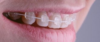
- Expansion plates equipped with springs and expansion mechanism. They are a more gentle option and are used to correct the jaw during the period of milk or early replacement teeth.
This device consists of a base that exactly follows the arc of the sky. The device has a built-in small mechanism that allows for minor correction of the jaw shape.The plate is attached to the supporting teeth using metal clasps, which allows it to be removed at any time.
Wearing this device allows you to correct the shape of the jaw arch by expanding the palatal suture and directly transforming the bone tissue in the area of the alveolar bone, due to the tight fit of the base of the plate.
These devices are capable of restoring the normal shape of the jaw arch only in childhood. Surgical methods are most often used to treat adults.
Surgical methods
Surgical correction of the arch can act not only as an independent method . It is often used in conjunction with hardware treatment.
The following methods are used for surgical treatment :
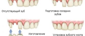
- Tooth extraction .
This method allows you to obtain free space, due to which expansion will be possible. Typically, premolars are suitable for extraction. To obtain a positive treatment result, it may be necessary to remove 2 teeth to ensure uniform jaw growth on both sides. - Perforation of the jaw bone .
To do this, the mucous membrane is peeled off, after which, using a dental bur, through holes are made in the bone at an equal distance from each other. The holes are connected with a small groove. Then an expansion mechanism is installed, due to which the jaw expands, and the perforated area is filled with new bone tissue.
Causes
The most difficult reason in terms of prevention and treatment for a narrowing of the upper or lower jaw by 2 teeth or more (II degree and higher) is unfavorable heredity, since most of the structural features of the skull are determined at the genetic level. But other factors related to childhood also make a significant contribution:
- delay in introducing complementary foods;
- habit of thumb or pacifier sucking;
- early loss of baby teeth, especially molars or several units in a row on one side (the lack of chewing load in this area interferes with bone development);
- lack of solid food in the diet;
- metabolic disorders - rickets, type I diabetes mellitus, etc.;
- parafunction (unjustified increased activity of a nervous nature) of masticatory or facial muscles, including bruxism, involuntary movements of the tongue, etc.;
- rhinitis, sinusitis, tonsillitis, polyps and other diseases that force the child to breathe more often through the mouth than through the nose;
- injuries.
It can also be provoked by a cleft palate, caused by a delay in intrauterine development.
Why does it happen?
Numerous clinical observations have made it possible to identify the causes of jaw narrowing .
The main ones include:
- Late transition of the child to solid food. Chewing hard foods promotes the development of jaw bones, which is important during the period of active growth of a child.
- Premature removal or loss of baby teeth. In this case, the absence of chewing activity and the release of dysfunctional space play a role.
- Disruption of metabolic processes leading to changes in the structure of bone tissue.
- General pathologies that provoke the appearance of constant nasal breathing or impair the functions of speech and swallowing.
- Having bad habits. For example, constant sucking of a pacifier or fingers. This group of reasons also includes a child falling asleep with a bottle in his mouth or being later weaned from it.
- Anomalies in the development of the muscular apparatus of the jaw and tongue.
- Genetic predisposition.
Diagnostics
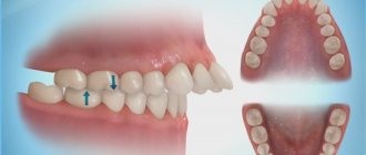
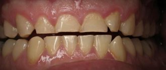
It is not difficult to determine the narrowing of the jaw in a child or adult during an external examination. Its clear sign is the “pressed-in” dentition in certain areas or along the entire length with partially expanded, crowded units. Another characteristic feature is malocclusion.
At the same time, it is necessary to distinguish pathologies of jaw development from atypical orthodontic and dental situations such as supernumerary units, macrodentia, individual structural features of the skull, etc. They can also be combined. Therefore, a preliminary diagnosis made visually is confirmed with the help of one or more additional studies:
- According to Pon-Linder-Hunt , where the difference between the normal and existing width of the dental arch for a given patient is calculated using a special formula. The method requires taking several measurements directly in the oral cavity and taking into account different indicators for representatives of individual nations. But it gives the attending physician a fairly accurate answer to the question of whether several units will have to be removed to level the entire row.
- According to Howell-Gerber-Gerbst - another relatively complex mathematical scheme that is well suited for measuring the shape, length, width and other individual parameters of a row.
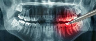
- Teleradiography is a type of X-ray examination that allows you to take a picture of the entire skull. It gives a detailed picture of the deviation itself and reveals its causes, if they are embedded in the structure of other bones.
- Anthropometry is an accurate three-dimensional cast, on which it is easier to take the necessary measurements, especially when the upper or lower jaw of a child is narrowed.
- X-ray - used to assess the condition of the upper palate and its suture, bone structure.
- Orthopantomography - a classic frontal image, is now increasingly being replaced by CT (computed tomography) due to its improved clarity and expanded capabilities - such as obtaining 3D images.
In combination, these methods allow the orthodontist to study the current condition of the bone, the degree and direction of deformation when narrowing the upper and/or lower jaw.
When choosing a correction method, it is also important that they provide an opportunity to evaluate the resource for moving teeth into the correct position and straightening the bite.
Reasons for formation
A child’s jaws are formed under the influence of the chewing muscles, tongue and facial muscles. Failure in their functioning can lead to pathological changes in the shape and dimensions of the arches.
Factors that provoke narrowing of the jaws:
- loss or removal of milk units;
- late introduction of solid foods into the diet;
- osteogenesis disorder;
- sucking of thumbs, tongue, cheeks;
- mouth breathing;
- genetic predisposition;
- general pathologies.
Diagnostic methods
Dental pathology is manifested by a variety of clinical symptoms that make diagnosis difficult. An experienced dentist is able to make the correct diagnosis after a clinical examination. Sometimes additional diagnostic measures are required:
- Radiography. A photograph of the palate allows you to assess the condition of the palatal suture.
- Teleradiography. Lateral shot of the face and jaw.
- Orthopantomography. Panoramic photo of the dentofacial apparatus.
- Anthropometry. Study of a model of jaws made of polymer or gypsum.
- Pon's method. Determination of the individual norm of the width of the dental arches.
- Howley-Herbst diagram. A graphical method that consists in constructing a jaw arch based on the width of the frontal and lateral incisor and canine.
Indications for expansion
The row expansion procedure is advisable in certain clinical situations:
- Narrowing of the HF row with symmetrical crossbite.
- Defects of the face and the relationship of its parts.
- Incorrect position of units or groups.
- Anomalies of occlusion.
- Difficulty breathing through the nose due to the deepening of the palate.
- Injuries to the mucous membranes from incorrectly located elements.
- Inflammatory processes in the periodontium.
- Dysfunction of the dentofacial apparatus.
- Congenital maxillofacial defects.
- Limited row space.
Orthodontic devices
Correction is carried out using removable and non-removable mechanical devices. In children with a mixed occlusion and an alveolar type of narrowing of the upper jaw, removable structures are used to expand up to 4 mm.
For mechanical expansion, the screw is turned a quarter turn weekly. The expansion should not exceed 1 mm per month. The expansion of the arch occurs through the alveolar inclination of the masticatory units. There is no rupture of the palatal suture.
If expansion of more than 4 mm is required, the palatal suture must be opened.
In children under 16 years of age, orthodontic treatment using palatal suture rupture is effective in almost 100% of cases. The expansion process can be slow or fast. In the first case, the screw is activated once every three days, in the second - twice a day. It is prohibited to use the quick method in persons with weak periodontal disease.
Derichsweiler apparatus
A compact non-removable orthodontic structure consisting of a metal frame, a screw and bushings attached to the chewing units. The product is designed to correct the entire length of the HF. It is used in children at the end of the mixed dentition or in the permanent dentition. Has a strong effect on bone tissue.
Expansion plates
A gentle treatment option for correcting pathology in temporary or mixed dentition. The design fits tightly to the sky, reproducing its shape. It is connected to supporting units using clasps.
Operation
Surgical treatment is carried out in complex clinical situations and consists of three stages:
- Orthodontic treatment.
- Operation.
- Correction using orthodontic appliances.
Depending on the clinical indications, the surgeon performs tooth extraction or perforation of the jawbone.
Treatment in children
The optimal age for correcting a narrowed jaw in children is from 6 to 15 years. For violations limited to the apical base and alveolar process (the area of root growth and placement of their holes), braces are recommended. They will align their row in conditions when the rest of the chewing apparatus is formed correctly.
If the pathology affects the entire jaw or palate, removable or permanent plates are prescribed. They consist of a plastic or silicone base with expansion screws in the center. They are fixed using a system of arms, brackets and locks to give the correct position to the displaced units. If it is necessary to correct a deep bite, cross bite, etc., two-jaw structures with cheek shields are used.
In each individual case, the elements of the plate are determined and adjusted individually, using casts. Only the rigid frame and screws remain unchanged, which must be expanded by approximately 0.25 mm every 4 days. The advantages of the plates are:
- wide frame color options;
- low visibility to others;
- mild impact on target structures.
Their low cost may also be an important argument. It rarely exceeds 10 thousand rubles, while the price of individual brace systems varies between 50-100,000 (ligatures made of plastic or precious metals, respectively). Plus, removable models are easy to care for and make it easier to maintain oral hygiene after eating. But when prescribing such plates to children, more control is required from parents.
The time of wearing them should be at least 20 hours a day, and the child is unlikely to comply with it himself due to the discomfort that persists even after the appearance of the habit.
Treatment options in adults
For patients over 18 years of age or with complex curvatures, more or less extensive surgical solutions are recommended. The simplest methods of treating narrowing of the upper jaw in adults:
- Installation of the Derichsweiler apparatus - a beam screw design for the upper jaw only. He breaks the palatal suture and sets its bones in a new, wider position. Then the teeth are straightened with braces.
- Normalization of the bite by removing several units in a row (in the vast majority of cases, the first premolars, 1 on each side). The method allows you to correct the bite and aesthetics of the smile without treating the narrowing of the jaw itself, if it is impossible or not necessary. Extracted premolars are not always redundant. But due to them, space is freed up for the remaining ones, and their location is again leveled with the help of braces.
In the case of the lower jaw, the layer of gum tissue is lifted, the bone is drilled in several places and the holes are connected with grooves. An expander is installed in them, with which the patient with a narrowing will need to go through several years, and the mucous membrane is returned to its previous position. This procedure is called jaw perforation.
General overview
By narrowing we mean a condition of the dentition characterized by an altered anatomical shape. With natural development, the upper section has an ellipsoidal structure, and the lower section has a parabolic structure. Any deviation from the norm is considered an anomaly requiring orthodontic correction.
Geometric specificity causes reduced transverse dimensions with narrowing, expressed in one- or two-sided form. The consequence of the development of the anomaly is an increase in the palatal depth, leading to a violation of occlusion. As a rule, pathology is detected on the maxillary arch, which is caused by the peculiarity of the bone structure, which has less density.
The negative consequences that arise during the long course of the pathology include not only violations of the functionality and aesthetics of the jaw, but also such factors as:
- Difficulties in the respiratory process associated with contraction of the nasal cavity;
- Disorders of diction and articulation caused by limited space for the tongue;
- Problems with the gastrointestinal tract caused by poor quality food processing.
The choice of treatment method depends on the indications of the clinical picture, allowing one to determine the type of anomaly.