Modern dentistry uses the latest technologies in the treatment of certain pathologies. Recently, electroodontodiagnosis (EDD) , with the help of which it is possible to determine the current state of all the nerve elements of the pulp of any tooth. The basic principle is based on the use of weak electric current. It should be emphasized that EDI gives an idea not so much about the state of the dental pulp itself, but rather characterizes the integrity and functionality of its sensitive nervous system. As is known, during various pathological processes in the hard tissues and pulp of the tooth, not only the histological structure and hemodynamic processes in the pulp change, but also degenerative processes occur in the nerve receptors, which is manifested by a change in their electrical excitability. At the same time, it must be remembered that changes in EOM indicators can occur under various pathological conditions of the periodontal tissues and sensory nerves of the maxillofacial region.
The use of electric current is based on the well-known fact that every living tissue is characterized by excitability, or the ability to become excited under the influence of a stimulus. The minimum strength of stimulation that causes excitation is called threshold. The use of electric current for diagnostic purposes has become most widespread, since its strength and duration are easily dosed, and it can be used repeatedly without fear of causing damage.
There are a number of basic rules when using EDI:
- all physiotherapeutic procedures should be carried out only under the supervision of a doctor, who will actually write out the prescription;
- you must strictly follow all the requirements and instructions of the doctors that precede the first procedure;
- as a rule, EDI cannot be performed on a full or empty stomach; the best option would be 40-60 minutes after a meal;
- During the entire procedure, the patient is prohibited from talking, moving, sleeping, reading, etc.;
- After completing the diagnosis, the patient must rest for 30-40 minutes.
When is an electroodontometric study necessary?
Electroodontometry in dentistry is prescribed in the following cases:
- differential diagnosis of caries depth;
- diagnosis of pulpitis;
- identification of tumors on the roots of the tooth;
- injury to the maxillofacial region;
- inflammatory process of the maxillary sinus;
- inflammatory destructive bone disease;
- chronic focal or hematogenous anaerobic infection;
- jaw tumors of various origins;
- inflammation and damage to peripheral nerves;
- radiation damage to tissues;
- treatment with orthodontic appliances.
Technique and methodology of electroodontodiagnosis
1. Prepare the device for operation:
- connect the active and passive electrodes to the corresponding keys “A” and “P”;
- ground;
- connect to the network;
- press the “On” key (the signal light lights up) “50 or 200”.
2. Prepare the patient for the procedure:
- make you sit comfortably;
- explain possible sensations during electroodonto diagnostics;
- put a rubber mat on the floor to isolate the patient’s and doctor’s chairs;
- prepare the tooth for examination.
For examination, the tooth must be isolated from saliva and dried with a cotton ball in the direction from the cutting edge to the equator (alcohol or ether cannot be used). If there are dental deposits, they must be removed. If the teeth are carious, then it is necessary to remove the softened dentin and dry the cavity. For accurate diagnosis, if an amalgam filling is present, it is removed, because An amalgam filling is a good conductor of electric current, through which the electric current ramifies well. To avoid current leakage when testing the excitability of a tooth with a filling in contact with an adjacent filling, it is necessary to insert a celluloid plate smeared with Vaseline between them. Arrange the electrodes depending on the device used. Thus, when working with the OD-2m device, the passive electrode is placed together with a moistened pad on the back of the hand and fixed with a bandage; when working with the EOM-1 device, it is given to the patient’s hand.
The active electrode is placed on sensitive points:
- the middle of the cutting edge of the front teeth;
- the tip of the anterior tubercle at the premolars;
- the tip of the anterior buccal cusp at the molars;
- from the bottom of the carious cavity at 3-4 points.
3. Carry out the procedure:
— press the “50-200” keys (switching ranges), and the “50” or “200” signal light lights up. Start research on the 50 µA range. When working with the EOM-3 device, after placing the electrodes on the patient, the nurse smoothly and slowly moves the potentiometer handle to the right until a sensation appears in the tooth (warmth, burning, jolt), which the patient notifies with the sound “A-A”. The nurse registers the threshold current strength and releases the potentiometer knob, turning off the “Network” key.
When working with the EOM-1 device, after placing the electrodes, the patient presses the switch button and the pulses enter the patient circuit (the doctor’s hand holding the active electrode must be in a rubber glove). When minimal sensations appear in the tooth, the patient removes his thumb from the button and opens the circuit (before each study, the arrow returns to zero). The doctor records the threshold current on a milliammeter scale. Electrical excitability studies cannot be carried out from a filling adjacent to the gum; care must be taken to ensure that the active electrode holder does not come into contact with the mucous membrane. During the procedure, the teeth are periodically dried, because they are moistened by breathing.
Electroodontodiagnostics in modern dentistry
Electroodontodiagnosis (EDD) is a method of dental research based on determining the threshold excitation of pain and tactile receptors in the dental pulp when an electric current passes through it. The process of studying the electrical excitability of teeth is called electroodontometry (EOM). The current generated by EDI devices and used for EOM is called diagnostic current. It should be emphasized that EDI gives an idea not so much about the state of the dental pulp itself, but rather characterizes the integrity and functionality of its sensitive nervous system. As is known, during various pathological processes in the hard tissues and pulp of the tooth, not only the histological structure and hemodynamic processes in the pulp change, but also degenerative processes occur in the nerve receptors, which is manifested by a change in their electrical excitability. At the same time, it must be remembered that changes in EOM indicators can occur under various pathological conditions of the periodontal tissues and sensory nerves of the maxillofacial region.
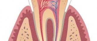
Normally, the dental pulp reacts to the electric current passing through it with minor pain, a tingling sensation, a feeling of a slight jolt, a weak electric shock, etc. The high sensitivity of the pulp to the action of irritants is explained by the large number of sensory nerve endings located in the subodontoblastic nerve plexus of Rashkov, the odontoblastic layer, and predentin.
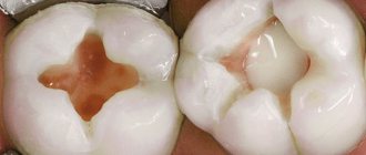
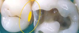
Dental caries , as the process progresses and the carious cavity deepens, causes the development of changes in the pulp, leading to a decrease in the sensitivity of nerve receptors: deposition of replacement dentin, changes in the odontoblast layer, initial degenerative processes in the nerve elements. The listed phenomena can gradually lead to a slight decrease in EOM indicators.
Acute forms of pulpitis are accompanied by severe pain, however, EOM indicators, as a rule, decrease slightly, and sometimes remain at the level of the physiological norm. This is due to the fact that the sensitivity of nerve receptors is influenced, first of all, by the duration of the pathological process and the degree of dystrophic changes in the dental pulp, and not by the severity of inflammatory phenomena. As is known, in acute forms of pulpitis, significant degenerative processes in the nerve elements of the pulp do not occur due to the transience of the process. At the same time, a significant decrease in the electrical excitability of the pulp and the absence of positive dynamics in EOM indicators during therapy (for example, with a biological treatment method) indicate the irreversibility of the pathological process and the ineffectiveness of the ongoing therapeutic measures, which is an indication for the use of extirpative treatment methods.
Chronic forms of pulpitis occur with irreversible atrophy of the cellular elements of the pulp, its replacement with coarse fibrous connective tissue, progressive dystrophic changes in the nerve fibers, and a change in the excitability threshold of the nerve receptors of the pulp. This leads to a significant, 5-6 times, increase in EOM indicators compared to the physiological norm. The decrease in electrical excitability of the pulp is even more pronounced with the death of its coronal part - chronic gangrenous pulpitis. It should be emphasized that during exacerbations of chronic forms of pulpitis, EOM indicators do not change, remaining at a level corresponding to the degree of dystrophic changes in the nervous apparatus of the pulp. EOM also provides valuable diagnostic information for “residual” pulpitis.
Periodontitis is characterized by total necrosis of the dental pulp. In this case, the sensitive nerve endings of the periodontium react to irritation by electric current. The diagnostic current values increase significantly - usually more than 10 times compared to the physiological norm. Often, the subjective sensations of patients during EOM also change - tactile sensations predominate: blow, push, etc. In some cases, there is no reaction to the diagnostic current at all.
Non-carious lesions of hard dental tissues , if there are no secondary inflammatory-dystrophic processes in the pulp, are accompanied by only minor changes in electrical excitability. With pathological abrasion of hard dental tissues, even with significant loss of enamel and dentin (III degree according to Bracco), EOM indicators increase only 1.5–2 times, and in the initial stages of the disease remain within normal limits, which is evidence of the absence of serious pathological processes in pulp and is associated mainly with changes in the electrical conductivity of hard tissues. With wedge-shaped tooth defects, EOM indicators increase 2–3 times compared to the norm, which is quite understandable from the point of view of the dynamics and severity of dystrophic processes occurring in the dental pulp in this form of pathology. With hyperesthesia of the hard tissues of the teeth, the electrical excitability of the pulp is either within the physiological norm, or, in severe forms of this pathology, slightly increases.
Periodontal diseases may be accompanied by secondary degenerative processes in the dental pulp. At the same time, there was no unidirectional trend in changes in EOM indicators: the electrical excitability of the dental pulp in this category of patients can be either increased or decreased or be within the physiological norm. In this regard, in periodontal patients, EDI has a relatively low diagnostic value and is used only to identify possible complications, such as retrograde pulpitis.
Traumatic, inflammatory and oncological processes in the maxillofacial region quite often occur with damage to the nerves that provide sensitive innervation. In this case, the EOM indicators of one or several adjacent teeth may change. Such manifestations are most typical for traumatic injuries of teeth and jaws, diffuse inflammatory processes (sinusitis, osteomyelitis of the jaw), tumors, neuritis of the branches of the trigeminal nerve, side effects of radiation therapy for diseases of the maxillofacial area. At the same time, it was noted that in pain syndromes of central origin, for example, in trigeminal neuralgia, changes in the electrical excitability of dental pulp receptors do not occur, which is an important diagnostic criterion.
Orthodontic treatment is associated with fairly large non-physiological forces acting on the teeth for a long time. It is believed that a decrease in the electrical excitability of a tooth during the active stage of orthodontic treatment indicates excessive load and the danger of developing dystrophic changes in the dental pulp, up to its death.
Temporary (baby) teeth are not recommended to be examined using EOM. This is due, first of all, to the difficulty of adequate contact between a doctor and a child patient, the difficulty of obtaining reliable “feedback” when taking measurements and, as a consequence, the low degree of reliability of the results obtained.
Teeth that are in the stage of root formation , due to the structural features and development of the sensory apparatus of the pulp, also pose a significant problem for EOM research. During the initial period of tooth eruption, electrical excitability is absent or sharply reduced. As the tooth develops, electrical excitability increases and reaches normal only by the time the roots are fully formed. In this regard, in this category of patients, the indicators of electrical excitability of teeth that are at the same stage of root formation as that of the tooth under study are taken as the physiological norm. There is also evidence that the above-described pattern of changes in EOM indicators makes it possible to monitor the dynamics of tooth development from the moment of its eruption to the complete formation of roots.
One of the most modern, functional, ergonomic and informative devices for EDI, in our opinion, is the PulpEst device, developed and manufactured by Geosoft-Dent. This device generates a pulsed diagnostic current with the following characteristics: frequency – 3 pulses/s; amplitude – from 0 to 180 V.
EDI is one of the most reliable clinical research methods, allowing the dentist to obtain important diagnostic information about the condition of the dental pulp in the absence of pronounced clinical and radiological symptoms, to monitor the dynamics of the pathological process, and to evaluate the effectiveness of the treatment. There is also no doubt that the value of this method is often overestimated, which leads to annoying diagnostic errors.
This is especially evident when specific digital values of EOM are strictly associated with specific dental diagnoses. With a competent and scientifically based approach, combined with technologically correct implementation and adequate logistical support, EDI research can take its rightful place in dental practice, becoming an invaluable aid in difficult clinical situations.
The main reasons for false-positive reactions during EDI
1. Contact of the conductor/electrode with an extensive metal restoration (bridge, class II filling) or with the gum, allowing current to pass through the periodontium.
2. The patient's anxiety when he has not been correctly explained what to expect. An agitated, nervous or fearful patient may raise their hand as soon as they think the device is on or when asked if they feel anything.
3. Wet (colliquation) necrosis of the pulp. (Current may pass through the periodontium and the patient may slowly raise his arm at near maximum readings.)
4. Lack of isolation from saliva.
Electroondometric diagnostic procedure
Before the electroodontic diagnostic procedure, the doctor must explain to the patient the essence of the method and the purpose of its use, warn about possible unpleasant sensations and describe the technique. The patient must understand what actions he needs to perform when a pain signal occurs. An important condition for using the device is its disinfection and sterilization.
Electroondometry stages:
- Preliminary cleaning of teeth from plaque and stones to obtain a more accurate result.
- Attaching a neutral electrode to the patient. It may take the form of a part of a diving apparatus hose. The electrode does not touch the area being analyzed.
- Drying the tooth with cotton swabs and separating it from saliva with napkins or gauze.
- Applying a conductor gel to the active electrode for better contact with tooth enamel. A cap can act as a conductor, which must first be moistened.
- Place the electrode on sensitive teeth and turn on the current supply. Monitoring by the doctor to ensure that the mucous membrane does not come into contact with the active electrode.
- If pain occurs, the patient gives a hand signal to the dentist. At this moment, the current supply stops, and the result obtained is recorded.
- Repeat the procedure to obtain the most accurate data.
What is a temperature test and a cold test?
At the same time, excellent diagnostic data is provided by a temperature test - an examination of the condition of the tooth using a hot or cold temperature stimulus. When a certain plus or minus temperature is reached, the neurovascular bundle of a living tooth gives a response. The duration of this reaction and the speed of its onset can say a lot about the diagnosis and treatment tactics.
If a patient complains of sensitivity from hot food or drinks, then a heat test . It involves applying heated gutta-percha to a suspicious tooth. The thermal effect should be short-term - no longer than 5 seconds, otherwise the tooth may be damaged. The heat test is not a typical test, but it can be used as a diagnostic test.
From a clinical point of view a cold test ; it is also the simplest and most reliable method for assessing the vitality of a tooth (that is, whether the tooth is alive or not). Sometimes the cold test is called the “freeze test” in English.
What is a temperature test and a cold test?
How is the freeze test carried out?
The freeze test requires a substance with a sub-zero temperature. If you simply pour water from a vacuum cleaner on your tooth, this, unfortunately, will not provide reliable information. The tooth may react, but the doctor will not be able to assess this reaction.
The refrigerant can be ice (0°C), chloroethane (-5°C), frozen carbon dioxide/dry ice (-75°C) or a special refrigerant spray. One of the most popular and effective sprays is ENDO-FROST, its temperature is -50°C. It has been proven that at a temperature of -45-50°C, the accuracy of the cold test is 85%, and this is quite reliable for an additional diagnostic method.
How is the freeze test carried out?
Freeze test technology
The doctor dries the tooth of interest, isolates it from saliva, and touches the enamel with a small cotton ball containing a cooling solution. That is, there is no need to spray the tooth itself with a spray - the point is to apply the cotton wool in a targeted manner. First, it is better to apply a cotton ball to obviously healthy teeth to understand what a person’s normal reaction is. Then the test is carried out on the suspected causative tooth.
As soon as the tooth reacts with pain, the patient raises his hand up, and the doctor removes the cotton wool. And as soon as the pain goes away, the patient lowers his hand down. The doctor needs to detect the speed of onset of pain and its duration.
Freeze test technology
Analysis of test results:
- A healthy tooth means a quick pain reaction and quick (3-5 seconds) pain relief.
- Reversible inflammation in the pulp – pain reaction up to 15 seconds. This happens when a tooth aches after treatment of deep caries or preparation of a living tooth (without prior root canal treatment) under a metal-ceramic crown.
- Pulpitis is a long or rapid pain response, but a long passage of pain is required (more than 30 seconds). The tooth is stuck. The degree of pain varies from person to person: different people may either ache or ache quite severely - this does not indicate the depth of the inflammation. The key indicator is still the time of pain reaction.
- Periodontitis - the tooth does not hurt at all, because the “nerve” has died.
For diagnosis, it is useful to compare with an antagonist - that is, with a tooth on the opposite side (above or below the causative one).
There is a category of patients who come to an appointment with a bottle of cold water and constantly take small sips from it because it relieves pain. In such patients, the use of the freeze test relieves the pain symptom. That is, for the doctor this will confirm the diagnosis of irreversible pulpitis and a signal for root canal treatment.
Analysis of test results
When the freeze test is not used
The cold test will not be effective when performed on a tooth with a solid composite filling, a metal-ceramic crown, a zirconium crown, or a CEREC crown. After all, to get a response from the tooth, the cooled cotton wool must be applied directly to the enamel. And when there is no way to touch the enamel, then the test will not work.
Is the freeze test harmful to teeth?
Clinical studies have refuted concerns about the possible negative effects of refrigerant on enamel and on porcelain veneers and crowns. The use of the freeze test is absolutely safe and does not cause any harm to teeth and artificial restorations.
Is the freeze test harmful to teeth?
Temperature tests are a modern, simple and safe way to understand why a tooth hurts after caries treatment, filling replacement or root canal treatment. Dentist therapists from the KANO clinic network will help you deal with these problems!
When is the freeze test performed?
It is useful to do a freeze test in cases where it is not clear which tooth is causing pain and for what reason: due to a poorly treated tooth canal or for another reason.
Freeze test is used:
- If the caries is so deep that it may turn out to be pulpitis. After all, the tactics for treating caries and pulpitis are radically different! • If there is any doubt whether pulpitis has turned into periodontitis.
- With acute pulpitis, the patient often cannot determine which tooth is the cause of suffering, because the pain “radiates” to the entire half of the head. And the cold test is an objective indicator of a diseased tooth.
When is the freeze test performed?