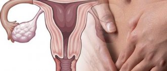
Candidiasis or thrush
is a fungal, infectious disease caused by opportunistic fungi of the genus Candida. Microorganisms are present in most healthy people. A person may encounter them for the first time in the womb, during childbirth, or during breastfeeding. Candidiasis develops against the background of reduced immunity, when the body can no longer restrain the active proliferation of the fungus.
The infection affects the mucous membranes of the mouth and genitals. It can develop on smooth skin, nails, and in the intestines, as a type of dysbacteriosis. There is visceral (systemic) candidiasis that affects internal organs. Despite the fact that Candida fungi are present in many people, and the disease itself is considered a neglected disease, it must be treated even if there are no external manifestations.
Causes of candidiasis
- Frequent damage to the skin and mucous membranes, for example, due to illness, working with aggressive chemicals, dry skin, age-related changes.
- Prolonged exposure to water, humid and warm environments. This includes a climate that is not suitable for your skin type, as well as occupational hazards.
- Mechanical damage to the skin and mucous membranes: injection drug addiction, long-term surgical interventions, catheters, shunts, endotracheal tubes.
After entering the body, the fungus may not manifest itself for a long time, since its reproduction will be suppressed by the immune system. In some cases, natural defenses weaken, leading to candidiasis (thrush). Among the factors that contribute to the proliferation of Candida:
- Chronic, endocrine and immune system diseases (diabetes, HIV).
- Metabolic disorders or taking medications that disrupt the natural microflora (antibiotics, hormonal contraceptives).
- Unbalanced diet, lack of sleep, stress and depression.
The fungus can be found in raw meat, unpasteurized dairy products, and, in more rare cases, on fresh vegetables and fruits. You can become infected with candidiasis from animals: dogs, calves, poultry.
1.General information
In order not to be interrupted by terminological references in the future, we will stipulate the following as a kind of preamble.
Opportunistic is an infection that occurs, literally, “at an opportunity,” “if possible,” i.e. under a certain combination of conditions favorable for the opportunistic microorganism. In turn, conditional pathogenicity implies asymptomatic parasitism or commensalism (harmless coexistence) of any microculture in the host’s body - as long as the host is healthy and immunocompetent - and the ability of this culture to undergo rapid pathogenic activation with a weakening of the immune defense, a significant change in the structure of the microbiome and some other circumstances.
Mycosis is a systemic infection by fungal cultures. Candidiasis is an extremely widespread (in fact, the most common) variant of mycosis, caused by yeast-like opportunistic fungi of the numerous genus Candida, most often Candida ablicans (the causative agent, in particular, of urogenital “thrush”).
Thus, pharyngeal candidiasis is an opportunistic mycosis caused by the activation of Candida and predominantly localized in one of the ENT organs.
A must read! Help with treatment and hospitalization!
Risk factors for development
The likelihood of infection increases with casual sexual intercourse, uncontrolled use of antibiotics and drugs that disrupt the natural microflora. Foods with large amounts of sugar and carbohydrates create a favorable environment for fungal growth. Excessive sweating also leads to an exacerbation of candidiasis, so it is necessary to wear cotton underwear that allows the skin to breathe and moisture to evaporate.
At the same time, excessive cleanliness can also cause harm. We are talking about douching. It should not be used as a method of contraception, since it is not effective, and also as a means of hygiene, because it leads to the leaching of the protective flora. If you experience discomfort, you should first consult a doctor.
Classification of the disease
Based on the depth of damage, candidiasis is divided into superficial and systemic forms. The first appear on visible parts of the body: mucous membranes, skin and its appendages. The second, systemic, are combined forms of the disease affecting internal organs. Without proper treatment, systemic (visceral) candidiasis can lead to the development of candidal sepsis. Most often, the fungus affects:
- gastrointestinal tract (esophagus, stomach, intestines);
- urinary organs (bladder, urinary tract, kidneys);
- lower respiratory tract (trachea, bronchi, lungs).
The disease is also classified based on the rate of spread of infection and the presence of relapses. Candidiasis of the liver and spleen often takes a chronic form. The acute, generalized form is asymptomatic candidemia, candida septicemia, candida thrombophlebitis, myositis, arthritis, cerebral candidiasis and other forms.
Classification of candida lesions according to ICD-10
- Candidiasis.
- Candidal stomatitis.
- Pulmonary candidiasis.
- Candidiasis of the skin and nails.
- Candidiasis of the vulva and vagina.
- Candidiasis of other urogenital locations.
- Candidal meningitis.
- Candidal endocarditis.
- Candidal septicemia.
- Candidiasis of other locations.
- Candidiasis, unspecified.
Officially, the diagnosis is made in accordance with ICD-10. In practical healthcare, such a classification is not entirely convenient, so they use the classification of deep candidiasis, which is a deeper list. When making a diagnosis, first indicate the name and form of the infection with a description of all localization points.
Skin treatment is carried out by a pediatrician in children and by a dermatologist in adults. Therapy is also carried out by doctors of other specializations, depending on the affected organ. For example, this could be a dentist, neurologist, gynecologist, pulmonologist.
Publications in the media
Candidiasis is an opportunistic mycosis that occurs with lesions of the mucous membranes and skin; in patients with severe immunodeficiency, disseminated forms are possible, most often affecting the lungs and gastrointestinal tract.
Etiology. The causative agents are yeast-like fungi of the genus Candida. 90% of lesions are caused by Candida albicans (which also includes C. stellatoidea). Other pathogens - C. tropicalis, C. krusei, C. lusitaniae, C. parapsilosis, C. kefyr (formerly C. pseudotropicalis), C. guillermondii, C. (Torulopsis) glabrata and very rarely - C. rugosa, C. utilis , C. lipolytica, C. zeylanoides.
Risk factors • Hematogenous disseminated candidiasis •• Neutropenia •• Antibiotic therapy •• Long-term catheterization of blood vessels •• Previous hemodialysis • Mucocutaneous candidiasis •• Immunodeficiency conditions •• Antibiotic therapy •• Douching and chemical irritation of the vagina •• Vaginitis •• Wearing dentures • • Long-term use of GCs •• Taking oral contraceptives •• Hyperglycemia.
Epidemiology. Candida is widespread in nature, mainly on plant substrates, various household items, especially children's toys. Candida albicans is normally present in the mouth, gastrointestinal tract, vagina and sometimes on the skin. Candidiasis is registered only in humans. The incidence increased significantly with the introduction of antibiotics and is currently progressing. Candida is one of the most common causative agents of opportunistic mycoses. Any immunodeficiency state and disruption of normal microbial cenosis lead to the onset of the disease. The possibility of transmission of the pathogen through household contacts has not been determined, but children become infected when passing through the birth canal or during breastfeeding. Urogenital candidiasis is transmitted sexually.
Pathogenesis. With disseminated lesions, the formation of foci of necrosis and neutrophilic inflammatory infiltration is noted. The development of candidiasis is facilitated by damage to the skin, increased sweating, and maceration. Dry, intact skin is resistant to Candida albicans. Excessive growth of Candida albicans is facilitated by disturbances in the microbial cenosis of the body caused by inadequate use of broad-spectrum antibiotics or changes in the microenvironment. The development of candidiasis is facilitated by metabolic and hormonal disorders (for example, diabetes, pregnancy, and the use of oral contraceptives). Immunodeficiency states and the use of immunosuppressants (for example, GCs) can cause fulminant forms and chronic candidiasis of the skin and mucous membranes.
Genetic aspects. Candidiasis is a companion to numerous inherited immunodeficiencies. Examples: • Familial chronic candidiasis (114580, Â) is constantly accompanied by viral infections, alopecia, tooth loss are characteristic • Familial chronic mucocutaneous candidiasis (*212050, r) affects the skin, nails, characterized by constant thrush, iron deficiency • Autoimmune polyglandular type syndrome I • Hyperimmunoglobulinemia syndrome E (147060, Â) with a defect in the chemotaxis of neutrophil leukocytes • Jobe's syndrome.
Clinical picture
• Superficial candidiasis develops in areas of the skin with relatively high temperature and humidity, with maceration of the skin associated with regular contact with water •• Candidal intertrigo is observed with diaper rash: erythematous or vesiculopustular rashes with maceration (usually in the area of large skin folds in children ), which leads to the development of erosions; whitish necrotic areas of the epithelium with ulcerated edges •• Diaper dermatitis is most often caused by Candida albicans: scaly rash or vesiculopustular rash accompanied by inflammation and intense itching •• Paronychia and onychia are usually caused by Candida albicans; most often, lesions are observed during maceration of the hands and feet caused by constant contact with water (in dishwashers and laundresses); characterized by thickening and discoloration of the nail plates, less often - nail loss.
• Mucocutaneous candidiasis is most often observed in the oral cavity and vagina. • Oral candidiasis (thrush) is a characteristic consequence of taking broad-spectrum antibiotics or immunodeficiency states. Typical manifestations are white or yellowish plaques on the surface of the mucous membrane; they are painless and do not merge. The lesions are often associated with diffuse erythema, thick dark brown deposits, deep fissures, and increased dryness of the mucous membrane. •• Vulvovaginal candidiasis caused by Candida albicans is widespread among women taking oral or intrauterine contraceptives, or who are in the third trimester of pregnancy (the condition is indirect). immunosuppressive effects of high concentrations of progesterone and serum a-globulin-related factor). It may be asymptomatic or cause intense itching or discomfort. It manifests itself as discharge such as leucorrhoea or films of varying thickness in the form of “pressed cheese”. Reddish spots are observed on the vaginal mucosa, labia, vulva and other areas of the perineum • Chronic mucocutaneous candidiasis is a rare pathology mediated by defects in T-lymphocytes; possible damage to the skin (including the scalp), mucous membranes (cheilitis, esophagitis), onychia and paronychia. In the most severe cases, granulomatous cutaneous candidiasis is observed with the appearance of infiltrates with unclear contours on the skin and nails, which are later covered with serous-sanguinary crusts, permeated with mycelium.
• Disseminated candidiasis (candidal sepsis, systemic candidiasis) is characterized by a severe course, high fever of the wrong type and damage to various organs (lungs, kidneys, brain, gastrointestinal tract, etc.). Dissemination of superficial candidiasis is rarely observed •• Candidal endocarditis. Symptoms: high body temperature, pain in the heart, heart murmurs, enlarged spleen, anemia •• Pulmonary lesions are accompanied by the development of infiltrates, including pseudomycelium of the pathogen, sometimes it grows into blood vessels. Symptoms: severe cough, initially dry, then with a small amount of viscous mucopurulent sputum, sometimes streaked with blood. X-ray infiltrates are more often detected in the lower lobes, disintegration with the formation of cavities and involvement of the pleura in the process is often observed •• Candidal septicemia is similar to sepsis of other etiologies •• Eye lesions: candidal retinitis and candidal panophthalmitis •• Candidal meningitis •• With candidiasis of the gastrointestinal tract, pain is noted in abdomen, intestinal bloating, diarrhea with blood in the stool •• Hepatic candidiasis is a combined granulomatous lesion of the liver and spleen with invasive growth of Candida albicans in people with immunodeficiency conditions. The main clinical sign is fever. Signs of oropharyngeal candidiasis are identified; pain or tenderness is possible on palpation in the right hypochondrium. Changes in liver function tests: moderate increase in bilirubin content and enzyme activity.
Diagnosis • Disseminated candidiasis •• Isolation of the pathogen from the blood (cultures from lysates or centrifugates are more preferable), cerebrospinal fluid, pericardial fluid. The diagnosis is more likely when the pathogen is isolated from several areas of the body •• If isolation results are negative, biopsy or aspiration of samples from the lesions •• Laparotomy or laparoscopy for liver candidiasis: small white nodules are found, not exceeding 5 mm in diameter • Mucocutaneous candidiasis • • Microscopy of clinical material treated with 10% KOH solution or Gram-stained (Gram-positive yeast cells are detected) •• Isolation of the pathogen is carried out by inoculation on conventional mycological media or blood agar •• X-ray with barium sulfate for candidal esophagitis: “cobblestone” syndrome is detected » and, less commonly, a fistula or dilatation of the esophagus (due to denervation) •• For esophagitis - endoscopic biopsy. The detection of thrush and dysphagia in HIV-infected people with a decrease in the severity of manifestations when taking antifungal agents allows us to establish a preliminary diagnosis of candidal esophagitis.
Differential diagnosis • Hematogenous disseminated candidiasis is differentiated from various cryptogenic bacterial infections, as well as from opportunistic infections in patients with neutropenia • Mucocutaneous candidiasis is differentiated from hairy leukoplakia and lesions caused by yeast or other yeast-like fungi.
TREATMENT
Mode • For disseminated candidiasis - inpatient • For mucocutaneous candidiasis - outpatient.
Management tactics • Disseminated candidiasis •• Fluid replacement and correction of electrolyte balance •• In severe lesions - support of hemodynamics and respiratory function •• In patients receiving daily amphotericin B, blood, serum electrolyte and creatinine levels should be examined at least twice a week. When isolating a blood culture, blood cultures must be repeated until a negative result appears • Examination of children and patients with severe immunodeficiency at each visit to the doctor with an appropriate physical examination and collection of a detailed medical history.
Drugs of choice
• For disseminated candidiasis •• Fluconazole. 400 mg IV on the first day, then 200 mg/day for 1 week, then at the same dose IV or orally for at least 2 weeks after clinical recovery and normalization of bacteriological parameters. For hematogenous disseminated candidiasis, the drug is as effective as amphotericin B, but less toxic for patients without neutropenia, HIV infection and severe immunosuppression after organ transplantation •• Amphotericin B is an alternative drug for hematogenous disseminated candidiasis and the drug of choice for patients with neutropenia and immunosuppression . Start with a test dose of 1 mg, then it is gradually increased to the therapeutic dose (0.3–0.7 mg/kg/day). Sometimes the entire therapeutic dose is administered after a test dose, for example in critically ill patients. The duration of treatment depends on the patient's condition and the form of hematogenous disseminated candidiasis. Treatment is usually continued for 2–10 weeks. The total dose of the drug per course of treatment is 0.2–2.0 g.
• For mucocutaneous candidiasis •• For vulvovaginal candidiasis ••• Miconazole - daily before bedtime intravaginally in the form of a 2% cream or suppository of 100 mg (for 7 days) ••• Clotrimazole daily before bedtime intravaginally in the form of a suppository of 100 mg for 6–7 days or 200 mg for 3 days, or in the form of 1% cream for 6–7 days ••• Nystatin in the form of cream, 100,000 units/g 1 r/day or intravaginal tablets, 100,000 U/g 2 times a day for 7 days ••• Fluconazole orally 150 mg 1 time / day twice with an interval of 7 days (or one tablet 450 mg once) •• For candidiasis of the oral cavity and pharynx ••• Clotrimazole: tablet (lozenge) 10 mg, keep in mouth for 20 minutes, 5 times a day for 7–14 days (48 hours after the disappearance of thrush) ••• Lozenges with nystatin, 1–2 pieces, 4–5 times a day for 7–14 day (48 hours after the disappearance of thrush) ••• Nystatin suspension for oral administration, 5–10 ml, before swallowing, rinse your mouth for 20 minutes 4–5 times a day (treatment), 2–5 times a day (prevention of relapse) .
• For candidal esophagitis • Ketoconazole orally 200–400 mg 4 times/day for 14–21 days •• or fluconazole orally: first dose 200 mg, then 100 mg 4 times/day for 10–21 days.
• For gastrointestinal candidiasis - fluconazole 200 mg orally 4 times a day for 14–21 days.
Alternative drugs • For disseminated candidiasis •• Fluconazole - for infection caused by Candida lustaniae, resistant to amphotericin B •• Amphotericin B - for infection with Candida krusei, resistant to fluconazole •• Other synthetic antifungals - imidazole and triazole derivatives, depending on their activity and safety • For mucocutaneous candidiasis •• For vulvovaginal candidiasis ••• Fluconazole 150 mg 1 time per day twice with an interval of 7 days ••• Any antifungal creams or suppositories to prevent relapse can be used every month a few days before menstruation — for women at risk (with an IUD, as well as for those taking oral contraceptives, antibiotics) •• For candidiasis of the oral cavity and pharynx ••• Ketoconazole 200–400 mg orally 4 times a day for 14–21 days •• • Fluconazole 50-200 mg orally 4 times a day for 14-21 days • For candidal esophagitis - amphotericin B (doses vary).
Complications • With disseminated candidiasis •• Pyelonephritis •• Endophthalmitis •• Endocarditis, myocarditis, pericarditis •• Arthritis, chondritis, osteomyelitis •• Pneumonia •• CNS infection • With mucocutaneous candidiasis •• Patients with immunodeficiency often develop severe complications. The severity of complications in immunodeficiency depends on the degree of impaired immune status (a widely used marker is the number of CD4+ cells). Moderate suppression of the immune system (CD4+ cell count 200–500/μl) can lead to the development of chronic candidiasis. In severe immunosuppression (CD4+ cells less than 100/μl), thrush can cause damage to any system of the body, especially the kidneys (candiduria).
Forecast. The mortality rate of patients with hematogenous disseminated candidiasis is 40–75%, mortality from candidemia is 15–37%.
Prevention • Disseminated candidiasis. The use of fluconazole at a dose of 400 mg/day in patients who have undergone bone marrow transplantation or undergoing treatment for acute leukemia reduces the incidence of candidiasis • Mucocutaneous candidiasis: wearing cotton underwear.
Synonyms • Thrush • Candidiasis • Yeast mycosis • Moniliasis • Oidiomycosis.
ICD-10 • B37 Candidiasis
Notes • Most candidal infections are associated with endogenous flora • Person-to-person transmission is rare • Candidal vaginitis can be sexually transmitted (rare) • Oral candidal leukoplakia may be a precancerous condition (rare) • In patients with severe immunodeficiency, esophageal candidiasis may appear together with other infectious (eg, HSV, CMV) esophagitis • Skin tests, often used to diagnose or rule out anergy, are positive in 70–85% of individuals • Angular cheilitis (zaeda, angulitis) - inflammation and fissure in the area of the corner of the mouth; predisposing factors - decreased bite height (when wearing removable dentures), malnutrition, allergic dermatitis or fungal infection (Candida albicans).
Symptoms of candidiasis
The symptoms of the disease are directly related to the affected organ. Superficial forms have obvious signs, while systemic forms can be asymptomatic or be similar to other diseases. In the presence of chronic diseases (diabetes mellitus, HIV, AIDS, leukemia and other types of oncology), candidiasis may not be detected at all. Since its symptoms will be perceived by the patient as an unhealthy state due to the existing disease. To make matters worse, Candida susceptibility testing is not considered a routine procedure in many teaching hospitals and laboratories.
Candidiasis of the skin and nails
The infection is expressed in the form of allergic rashes (candidamycids), erythematous-squamous spots, urticarial and bullous type rashes. Nail pathologies are divided into:
- parochinia - purulent inflammation of the periungual and subungual spaces, absence of eponychium (skin at the base of the nail plate);
- onychia - purulent inflammation of the nail fold, deformation of the nail with discoloration and brittleness.
Candidal skin lesions may be accompanied by fever, inflammation and swelling of the lesions.
Candidiasis (thrush) of the oral mucosa
The fungus can affect both individual areas of the oral cavity and completely all mucous membranes: lips, gums, cheeks, palate, tongue, tonsils, uvula and pharynx. This is expressed in the presence of a white coating, compared to a curd mass. Swelling and redness may appear. There is often pain when swallowing, talking, or palpating. Saliva becomes viscous and bad breath appears.
Thrush may be accompanied by candidal cheilitis - damage to the lips and corners of the mouth. Painful cracks appear, covered with a white coating, and pieces of skin peel off from the lips. The disease is characterized by a long course with the possible addition of bacterial infections.
Intestinal candidiasis
Intestinal infection with fungi of the genus Candida can be an independent disease or develop as a result of damage to the oral cavity. Expressed as severe dysbacteriosis with the following symptoms:
- diarrhea;
- excessive gas formation;
- nagging pain in the rectum;
- an admixture of white flakes in the stool.
In most cases, symptoms are sluggish or completely absent. The disease is dangerous because the body does not receive enough vitamins and microelements, which is especially dangerous for a growing child.
Candidiasis of the genitourinary system (urogenital candidiasis)
Unlike sexually transmitted diseases, candidiasis of the genitourinary system is often hidden and asymptomatic. The fungus is discovered during a planned infection or against the background of other diseases. Symptoms begin to appear against the background of declining immunity, ongoing pathological processes of microflora destabilization and re-infection. In women, candidiasis manifests itself as follows:
- itching and burning of the external genitalia;
- swelling and redness;
- white, cheesy discharge;
- pain during sexual intercourse.
In men, the symptoms are similar:
- itching and burning of the head of the penis;
- white plaque and discharge:
- pain during sexual intercourse and urination.
Fungal infections of the bladder, excretory tract and kidneys are characterized by frequent urination, including false urges, pain in the suprapubic region. Candidiasis can be accompanied by bacterial infections. Complications include: cystitis, pyelonephritis, necrosis of the papillae, abscesses, formation of mycelium in the renal pelvis. Without proper treatment, against the background of chronic diseases, there is a risk of developing kidney failure.
3. Symptoms and diagnosis
As a rule, pharyngeal candidiasis occurs with few symptoms or with moderate severity of clinical manifestations. Typical symptoms include inflammation of the tonsils and mucous membranes, a dirty white coating, low-grade fever, mild malaise, discomfort or pain when swallowing.
Granulomatous, atrophic, hyperplastic, erosive-ulcerative forms are relatively less common, requiring differential diagnosis, in particular, with tuberculosis.
Candidiasis of the pharynx is easily chronic and occurs, as a rule, in waves.
The prognosis worsens when the mycotic infection spreads to adjacent and internal organs.
If oropharyngeal candidiasis is suspected, a smear is taken from the affected surface, then a culture is performed on a nutrient medium; Recently, genetic identification (PCR) and serological tests (detection of specific antibodies) have been increasingly used.
About our clinic Chistye Prudy metro station Medintercom page!
Diagnosis of candidiasis
The study is based on the isolation and identification of a species of fungus of the genus Candida. Currently, there are about 150 species, differing in morphological and biochemical properties. The most common fungus is C. albicans, accounting for up to 80% of cases of candidiasis of the digestive tract and up to 70% of genital infections. Before prescribing treatment, it is also necessary to determine the sensitivity of the isolated strain to antimycotics (antifungal medications): amphotericin B, voriconazole, itraconazole, fluconazole, flucytosine.
Microscopic diagnostics
1. Microscopy of a smear is taken from the area of the affected mucosa. Allows for comparative characterization of blastospores and pseudomycelia. During the study, fixed and native preparations that stain microorganisms are used. To increase the information content, pseudomycelia of cells are treated with dyes. The contrast in staining of microorganisms makes it easy to distinguish candida from other forms, including bacteria, under a microscope.
2. Bacterial culture allows you to identify the causative agent of infection and determine its concentration. The analysis is used to monitor the effectiveness of treatment, as well as to identify the sensitivity of candida fungi to various antimycotics.
Diagnostics by ELISA and PCP
1. Enzyme-linked immunosorbent assay (ELISA) is based on the determination of antibodies that are produced by the immune system in response to foreign substances in the blood. This technique allows you to identify the pathogen and the degree of its development, establishing whether the disease occurs in an acute or chronic stage.
2. Polymerase chain reaction (PCR) is a highly sensitive test that allows you to directly detect the infectious agent. Thanks to it, it is possible to differentiate Candida fungi with pseudomycelium from those that do not have it. These data are important for subsequent interpretation of results and deeper diagnosis.
Treatment of candidiasis
Treatment of candidiasis is aimed at eliminating factors that contribute to the occurrence of candidiasis. If the skin is affected, local treatment is carried out using an open method using antifungal ointments.
The attending physician prescribes systemic and local medications for this disease. Local agents are not absorbed into the blood - they act only on the mucous membrane affected by the Candida fungus. They stop the reproduction and growth of fungi, relieve discomfort and restore affected tissue.
Interpretation of results
If there are clear signs of candidiasis (thrush), and during a laboratory test blastospores and pseudomycelia of the fungus were identified, the study ends here. A diagnosis is made and treatment begins.
If a microscopic examination gives a negative result, this does not indicate the absence of infection. The disease can occur in a latent chronic form. It is necessary to carry out a number of other tests, for example, microscopic examination of scrapings, determination of Candida DNA in scrapings, urine, and prostate secretions. Also prescribed:
- Clinical blood test.
- Test for HIV infection.
- Determination of trace element reserves in the body.
- Test for glucose and carbohydrate metabolism metabolites.
These laboratory tests can help identify conditions that may be causing the fungus to grow.
Advantages of taking tests at JSC "SZDCM"
- Own laboratory with the latest diagnostic equipment.
- Convenient location of terminals within transport accessibility from anywhere in the city.
- Qualified laboratory technicians and friendly staff.
- Fast analysis and several options for obtaining results. Choose the one that is most convenient for you.
Medical centers and laboratory terminals of the North-Western Center for Evidence-Based Medicine are located in St. Petersburg, Leningrad region, Veliky Novgorod, Okulovka, Kaliningrad and Pskov.
Analyzes
- Bacteriological study for opportunistic pathogenic flora (OPF)
- NC yeasts of the genus Candida: C.albicans, C.krusei, C.glabrata
- Study of the biocenosis of the urogenital tract in women (“Femoflor 13 - screening”)
- Candida albicans
- Mycoses: identification of clinically significant fungi with determination of sensitivity to antimycotic drugs (only for fungi of the genus Candida and Cryptococcus neoformans)
- Specific immunoglobulin E - Candida albicans
go to analyzes


