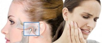
Pathological processes leading to disruption of the natural functional state of the temporomandibular joint are usually accompanied by characteristic symptoms. Signs indicating the presence of problems with the TMJ include pain in the back of the head and neck, attacks of uncontrolled teeth grinding, and heaviness syndrome in the problem area. The continued development of diseases, in the absence of medical intervention, entails more serious consequences - restrictions on mobility, difficulties with chewing food and opening the mouth, severe violation of occlusion and bite. One of the relaxation methods recommended when excess tension is detected is massage of the masticatory muscles of the jaw and facial tissues.
Features of the masticatory muscles:
- All chewing muscles are attached to the lower jaw (mandibula);
- All masticatory muscles elevate the lower jaw, performing chewing and facilitating the correct pronunciation of words;
- The chewing muscles, unlike the facial muscles, have fascia.
These features are not indicated in atlases and textbooks, and, as a rule, they are not required to be mentioned in tests. I brought them specifically to make it easier for you to navigate - as soon as we talk about any masticatory muscle, you immediately understand that it attaches to the lower jaw and lifts it. That is, one name of a muscle group is enough to remember the function and place of attachment.
Symptoms of TMJ problems
The disease manifests itself with the following symptoms:
- Painful sensations occur on palpation.
- When opening the mouth, the patient feels a sharp pain.
- Due to pain, a person experiences difficulty eating and communicating.
- Breathing through the mouth is difficult.
- To the touch, the chewing muscles are compacted and slightly increased in volume.
- Facial asymmetry with unilateral spasm.
- Low-grade fever.
- Headache.
The gnathologist
carries out differential diagnosis with a fracture or dislocation of the jaw, as well as with an infectious lesion.
There are three degrees of disease progression: mild (the patient is able to open his mouth four centimeters), moderate (the mouth opens two centimeters), severe (the patient cannot open his mouth more than one centimeter).
Anatomy of the masticatory muscles:
I. Temporal muscle (musculus temporalis)
The largest and most visible muscle of mastication. Unlike all other chewing muscles, its abdomen is located on the cranial vault (fornix cranii).
In green I highlighted the outline of the temporal muscle itself, in blue - the outline of the powerful tendon with which the muscle is attached to the coronoid process (processus coronoideus) of the lower jaw. I outlined the coronoid process in yellow, I hope you remember it from craniology. When you show the location of the temporal muscle during the test, you should outline the entire contour, that is, both the muscle and its tendon.
The temporal muscle is located in the temporal fossa (fossa temporalis). If you take a dissected skull, or even a high-quality artificial specimen, you will see impressions on the sides. These impressions are shallow, but occupy a large area. It is these depressions that are called the temporal fossae. In the picture from Wikipedia, the temporal fossa is highlighted with a black frame:
Origin: The temporalis muscle begins on the temporal surface of the frontal bone, as well as on the squamous surface of the temporal bone.
Attachment: The temporalis muscle attaches to the coronoid process of the mandible (the sharpest process of the mandible). Don't forget that the chewing muscles start from the stationary part and are attached to the bone that they move.
Function: the temporalis muscle closes the mouth, raising the lower jaw until the teeth are closed. Also, if you push your lower jaw forward, like a drawer in a table, it is the temporalis muscle that will push it back to its normal position.
II. Chewing muscle (musculus masseter)
It is also a very noticeable and large muscle. If we look at a tablet with an image in profile, it is impossible to confuse the masticatory muscle with anything else. See:
The chewing muscle is extremely important for survival, so our distant ancestors developed this muscle to be very powerful and strong. The masticatory muscle consists of two parts - superficial (pars superficialis) and deep (pars profunda). You can easily distinguish the superficial part - it is attached to the anterior part of the zygomatic arch (arcus zygomaticus). The deep part is also attached to the zygomatic arch, only not in front, but in the back.
I highlighted the superficial part with a green outline, and the deep part with a blue outline:
Origin: Both parts of the masticatory muscle begin from the zygomatic arch.
Attachment: both parts of the masticatory muscle are attached to a special place on the lower jaw - to the masticatory tuberosity (tuberositas masseterica). Let me remind you that the chewing tuberosity is located on the outer surface of the branch of the lower jaw (ramus mandibulae).
Function: raise the lower jaw, chewing and helping to pronounce words. Also, the superficial part of the masseter muscle helps push the jaw forward. We will push it back using the temporal muscle, as you remember.
III. Lateral pterygoid muscle (musculus pterygoideus lateralis).
Now we need a tablet with a skull, which we look at as if from inside the oral cavity (cavitas oris). Something like this:
We turned the skull with the back of the head towards us and sawed it in the frontal plane so that we could look from the inside at the oral cavity, gums and teeth.
Let's look at the lateral pterygoid muscle. Here we can see both lateral pterygoid muscles, so I've highlighted both:
The lateral masseter muscle has two parts - upper and lower.
a) Upper part.
Origin: Infratemporal crest of the greater wing of the sphenoid bone. Let's remember the sphenoid bone (os sphenoidale) - it has a body (corpus), large wings (alae majores) and small wings (alae minores), as well as pterygoid processes (processus pterygoidei).
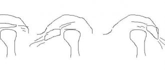
For some reason, many people like to confuse large wings and pterygoid processes. Let's look at a simplified diagram (front view) so as not to confuse these anatomical formations:
We are interested in the large wing. It is on this that the small infratemporal crest (crista infratemporalis) is located. This is where the origin of the superior portion of the lateral pterygoid muscle is located:
The upper part of the lateral pterygoid muscle is attached to the temporomandibular joint (articulatio temporomandibularis).
b) The lower part of the lateral pterygoid muscle also begins with the sphenoid bone. Now we need to shift our attention from the large wing a little lower - to the pterygoid process (processus pterygoideus). It seems to split into two plates - medial and lateral. Look at the penultimate picture - there I highlighted the pterygoid processes in brown.
Let's find the lateral plate (lamina lateralis) of the pterygoid process. This is, in general, not difficult:
It is from this plate that the lower part of the lateral masseter muscle begins.
So, the beginning: the lateral muscle (its lower part) begins with the lateral plate. Great memory, by the way.
Attachment: pterygoid fossa (fovea pterygoidea) of the lower jaw. Not a particularly well-known anatomical formation, so I found this diagram:
Our pterygoid dimple is located at number 4.
The function of the lateral pterygoid muscle is to raise the jaw (like all masticatory muscles), as well as to abduct the lower jaw in the direction opposite to the muscle. For example, the left lateral pterygoid muscle, when contracted, moves the lower jaw to the right side.
IV. Medial pterygoid muscle (musculus pterygoideus medialis). Let's take a tablet we already know to look at this muscle:
Beginning: Pterygoid fossa (fossa pterygoidea) of the sphenoid bone. By the way, the lower jaw also has, as you remember, a pterygoid fossa, only it is called fovea pterygoidea, so as not to be confused. Ancient anatomists are very prudent guys, definitely.
To be honest, I find it difficult to show it on a tablet. I don’t have a skull at hand at the time of writing this article, but the respected Sinelnikovs indicate in their atlas that “... the medial and lateral plates limit the pterygoid fossa.” Moreover, in the illustration closest to the text, the fossa pterygoidea is indicated on the lateral plate (two pictures ago I highlighted the lateral plates of the pterygoid processes in red). The Sinelnikovs are right in any case - they are the kings of anatomy, after all. I advise you, dear readers, on occasion to ask your teachers about the location of this hole.
In general, speaking about the beginning of this muscle, we focus on the lateral plates of the pterygoid processes of the sphenoid bone and clarify the fossa with the teachers.
Insertion: pterygoid tuberosity of the lower jaw. There won't be any difficulties here anymore, here's where this place is:
Function: Raises the mandible, as well as “pushes” the mandible to the opposite side (i.e., similar to the lateral pterygoid muscle).
General overview
The masticatory muscles are located on the lateral areas of the skull, and are located in pairs, four on each side. The masseter muscle itself, as well as the temporal muscle, are characterized by a superficial location, while the pterygoid muscles (medial and lateral) are located in the infratemporal cavity. Starting on the cranial bones, they connect to the mandibular region, and set the jaw in motion by contracting and relaxing.
The functionality of the anatomical elements under consideration is difficult to overestimate, since they are involved not only in the processes of food processing, but also in breathing, as well as in the implementation of speech function. Massage of the masticatory muscles for TMJ dysfunction allows you to relieve overstrain, relieve pain and restore jaw mobility.
Lexical minimum
- Mandibula;
- Musculus temporalis;
- Fornix cranii;
- Processus coronoideus;
- Fossa temporalis;
- Musculus masseter;
- Arcus zygomaticus;
- Tuberositas masseterica;
- Ramus mandibulae;
- Musculus pterygoideus lateralis;
- Cavitas oris;
- Os sphenoidale;
- Alae majores;
- Alae minores;
- Processus pterygoidei;
- Crista infratemporalis;
- Articulatio temporomandibularis;
- Lamina lateralis;
- Fovea pterygoidea;
- Musculus pterygoideus medialis;
- Fossa pterygoidea.
Diagnostics
The patient experiences pain from jaw movement. In this situation, it is necessary to consult a doctor as soon as possible to begin treatment. It is necessary to find out how to relieve jaw muscle spasm in order to restore health.
During the examination and history taking, the doctor observes the patient, paying attention to his posture and head tilt, neck movements, facial expressions, and facial expressions. The state of the muscles during speaking and swallowing is determined. The presence of the corneal reflex is checked, and the amplitude of movements of the lower jaw is determined. The doctor finds out whether there are problems with the spine.
The patient is examined using diagnostic equipment, an occlusiogram is prescribed to determine the type of bite, and its dynamics and statics are measured. If necessary, an X-ray of the TMJ and an orthopantomogram of the jaws are prescribed, and an electromyographic study is performed.
Consequences of jaw spasm
Trismus is unpleasant for a person not only due to pain. When opening and closing the mouth is painful, the normal process of eating is disrupted and the gastrointestinal tract suffers. When the jaw spasms, breathing is impaired, which leads to constant oxygen starvation of the brain and headaches. This affects the composition of the blood and the functioning of the cardiovascular system.
Trismus caused by infections or tumors causes serious health problems. In this case, long-term serious treatment is required. In the initial stages, pain is completely relieved. For systemic diseases, when trismus is a secondary symptom, complex therapy is necessary.
Prevention of hypertonicity of the TMJ muscles
Preventive measures against hypertonicity of the lower jaw muscles will help prevent the disease with an integrated approach. It is much easier to follow the general recommendations of doctors than to face a painful spasm of the TMJ. Prevention includes the following activities:
- Preventive visits to the doctor every six months.
- Careful systematic oral hygiene.
- Treatment of caries at its first symptoms.
- Orthodontic treatment for malocclusion pathologies.
- Disinfection of food products.
- Antitetanus therapy for animal bites.
- A set of necessary vaccinations.
- Do not open your mouth wide (jaw dislocations often occur when trying to bite off a large fruit).
- Do not overuse solid foods and do not chew nuts with your teeth.
- Avoid stressful situations, react less to stress.