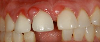
A 60-year-old patient came to the Research Center clinic with a complaint that she had mobility of the front tooth of the central incisor. Purulent discharge appeared from the gums in the area of the front teeth from the fistulous tract. From her story it became clear that the patient had previously had metal-free structures installed, crowns on her front teeth, the so-called. central incisors. She had them done in another clinic, where before installing crowns she had intra-canal teeth whitening
, which may have triggered a number of pathological processes.
The decision was made based on the CT scan:
- remove 2 central incisors, remove two upper front teeth
- and install implants with immediate loading.
Immediate loading
means that prosthetics of the upper anterior teeth will be performed on our patient also on the day of surgery.
It was also decided for the patient to leave and use as temporary crowns the veneers of those crowns that she had on her front teeth at the time of her visit to our clinic.
Therefore, the teeth, two central incisors, were removed very carefully. The photograph clearly shows that both incisors have a lesion
: the root part of the teeth was destroyed, eaten away, and these teeth could not be saved.
The anterior teeth were removed atraumatically,
with preservation of holes. And - with the preservation of the old crowns as temporary prostheses for the upper front teeth, which were useful to us for securing them to the installed implants in the future.
In this photo you can see the sockets of the teeth
. I can note that the removal of the upper front teeth was atraumatic, but in the area of tooth 11 (the first incisor of the first segment), due to a fistulous tract on the gum, severe destruction of the root occurred, as a result of which the cortical plate was lost. When such a fistulous tract occurs in the area of the front teeth, it means that the process has already started, a lot of unpleasant things have already happened. In this case, the cortical plate was destroyed. Before the removal of the front teeth, at the planning stage, individual templates were made for the patient, with the help of which the implants were subsequently installed.
How does the chronic form of granulating periodontitis occur?
A problem tooth always has a filling or a pathological carious cavity, which causes pain when probed. There is no reaction to high or low temperatures, and painless percussion is also observed. Sometimes granulation tissues grow from the source of destruction in the peri-apical region into the root canals, which causes bleeding and slight pain upon probing.
A mandatory sign is a fistula through which granulations bulge and pus is released. The mucous membrane is swollen, hyperemic, and has a cyanotic tint. One of the most dangerous complications is the death of the growth plate and stopping further bone formation.
X-ray signs
X-ray images clearly show the destruction of the cortical alveolar plate and a noticeable focus of resorption of spongy bone tissue with unclear contours at the apex. Destruction also occurs in the area of bifurcation of molars. Pathological root resorption in permanent teeth is much less common than in temporary teeth.
Differential diagnosis
It is important to distinguish the disease from such diseases with similar symptoms:
- chronic fibrous and gangrenous pulpitis;
- deep chronic caries;
- pulpitis, with a history of complicated focal periodontitis.
Let's start implanting the upper front teeth
a highly accurate surgical template
, individually made based on the CBCT image and using the 3D Shape intraoral scanner . Under the Straumann Bone Level SLActive implantation system, as part of a complete protocol for implantation of the upper anterior incisors.
Here you can observe the result of one-stage implantation of the front teeth; Straumann implants were installed. A bed for the implants was formed, which were installed in a strictly necessary position so that we could do aesthetic work not only on implantation, but also on further prosthetics of the upper anterior incisors.
You can see Straumann implants installed in exactly the right position
, as we planned. They are presented with the template removed. Bone biomaterial was placed in the sockets, which prevents the socket from collapsing in order to preserve the outer contour of the gum. In addition, in the area of the tooth that had a fistulous tract (tooth 11), in the photo it is on the right, a collagen membrane was sutured. The membrane was tunneled and secured with a single suture to allow bone to form and mature normally in the socket.
Here you see the same implants, but in front view. You can clearly see how the collagen membrane was fixed, and it should be noted that in order for the work to be aesthetic on the one hand, and minimally traumatic on the other, mucosal detachment was carried out using the tunnel method
with further installation of the membrane.
Features of the course of chronic granulomatous periodontitis
This pathology develops only after the formation of roots and periodontium is complete. Growth to the sides and deeper into the granulation tissue is limited from the unaffected bone by a connective tissue capsule. Its fibers are extensively woven into the periodontium. Since the surrounding bone is dense, the lesion is clearly defined on x-ray.
The disease is also asymptomatic, with the exception of rare pain when pressing, and is accompanied by a change in tooth color. Probing and percussion do not cause pain, there is no reaction to temperature stimuli. The tooth may be intact or may have a carious cavity or filling.
X-ray signs
- Destruction of the cortical plate of the alveolus in the area of the apex of the roots of the damaged tooth.
- The clearing segment of the bone tissue has a round or oval shape and perfect outline, and its diameter does not exceed 4-5 mm.
Differential diagnosis
It is necessary to distinguish granulomatous periodontitis from the following diseases:
- deep chronic caries (treatment of caries in children is accompanied by pain during preparation of the enamel-dentin junction, reaction to cold);
- chronic gangrenous and fibrous pulpitis, complicated by focal periodontitis (sharp pain on probing);
- fibrous and granulating chronic periodontitis (diagnosed using x-ray);
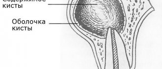
- cystogranuloma and radicular cyst (in this case, the focus of destruction will be more than 5-8 mm).
Structure
The anatomical structure of the crest includes the base of the process with alveoli, separated by septa. This is where teeth develop and grow. The ridge consists of two walls - the outer one on the side of the cheek and the inner one, turned towards the tongue. The surface is lamellar, it is adapted to support row units of different types, differing in structure and loads.
The process of the maxilla is located between the plates, it acts as an integral part of the cancellous bone. Between the individual alveoli of the structure there are partitions that separate the tooth sockets. At the same time, the cells of the area constantly take part in the formation and resorption of bone, this process is compensatory, in this case it proceeds normally. The features of the ridge allow it to adapt to different conditions, which makes the functioning of the dentition correct.
The crest of the lower jaw has an unpaired and symmetrical structure. Externally, the site forms an arc with a main center and two branches. Alveolar axons are located on each side in an amount of 8 pieces. The sockets can have different shapes and sizes, depending on the position of the units, but most often they are cone-shaped alveoli. For areas where teeth with multiple roots develop, there are partitions that separate the canals.
Features of the clinical picture of chronic fibrous periodontitis
This form is rare in children. In the apical zone of the roots, coarse fibrous connective tissue is formed, replacing the periodontium. Such fibrosis occurs after acute periodontal inflammation or after injury. Sometimes it develops after pulpitis or other forms of chronic periodontitis. The gap sometimes narrows due to excess cementosis.
The disease is also asymptomatic, without pain. The tooth can be intact or filled, with painless percussion. On an x-ray, the periodontal fissure is unevenly widened or narrowed. To clarify the diagnosis, the age of the child is taken into account.
Periodontal ligament
The periodontal ligament is the connective tissue that surrounds the tooth and connects it to the inner wall of the alveolar bone.
It begins 1-1.5 mm below the enamel-cement junction.
It's hard to believe, but its width (on average) is only 0.2 mm. 0.2 millimeters, Karl! The clarification “on average” is explained not only by the individual characteristics of the periodontal ligament in different people, but also by changes in the load on the tooth. The relationship is direct: the greater the load, the wider the ligament.
The main components of the periodontal ligament are
- periodontal fibers;
- cells;
- intercellular (ground) substance;
- vessels, nerves.
Reminds me of something, doesn’t it? The connective tissue of the gums has a similar composition:
The similarity is not without reason, because the periodontal ligament is a continuation of the connective tissue of the gums with its own characteristics, thanks to which its unique function is realized.
A few words about each of the components of the periodontal ligament.
Acute periodontitis of permanent teeth
Acute periodontitis in children often occurs after a tooth injury or due to incorrect treatment of pulpitis.
- Serous acute periodontitis.
The pain in the causative tooth increases, but the general condition of the child is not disturbed. The tooth is intact or has a fractured crown. If periodontitis is of toxic origin, there are signs of carious cavity preparation. If the pulp is dead, probing is painless, there is no reaction to temperature. On the mucous membrane of the gums there are symptoms of minor inflammation. There are no signs on the X-ray yet. A day later, the serous phase turns into a purulent phase.
- Acute purulent periodontitis.
Accompanied by constant throbbing pain and sharp pain even with a slight touch. Body temperature rises and intoxication begins. The tooth is intact, previously treated or with a carious cavity, mobile due to the accumulation of pathological exudate in the periodontal gap. An abscess forms under the periosteum, and sometimes facial asymmetry develops. X-ray also does not show anything concrete, except for a lower clarity of the picture.
It is important to distinguish acute purulent periodontitis from the following diseases:
- acute diffuse pulpitis, with a history of perifocal periodontitis (painful probing);
- acute purulent pulpitis, complicated by perifocal periodontitis (pain intensifies with hot stimuli and calms down with cold stimuli);
- exacerbation of periodontitis (changes in the periapical zone);
- acute odontogenic periostitis and another no less insidious disease - osteomyelitis.
Exacerbation of chronic periodontitis in molars with incomplete root growth is more common than the acute form. The tooth constantly hurts, percussion is also painful. The mucous membrane of the alveolar process swells and turns red, the crown changes color, and sometimes a fistula appears. The X-ray shows destruction of the cortical alveolar plate, a segment of bone clearing and deformation of the periodontal fissure.
Now - the stage of prosthetics of the upper front teeth
In this photo you can see the fixation of temporary crowns
on previously installed Straumann implants. It can be seen that the crowns have settled exactly as they were originally. As you remember, we decided to use the patient's old crowns as temporary crowns.
screw-retained
temporary crowns
. The shafts extend exactly to the back surface of the teeth, as pre-planned in the computer program.
You can see what the patient received on the day of the operation, namely:
on the day of the operation the patient left with her teeth!
And almost no one around you will notice that any surgical procedures have been performed. The patient looks exactly the same as she came BEFORE the immediate implantation surgery.
Temporary crowns for the front teeth, as you can see in the photo, we left the old ones.
Since the accuracy of implantation was very high, i.e. Straumann implants were positioned in the bone, I would say, with filigree precision, so there was no need to make additional temporary prostheses. Here I would just like to note to readers of the article
, those who are planning implantation and surgery - do not worry about the fact that if your front teeth are implanted in the frontal area, you will have to walk without teeth for some time.
No, that's not true. After the operation, you will leave with your teeth on the same day
.
Final picture of the result of implantation and prosthetics
You can clearly see how the Straumann Bone Level implants stand and how deep they are.
Both implants and temporary structures (crowns) cost exactly as needed. They were delivered exactly as planned, which gives a positive result. And it gives us all the prerequisites that the result of a one-step implantation of the front teeth, performed by doctors of the German Implantology Center, will be highly aesthetic and durable. And it will delight our patient for many, many years.
Diagnostics and correction
The pathological condition can be detected during a routine examination. In this case, the doctor prescribes an x-ray, which allows you to clarify the diagnosis and determine what the treatment regimen will be. Various methods are used to restore the structure; indications for correction are:
- atrophic processes affecting the ridge;
- defects caused by injuries, chronic diseases, surgical interventions.
Treatment is prescribed depending on the degree and severity of the pathology, tissue condition and other factors. The correction process usually includes the following steps:
- administration of anesthesia (conduction is used);
- surface treatment using antiseptic agents;
- removal of bone tissue particles, including fragments, if the cause of the intervention was injury or gradual destruction of the ridge;
- eliminating remaining sharp edges;
- closing the wound, applying sutures and dressings.
The exact procedure depends on the reason for the surgery, for example, you want to reduce a dislocation rather than remove sections of bone. To do this, a complete study is first carried out. After completing treatment measures, the Patient is required to comply with certain actions:
- exclude physical activity during rehabilitation;
- cessation of smoking, alcoholic beverages, and other bad habits;
- correction of the diet, exclusion of solid and spicy foods for the period of healing and recovery;
- observe the rules of personal hygiene, take care of the oral cavity;
- rinse your mouth after every meal using antiseptic agents.
Correction of the appendix is a complex stage of prosthetics or elimination of various pathologies caused by injuries or developmental disorders. Treatment should only be carried out by a qualified doctor with experience in this field.
Possible pathologies and injuries
The most common problem encountered in dental practice is atrophy affecting the alveolar ridge. The reasons for this phenomenon:
- injuries, mechanical damage to the area;
- osteoporosis;
- lack of treatment in the form of prosthetics, if there are indications for this;
- fragility, weakening of the bone.
Before you begin installing the prosthesis, you need to correct the process. The bone thickens in the necessary places, the structure becomes reliable, durable, and able to withstand loads. The procedure for performing the operation depends on the condition of the tissues and the cause of the damage.
Most often, correction is needed as a result of damage to the appendix after injury. The most common ones include:
- fractures of the alveolar part;
- physiological aging, that is, natural processes;
- destruction of the ridge for various reasons.
This is not only impacts and other damage, but also a congenital weak bite, in which the tissues are subject to natural increased wear. If timely measures are not taken, there is a likely risk of tooth loss. Therefore, regular preventive examinations come first, especially in cases of bone fragility.