Cyanosis is a symptom characterized by a blue tint of the skin and mucous membranes, which occurs due to an increased level of carbhemoglobin in the blood. Cyanosis, which is associated with the entry of chemical compounds into the body or the deposition of any substances in the skin, is considered to be false cyanosis.
Cyanosis resulting from hypoxemia is considered true. Capillary blood in the presence of true cyanosis contains more than fifty g/l of reduced hemoglobin.
Cyanosis is a fairly common symptom of heart disease. The further a part of the body is from the heart muscle, the more intense its cyanosis.
Cyanosis of the fingers and toes, nasolabial triangle, and ears is called acrocyanosis. Acrocyanosis occurs when the level of reduced hemoglobin increases in the venous blood. Acrocyanosis occurs due to excessive absorption of oxygen by tissues when blood flow slows down. Central cyanosis is a widespread cyanosis. The cause of diffuse cyanosis is a lack of oxygen in the pulmonary circulation.
Patients with both peripheral and central cyanosis often come to the Yusupov Hospital. Cyanosis of the face brings aesthetic discomfort to patients, while cyanosis of the extremities may remain unnoticeable for a long time. By signing up for a consultation by phone or online, during an individual conversation you can find out the differences between cyanosis and acrocyanosis, learn about the possible causes of cyanosis, view photos of patients with cyanosis of the skin, photos of patients with cyanosis of the nasolabial triangle and calculate the financial side of the issue.
The Yusupov Hospital is engaged in scientific activities, diagnostics, treatment, rehabilitation and prevention. This is the only medical institution in Moscow of such a high level.
Triangle structure
If you go in order, says Tatyana Romanenko, moving from top to bottom along the arterial blood flow, then the first will be the facial artery (a. facialis). “It is a continuation of the external carotid artery (a. carotis externa). The facial artery enters the face, bending around the inner edge of the angle of the lower jaw at the level of the anterior edge of the masticatory muscle, after which it goes into the thickness of the muscles. On the face, it passes near the corner of the mouth, wing of the nose and anastomoses (connects) in the medial corner of the eye with the artery of the dorsum of the nose (a. dorsalis nasi), which is a branch of the ophthalmic artery (a. ophthalmica), belonging to the basin of the internal carotid artery (a. carotis interna),” notes Tatyana Romanenko.
The therapist also says that, having reached a place just below the corner of the mouth, the facial artery gives off a branch: the inferior labial artery (a. labialis inferior) and next to the corner of the mouth - the superior labial artery (a. labialis superior). They both hide in the thickness of the orbicularis oris muscle (m. orbicularis oris) and anastomose with the arteries of the same name on the opposite side. This creates a single anastomosis, consisting of four arteries of the lips (top and bottom), around the mouth.
Veins accompany it along the entire length of the facial artery and its branches, says the therapist. Top down:
- superior ophthalmic vein (v. ophthalmica superior).
The tributaries of the superior ophthalmic vein are:
- nasofrontal vein (v. nasofrontalis);
in the medial corner of the orbit it anastomoses with
- angular vein (v. angularis), which is the root of the facial vein;
- and the inferior ophthalmic vein (v. ophthalmica inferior); at the medial corner of the eye it flows into the angular vein.
From the medial outer corner of the orbit, the inferior ophthalmic vein goes deep into it, then divides into two trunks. One of them flows into the cavernous sinus (sinus cavernosus) or into the superior ophthalmic vein, the other passes through the inferior orbital fissure and flows into the cavernous sinus (sinus cavernosus), which lies at the base of the skull.
Marks of old age. Why does age-related pigmentation appear? More details
Introduction
So, a dangerous triangle of the face. We can describe this zone in this way: the apex of the triangle is located in the glabella area, its legs enclose the nasolabial folds and reach the base, which is located under the lower lip [Fig. 1].
Rice. 1. Dangerous triangle of the face.
The area within this zone is often corrected with filler: just think about glabellar lines, nasal hump correction, nasolabial folds and lip remodeling. The anatomical feature, or originality of this zone, which makes it so “insidious”, lies in its blood supply and especially in the topography of the arteries.
Zonal risks
Such detail is extremely important to understand the potential danger of the nasolabial area. “The rich vascularization (good blood supply) of this area, anasomoses (connections) between the vessels sharply increase the risk of the spread of any infection. Hence the frightening name that this area of the face received for a reason, since the infectious process in a very short time can spread to the sinuses (sinuses), then the meninges can be involved in it, and meningitis develops,” emphasizes Tatyana Romanenko.
The therapist also notes that if the process progresses unfavorably, thrombosis may develop. “This is an extremely dangerous situation, very often leading to death. Therefore, the phrase “Don’t crush pimples!”, which everyone often heard in adolescence from their mothers and grandmothers, should be taken very seriously. Modern medicine has already learned to cope with acne, including those that form in the area of the nasolabial triangle, using effective and safe methods. The main thing is to see a doctor in time,” says Tatyana Romanenko.
Complications can also arise during cosmetic procedures, in particular contouring, says Tatyana Romanenko. “There are risks of compression or embolism of the inferior or superior labial arteries, which can lead to ischemia and/or necrosis of the tissue supplying this area,” the doctor emphasizes.
The therapist also says that one of the most formidable and dangerous complications is blindness as a result of embolism of the ophthalmic artery (a. ophthalmica). “Embolization of this artery can occur due to the introduction of an embolus in any part of the facial artery. If it gets into the bed of the facial artery and some of its branches, the embolus with blood flow can be delivered to the place of blood supply to the eyeball through the ophthalmic artery. Such cases are rare, but the existing risks cannot be underestimated. Remember that the face is our beauty and a high-danger zone, so all manipulations should be performed only by professionals, and cleanliness and proper care will help you avoid health problems,” notes Tatyana Romanenko.
Sew a young face. How is the popular thread lift applied? More details
Signs of pneumonia in children
19.08.2020
Pneumonia , or pneumonia , is a condition that affects about six in 10 children. It is very important to protect a child from this disease, since not only the health, but also the life of the baby is at risk.
The essence of the disease is that the infection enters the alveoli - bubble-like processes that are located just below the bronchi. The danger is that it is in the alveoli that the process of exchange of carbon dioxide and oxygen occurs. And if this process is complicated by inflammation, then the normal functioning of the body while meeting all its needs is impossible.
As a result of the inflammatory process in the alveoli, the child develops oxygen starvation, that is, hypoxia. As a result, the main blow falls on the cardiovascular system - quite often, heart problems are a complication of pneumonia .
If we talk about the causes of pneumonia , then parents should know: the causes of this disease in adults and children are different. Most often, in adults, pneumonia develops independently, but in children, pneumonia is most often the result of some recent infection, for example, influenza or acute respiratory disease.
In the development and occurrence of this disease, an important role is played by immunity and his age: the younger the child’s age, the higher the risk of the disease. In addition, if the child is small, then it is much more difficult to diagnose and treat pneumonia .
Doctors have identified a number of diseases in which the risk of contracting pneumonia increases sufficiently, and the treatment of the disease itself is quite dulled. Among them are malnutrition, rickets, diseases associated with a deficiency of the immune system , anemia, diseases of the central nervous system heart defects .
Although pneumonia occurs due to one cause, doctors distinguish several types of pneumonia . This classification arose because pneumonia can affect different parts of the lungs . So, there are these types of pneumonia :
- focal pneumonia - inflammation of a small area (up to one centimeter in diameter) of the mucous membrane of the lung;
- segmental or polysegmental pneumonia - inflammation of an entire segment or several segments of the lungs ;
- lobar left-sided or right-sided pneumonia - inflammation of the right or left lobe of the lung.
To determine whether a child has pneumonia , you should look closely and listen to his well-being. You should pay attention to:
- breath. Increased breathing and shortness of breath in children can be a sure sign that various inflammatory processes are occurring the lungs
- Body temperature. Elevated body temperature is almost always one of the most important signs of inflammatory processes in the lungs ;
- skin retraction syndrome. It appears when the child breathes, and the texture of the skin between the ribs changes;
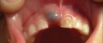
- blue discoloration of the nasolabial triangle.
Pneumonia is a serious illness, so you should not treat it yourself. That is, if your child has been diagnosed with pneumonia , immediately seek help from a specialist.
You should not use traditional medicine to cure a child’s illness on your own, because there may be no result, and time will be lost. The doctor will prescribe exactly the drugs that are suitable for your child: antipyretics, mucolytics that will help the child get rid of phlegm in the throat .
Based on the general condition of the child, the doctor can draw conclusions: should he undergo inpatient treatment, that is, in a hospital , or should he be treated at home. If the child’s condition is not very bad, then it is quite possible that the mother herself will be able to control his treatment, only from time to time showing the child to a specialist.
Keep an eye on your child, treat him if he gets sick, and let him grow up healthy and loved.
Published in Pulmonology Premium Clinic
Historical reference
To many, it seems far from them (or even a medical exaggeration) the situation when you can die due to damage in the area of the nasolabial triangle. In fact, history knows many examples when people died almost instantly. For example, the composer Scriabin . He was in England on tour when he developed a boil on his upper lip. Its appearance was accompanied by fever, headache and intoxication. Measures in the form of bandages with a special ointment seemed to have yielded results.
But almost a year later, when the composer had a small boil again, he simply tore it off with his hands. And after a while he felt bad. An ordinary pimple led to swelling, an increase in temperature to 40 degrees, and the development of infiltration. The composer developed a carbuncle. With such a pathology, fatal blood poisoning often developed. The composer's condition rapidly deteriorated, and pleurisy began. As a result, 7 days after he felt unwell, and a little less than two weeks after he accidentally popped a pimple on his face, Scriabin died.
There are many other examples of how intervention in such a controversial and sensitive area has caused serious health problems and deaths.
Causes of cyanosis
Whether constant cyanosis is characteristic of a particular disease or is transient in nature depends on the cause of its occurrence.
If after examination the cause of cyanosis of the skin cannot be determined, then it is called primary, or idiopathic.
Cyanosis may not have a pathological basis, but in order to protect the patient, it is necessary to undergo an examination and exclude organic pathology. Secondary cyanosis occurs as a result of some disease.
Central cyanosis most often occurs in respiratory failure, when there is high pressure in the pulmonary arteries, pathology of the bronchi, lungs, or if the patient has a congenital defect of the interventricular or interatrial septum. The most intense cyanosis will be in those areas where the skin is the thinnest; for example, cyanosis of the lips will be characterized by an almost purple color. A characteristic sign is also cyanosis of the nails.
Peripheral cyanosis is more often observed in patients with heart failure, as the speed of blood flow is significantly reduced. Areas of the body that are exposed to low temperatures have a bluish color.
Possible side effects
Basically, complications arise not so much due to rupture of a venous vessel, but due to interruption of arterial blood flow due to compression or when the drug is administered into the lumen of the vessel with subsequent embolization of the terminal branches with small fragments [Fig. 3].
Rice. 3. Reasons for the development of skin necrosis. Intravasal injection (top), vessel compression (bottom).
What could happen next? Stopping arterial flow leads to necrosis and tissue death in the blood supply zone of a given vessel. Cases of necrosis of the skin of the wing and tip of the nose, lip tissue and glabella area have been described [Fig. 4–5].
Rice. 4. Necrosis of the tip or wing of the nose.
Rice. 5. Zones of necrosis.
An even more serious risk factor is associated with the fact that the facial artery represents the communication between the external and internal carotid arteries. Its terminal branch, the angular artery, anastomoses with the ophthalmic artery, a branch of the internal carotid artery. It is this connection between the vessels that can lead to embolization of the ophthalmic artery with small fragments of filler and further penetration into the central retinal artery with a possible decrease in vision up to blindness [Fig. 6–7].
Rice. 6. Iatrogenic retinal artery occlusion caused by the injection of fillers.
Rice. 7. Microembolism of the ophthalmic artery: etiopathogenesis.