The new nickel titanium ProTaper represent a revolutionary advancement in the field of root canal treatment. ProTaper files were designed specifically to meet certain criteria - they must have superior flexibility, unparalleled efficiency and high security. The unique design features of ProTaper allow clinicians to more consistently create a smooth, tapered shape in highly curved canals and canals with anatomical challenges.
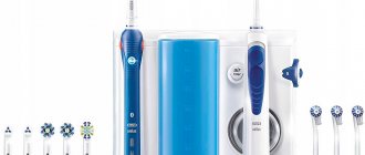
Protapers are a new version of the world's most popular endodontic nickel titanium instrument system, meeting the requirements of all dentists in all clinical situations.
Protapers are unique, ultra-flexible nickel-titanium files of a new generation that allow high-quality preparation of difficult root canals that are difficult to traditional instrumentation.
Composition: the cutting part of these tools is made of nickel-titanium alloy.
Nickel-titanium instruments should not be used to remove polymer pastes from root canals.
ADVANTAGES OF UNIVERSAL PROTAPERS
+ Easier
- uniform sequence of instruments regardless of the shape of the root canal
- simply remember the application protocol (color coding)
- Root canal drying and obturation products are specially designed to work with the ProTaper system and are color coded
+ Faster
- in most cases only 3 tools are needed
- high cutting efficiency
+ More efficient
- increased apical taper for better root canal preparation
- Better removal of dentinal debris thanks to the unique “multiple taper” of the instruments
+ Safer
- rounded guide tip minimizes the likelihood of deviation from the channel
- There is a manual version of instruments for anatomically complex clinical situations, as well as for clinicians who prefer to use manual instruments due to better tactile control
Selecting Ni-Ti files
This technique uses specially designed ProTaper files with uneven taper; The set includes 6 tools - 3 for shaping (Shaping, hereinafter S) and 3 finishing (Finishing, hereinafter F), which simultaneously repeats and simplifies the Schilder technique. Each S-file usually has some smoothing of the cutting angle (increasing taper along the length of the cutting part); while the taper of the F-file in the apical part is fixed, and on the coronal 2/3 of the length, on the contrary, it is reduced. When used correctly in a step-by-step manner, ProTaper files (Dentsply Tulsa Dental; Tulsa, Oklahoma) are effective, reliable, and easy to use (Figure 5).
Rice. 5. Root canals of a maxillary molar treated with ProTaper instruments. ProTaper files replicate significant canal curvatures well and produce smooth, streamlined shapes (work courtesy of Dr. John West, Tacoma, Washington).
The ProTaper technique closely replicates Schilder's technology, in which pre-curved reamers sequentially cut dentin in a reciprocating motion. S-files with non-aggressive small tips are held in the center of the canal previously traversed with hand instruments. Thanks to the increasing taper, each successive file selectively cuts dentin with its most effective cutting edges, without affecting the apical region.
PROTAPERS F1 PROTAPERS F2 PROTAPERS F3
Finishers are designed to finalize the apical third of the root canal, as well as to level and widen the middle third of the canals. They, like shapers, include 3 tools: F1, F2, F3.
The tip diameter of the F1 pro taper is 0.20 mm, F2 0.25, F3 0.30. It is characteristic that all 3 tools in the interval D0-D3 have a fixed taper (.07, .08, .09%, respectively). After D3, although the tool diameter continues to increase, the taper decreases to 0.055, thereby increasing the flexibility of the prop taper.
1. Indications for use: Protapers are intended for use only in a clinical setting by specially trained specialists. Scope of application: formation and cleaning of the root canal system.
2. Contraindications: Not known.
3. Warnings: None known.
4. Precautions:
- Repeated disinfection cycles and sterilization increase the risk of file fracture.
- These instruments should not be immersed in sodium hypochlorite solution.
- Cleaning of Instruments: Strictly follow the cleaning instructions recommended by the manufacturer.
- Irrigate generously and frequently.
- Create an adequate carpet runner using hand files up to a minimum of ISO size 15.
- Use the instruments at a constant rotation speed of 150 – 350 rpm with light apical pressure.
- Clean the tool grooves as often as possible and inspect the file for signs of deformation or wear.
- For optimal performance, it is recommended to use torque-controlled devices.
- Use the shaping files (S1, S2 and Sx), working in separate sweeping movements to create a straight-line approach to the root canal.
- Use finishing files (F1, F2, F3, F4 and F5) without using sweeping motions.
- Use appropriate finishing files to “passively” pass the entire working length of the canal and, once reached, immediately remove the instrument.
5. Adverse reactions: To date, no adverse reactions have been reported.
Confirmation of clinical success
Clinical studies of the ProTaper technique, with an emphasis on the execution procedure, were conducted on the mesial roots of extracted mandibular molars using CT analysis. In this study, horizontal sections of different root levels were examined using CT sections and spatial imaging. Some results are shown in Fig. 9. Green represents the anatomical contours before instrumentation and red represents the resulting preparation shape.
Rice. 9a. Horizontal CT sections of the coronal 1/3 of the root. Pay attention to the successful relocation of channels at this level
Rice. 9b. Horizontal CT sections of the middle 1/3 of the root. Note the rounded, centered preparation of the ProTaper files
Rice. 9c. Horizontal CT sections of the apical 1/3 of the root. ProTaper preparation ideally includes the original canal diameter
Rice. 9d. Condition before and after applying ProTaper S1, S2 and F1 files. Smooth, streamlined and centered preparation shapes follow the internal anatomy of the canal
Results 9a-9d kindly provided by Dr. Lars Bergmans and BIOMAT Research Cluster, Catholic University of Leuven, Belgium
So, ProTaper S-files, thanks to easy lateral sliding and selective cutting of dentin during the reverse movement, demonstrate a number of advantages:
- instruments move freely in the root canal during most of their work;
- the lumens of the canals in the coronal part delicately expand away from the outer concavity of the root;
- The “shaving” method of processing ensures centering of the preparation in the root canal and maximum preservation of dentin;
- instruments are in physical contact with more than 90% of the inner walls of the canal.
PROTAPERS - STEP-BY-STEP INSTRUCTIONS FOR USE
- Create direct access to the root canal orifice.
- Constantly irrigate and check the patency of the carpet with an ISO 15 size hand file.
- Usage protocol:
- Locate the mouth.
- Passively use an ISO size 15 hand file until you feel resistance.
- Use the S1 shaping file, working in a sweeping motion until you reach the penetration depth of an ISO size 15 hand file.
- Repeat this sequence until you have determined the working length using an ISO size 15 hand file and have gone through the entire working length of the canal using the S1 tool.
- Use the S2 shaping file, working in a sweeping motion until you reach working length.
- Confirm the working length.
- Use the F1 finishing file (without sweeping movements), moving it deeper with each insertion until you reach working length.
- Calibrate the apical foramen using hand files.
- Use the appropriate finishing file (F2, F3, F4, F5) without sweeping movements to the full working length if additional expansion is needed or if the apical foramen is larger. If necessary, use Sx sweeping movements to remove ostial dentin and/or to create a wider coronal portion of the canal.
Protaper Universal - video, operating method
6. Disinfection, cleaning and sterilization: Clean the instruments and stand first. Then sterilize the instruments in the bags using an autoclave at 134ºC, 3 bar pressure for 18 minutes.
6.1 General recommendations
- Tools marked "disposable" are not intended to be reused.
- The user is responsible for the sterility of the product upon first use and each subsequent use, as well as for the use of damaged or contaminated instruments.
- For your own safety, please use personal protective equipment (gloves, goggles).
- Use disinfectant solutions with proven effectiveness (listed by the German Society for Hygiene and Microbiology; CE marked; approved by the German Inspectorate for Hygiene and Microbiology).
- quality of food products and medicines).
- Tungsten carbide burs, plastic stand, hand tools and nickel - titanium tools are destroyed by hydrogen peroxide (H2O2) solution.
- Nickel-titanium instruments are destroyed when immersed in a NaOCl solution for more than 5 minutes with a concentration of more than 5%.
- Aluminum instruments are destroyed in a solution of caustic soda with mercury salts.
- Do not use acidic (pH < 6) or alkaline (pH > solutions.
- After 5 processing cycles, markings may be destroyed
Traditional shaping techniques
The step-back technique initially uses hand-held files of small ISO sizes to traverse the full length of the root canal. Larger files are then used to make apical preparations until the selected master file reaches working length. The remaining apical third of the canal is expanded with successively larger files, and as the file size increases, its working part is shortened. Upon completion of the preparation of the apical 1/3 of the canal, the coronal 2/3 is expanded, and the entire internal surface of the treated root canal is smoothed. Unfortunately, this technique has a number of disadvantages, for example: blocking the canal, formation of ledges, perforations, transportation (oval deformations of the apical foramen along the outer curvature of the root - translator's note), often requiring additional procedures, such as re-treatment, surgery and removal.
The crown-down technique uses ISO instruments of varying tapers from larger to smaller sizes until working length is achieved. In general, the root canal is opened in stages - from the coronal to the apical part. Although this technique avoids many of the disadvantages of the step-back technique, a few notes must be made:
- each file aggressively penetrates dentin with its large, hard tip and, when rotated, forms a round profile in the center of the canal, while the cross section of the root canal most often has an irregular configuration; and, as a result, proper cleaning and disinfection of such root systems does not occur;
- the taper of the prepared canal quickly follows the uniform taper of the file, especially in long, narrow and curved canals, which can lead to tight contact of the instrument with the canal walls, blocking and breakage;
- the likelihood of breakage of smaller instruments increases when they become jammed in the apical third of the canal.
The pre-enlargement technique uses suitably curved ISO instruments of varying tapers from smaller to larger sizes. In cases of narrow and severely curved canals, instruments are initially selected to expand the coronal 2/3 of the canal. Each subsequent larger file is immersed to a shorter length relative to the previous instrument, cutting off dentin and creating a streamlined access in the coronal and middle parts of the canal. And although the instruments are used from smaller to larger, their flexible tips move freely and safely through the treated area of the root canal. It is important to note that depending on the degree of bend, any ISO file will cut a cavity larger than its taper suggests. Each instrument cuts dentin with its larger, stronger, more active cutting threads in an outward motion. Correct step-by-step implementation of the pre-enlargement technique facilitates access to the anatomically more complex apical third of the canal: the finishing files move freely into the root canal cavity and act predictably in the apical region. This technique places emphasis on widening the coronal and mid-portions of the canal and smoothing the transition to the apical third of the preparation. Finishing the channel is a matter of skill (Fig. 4).
Rice. 4. Endodontically treated first maxillary premolar with a branched system ending in three apical foramina
The growing popularity of the pre-expansion technique is understandable due to the predictable success of the result. However, the disadvantages of the method include the use of a large number of tools, several recapitulations with a set of files and examples, significant time costs and a complicated processing procedure. Let us next consider a clinical technique based on the effective principles of pre-enlargement technology with significant time savings.
PROTAPERS - RE-TREATMENT
D1+D2+D3: instruments specially designed for simple filling of root canals:
- the D1 instrument has an active tip, facilitating initial penetration into the root canal
- instruments have different lengths and taper of the working part for working in different parts of the root canal (coronal/middle/apical)
The sequence of using the tools is easy to remember
- From shortest to longest tool: D1 – D2 – D3
Easy identification
- dark gray handles no more than 11 mm long for better visibility
- instruments have one, two or three white rings respectively
PROTAPER D3 – FOR THE APICAAL PART OF THE CANAL
Rules for using ProTaper instruments to remove filling material from root canals
- instruments are inserted into the canal by applying light pressure in the apical direction
- regularly remove the instrument from the canal, check its working part and clean it
- if the instrument does not advance deeper, use a hand instrument to overcome resistance and confirm patency of the canal
Recommended speed:
- for removing gutta-percha or Thermafil/ProTaper® obturators: 500 – 700 rpm
- for removing pastes based on zinc oxide and eugenol: 250 – 300 rpm
You can buy ProTaper (ProTaper) machine S1, 25 mm, Dentsply, for the treatment of complex cases, blister (6 pcs) produced by Dentsply at ExDent.ru at a price of 3,500 rubles.
Protaper Next – ROOT CANAL PREPARATION
FIFTH GENERATION OF INSTRUMENTS
In the early days of modern endodontics, there were many concepts, strategies, and techniques for root canal preparation. Over the past decades, dozens of tools for passing and shaping channels have appeared. Despite the different shapes of instruments, their number and the many proposed techniques, endodontic treatment was approached with optimism, hoping for success.
A breakthrough in clinical endodontics occurred with the transition from the use of a long series of stainless steel instruments and multiple Gates Glidden reamers to the use of nickel titanium instruments for root canal preparation. Regardless of the techniques used, the goals of mechanical root canal treatment were brilliantly defined almost 40 years ago by Dr. Herbert Schilder (1). With the correct treatment algorithm, mechanical root canal debridement should meet the biological goals of canal preparation, three-dimensional disinfection and obturation (Figure 1). The purpose of this article is to demonstrate and compare new generations of endodontic nickel titanium instruments designed to improve root canal preparation. It is important that this article will define a new tool system that combines proven design features of the past with the latest innovations of the present, and the operating technique will also be described.
PREPARATION OF ROOT CANALS WITH NI-TI INSTRUMENTS
In 1988, Walia introduced Nitinol, a nickel-titanium alloy for root canal preparation that is 2-3 times more flexible than stainless steel alloy, and for the same instrument sizes. A revolutionary result of the use of instruments made of nickel-titanium alloy was the ability to machine curved canals using continuous rotation. By the mid-1990s, the first commercially available nickel-titanium rotary instruments appeared on the market (3). Below is a mechanical classification of each generation of instruments. Rather than describe the myriad of tool cross-sections that exist, we will differentiate between tools with passive and active cutting capabilities.
FIRST GENERATION
To appreciate the evolution of nickel-titanium tools, it is useful to know that the first generation of nickel-titanium tools had passive radial cutting edges and fixed tapers of 4% and 6% along the entire length of the active blades (Figure 2) (4). This generation required a variety of instruments to achieve dissection goals. In the mid to late 1990s, GT (Dentsply Tulsa Dental Specialties) instruments were introduced which had a fixed taper on each individual instrument of 6%, 8%, 10%, and 12%. The single most important design feature of the first generation of nickel-titanium rotary instruments was the passive radial edges, which allowed the instrument to remain centered in the curvature of the root canal during operation.
SECOND GENERATION
The second generation of nickel-titanium rotary instruments entered the market in 2001 (6). The main difference with this generation of instruments is that they have active cutting edges and only a few instruments are required to complete a complete root canal preparation (Figure 3). To avoid the locking cone and resulting screw-in effect associated with passive and active tapered NiTi instruments, EndoSequence (Brassler USA) and BioRaCe (FKG Dentaire) offer instrument lines with alternating contact points (7). Although this characteristic is intended to reduce the effect of the locking cone, these lines of tools have a fixed taper in the area of the active cutting part. A clinical breakthrough occurred when the ProTaper (Dentsply Tulsa Dental Specialties), which features multiple increasing and decreasing tapers throughout a single instrument, was introduced to the market. This revolutionary progressive taper shape limits the cutting action of each instrument to a specific portion of the canal, allowing the use of a shorter sequence of instruments to safely reproduce the Schilder canal shape (Figure 4) (8).
During this period, manufacturers began to actively seek methods to increase tool fracture resistance. Some manufacturers have used electropolishing on their tools to remove surface irregularities created by the traditional tool grinding process. However, clinical observations and scientific research have shown that electropolishing dulls cutting edges. As such, the observed benefits of electropolishing were offset by the need to apply unwanted pressure inside the root canal to advance the instrument to its full working length. Excessive internal pressure, especially when using fixed-taper tools, results in a locking cone effect, a screw-in effect, and excessive torque on the rotating tool during operation (9). To compensate for the shortcomings or ineffective operation of the tool as a result of electropolishing, other cross-sections of the tools were proposed and the rotation speeds, despite all the dangers, were increased.
THIRD GENERATION
Improvements in the metallurgy of nickel-titanium alloys have become the hallmark of the third generation of instruments for mechanical root canal preparation. In 2007, manufacturers focused on heating and cooling processes for alloys to reduce cyclic fatigue and improve safety when using nickel-titanium instruments in more curved root canals (10). The desired point of phase transition between martensite and austenite can be identified with the search for a clinically more optimal metal than nickel-titanium. This third generation of nickel-titanium instruments is characterized by a significant reduction in cyclic fatigue and, accordingly, instrument fracture. Examples of heat-treated instruments include Twisted File (SybronEndo), Hyflex (Coltene Whaledent), and GT, Vortex, and WaveOne (Dentsply Tulsa Dental Specialties).
FOURTH GENERATION
Another progressive movement in canal preparation is reciprocation, which can be defined as any repeated back-and-forth or up-and-down movement. This technology was first introduced in the late 1950s by French dentist Blanc. Today, M4 (SybronEndo), Endo Express (Essential Dental Systems), and Endo-Eze (Ultradent) are examples of systems where the clockwise rotation angle of the instrument is equal to the counterclockwise rotation angle of the instrument. Compared to full rotation systems, a reciprocal instrument that rotates at equal angles clockwise and counterclockwise requires more pressure into the root canal to move forward, does not cut as efficiently as a rotary instrument of the same size, and is more limited in removing sawdust and debris from the root canal. channel.
Based on these early experiences, reciprocation technology has steadily evolved, leading to the creation of the 4th generation of root canal preparation instruments. This generation of instruments and reciprocation technology are embodied in the much-anticipated one instrument. ReDent-Nova (Henry Schein) introduced the Self-Adaptive Instrument (SAF). This instrument has the shape of a compressible hollow tube; it is assumed that the instrument exerts uniform pressure on the dentinal walls, regardless of the cross-section of the canal. The SAF instrument is mechanically rotated by a tip that makes both short 0.4 mm vertical movements and vibration movements with constant irrigation (11). Another promising single tool technique is called One Shape (Micro Mega), which will be mentioned below in the chapter on 5th generation tools. By far the most popular single instrument concept is the WaveOne (Dentsply Tulsa Dental Specialties and Maillefer) and Reciproc (VDW) system. WaveOne combines the best design features of 2nd and 3rd generation tools with a reciprocal motor that rotates any tool back and forth at unequal angles. The angle of movement counterclockwise is 5 times greater than the angle of movement clockwise, and it is less than the elastic limit of the tool. After 3 cycles of counterclockwise and clockwise rotations, the tool will make a full circle or 360º rotation (Figure 5). This new reciprocal movement allows the instrument to advance easily, cut effectively and efficiently remove sawdust from the root canal (12).
FIFTH GENERATION
The fifth generation of canal preparation instruments differs in that the center of gravity and/or the center of rotation is shifted (Figure 6). When instruments with a similar shape are rotated, a mechanical wave of motion is generated that moves along the entire length of the instrument. Similar to the progressive taper of any ProTaper instrument, the offset center of gravity further minimizes friction between the instrument and dentin (13). Also, this design improves the removal of sawdust from the canal and improves the flexibility of the active part of the Protaper Next tool. The advantages of tools with a shifted center of gravity will be described later in this article. Trade names for instruments based on this technology are Revo-S, One Shape (Micro Mega) and ProTaper Next (Dentsply Tulsa Dental Specialties/Dentsply Maillefer). Today's safest, most effective, and simplest tool systems utilize the time-tested features of past generations of tools combined with the latest technological advances. Below is a brief technical description of the ProTaper Next system.
- PROTAPER. NEXT
The new ProTaper Next (PTN) system (Dentsply Tulsa Dental Specialties) features 5 instruments of varying lengths for root canal preparation, sequentially labeled X1, X2, X3, X4, and X5 (Figure 7). From 1 to 5 they are color coded - yellow, red, blue, double black and double yellow stripes on the handle, corresponding to sizes 17/04, 25/06, 30/07, 40/06, and 50/06, respectively. The taper listed is NOT fixed across the entire working portion of the ProTaper Next tool. Imagine that the ProTaper Next X1 and X2 instruments have both an increasing and decreasing taper throughout the same instrument, while the ProTaper Next X3, X4, and X5 instruments have a fixed taper for the first 3 mm of length and then a decreasing taper on the entire remaining active part. ProTaper Next tools are a combination of 3 significant design features such as increasing taper on the same tool, M-wire technology and an off-center section. Taking the ProTaper Next X1 as an example, it has a centered mass and axis of rotation over the first 3mm of length, whereas from 4-16mm the X1 has a displaced cross-section. From 1 to 11 mm, the X1 instrument increases its taper, starting at 4%, while from 12 to 16 mm there is a decreasing taper to improve instrument flexibility and preserve root dentin during root canal preparation procedures. ProTaper Next tools are used at 300 rpm. and torque from 2.0-5.2 N cm. However, the authors prefer a torque of 5.2 Ncm because this level of torque has been found to be safe as long as clinicians carefully create a carpet path and use gentle outward sweeping movements during progressive root canal preparation (14). In the ProTaper Next technique, all instruments are used exactly the same and the sequence of use always follows the ISO color coding and is always the same, regardless of the length, diameter, or curvature of the canal.
PROTAPER NEXT PREPARATION TECHNIQUE
The ProTaper Next preparation technique is extremely safe, effective and simple, provided the access cavity is good and a carpet is created. As with any root canal preparation procedure, great care must be taken to create straight access to each orifice. This involves widening, smoothing and finishing the internal axial walls. For estial access, the ProTaper system offers an additional instrument called the SX. The SX instrument is used in an outward sweeping motion to preliminarily widen the orifice, remove dentin triangles, move the coronal portion of the canal away from the outer curvature of the root, or create a more pronounced shape. Perhaps the greatest challenge in endodontic treatment is finding, following and predictably maintaining the shape of the root canal to the apex. Traversing and maintaining the shape of the canal using fine hand instruments requires mechanical strategy, an experienced sense of work, patience and desire. A small hand-held instrument is initially used to reconnoiter, widen and smooth the inner canal walls. Once the entire length of the root canal is passable with a hand instrument, a continuous rotation instrument can be used to create a carpet and widen the original canal lumen in preparation for shaping (15). To be clear, a channel is only prepared when it is empty and has a proven smooth and repeatable carpet. With an estimated working length and in the presence of a viscous chelating agent, insert the #10 file instrument into the root canal and see how easily the instrument moves toward the apex of the root canal. In shorter, wide and straight canals, the #10 instrument can be easily inserted to the full working length. Once the #10 instrument is confirmed to move freely to its full working length, the carpet can be further widened with either a #15 hand instrument or specially designed instruments such as PathFiles (Dentsply Tulsa Dental Specialties). The created carpet path provides sufficient space for machining to begin with the ProTaper Next X1 tool. In other cases, teeth requiring endodontic treatment may have longer, narrower, and more curved root canals (Figure 8a). In such situations, the #10 instrument often does not penetrate the entire length of the root canal. It is generally not necessary to use #06 and/or #08 hand instruments in an attempt to immediately reach the apical foramen. Simply and gently work the #10 hand instrument within any area of the root canal until the instrument is completely free. ProTaper Next instruments can be used to create any part of the root canal with a smooth and reproducible carpet.
Although there is a glide path and a sequence of instrumentation, the ultimate goal is to complete the entire working length of the root canal, establish the working length, and check for apical patency (Fig. 8b). Safe work with the canal can begin after checking the created carpet, when tool #10 does not stick at the working length and can slide and move again in the area of the lower third of the root canal. When the canal is prepared, the access cavity is generously filled with 6% NaOCl solution. You can begin canal preparation with the ProTaper Next X1. It should be emphasized that when working with ProTaper Next instruments, pumping or pecking movements directed inward are never used. In contrast, ProTaper Next tools are used in an outward sweeping motion.
It is important that this method of use will allow any ProTaper Next tool to move passively inwards, following a carpet path and penetrating the entire working length. Instrument X1 is inserted into the access cavity into the previously expanded orifice and prepared canal. Before meeting resistance, consciously begin sweeping outwards (Figure 8c). Sweeping creates lateral space and allows this tool to penetrate a few millimeters inside. Sweeping movements serve to improve the contact between the instrument and the dentin, especially in canals that have a non-standard cross-section.
Continue working with the ProTaper Next X1 along the main part of the root canal. As you immerse the tool every next few millimeters, remove it and examine it while simultaneously cleaning the blades. Before re-inserting the X1 instrument into the root canal, it is strategically important to irrigate and flush out large sawdust, and reinsert the #10 instrument to break up the residual sawdust and debris into solution, then re-irrigate to refresh the solution. In one or more passes you should reach the working length of the X1 tool. For thorough mechanical preparation purposes, irrigate, recapitulate, and re-irrigate after removing any rotating instrument.
Take the ProTaper Next X2 and let it sink into the root canal. Before resistance is encountered, apply a lateral sweeping motion away from the dentinal walls, which in turn will move the X2 instrument into the root canal passively and progressively. The X2 tool will easily follow the path created by the X1 tool, making further expansion and gradually moving along the entire length. If the tool gets stuck and stops moving, remove it, clean it and check the edges. Re-irrigate, recapitulate, and re-irrigate to meet channeling goals. Continue working with tool X2 until working length is reached; note that one or more approaches may be required, depending on the length, width and curvature of the canal (Figure 8d).
Once the ProTaper Next X2 has reached working length, it is removed. The created form can be considered final only if the grooves in the apical part of the instrument are visually filled with dentin. Alternatively, the apex size can be checked with a 25/02 hand instrument. If the #25 hand instrument sticks at the working length, the preparation is complete. If the 25/02 hand tool moves freely at working length, this simply means that the apical opening is wider than 0.25 mm. In this case, the apex can be calibrated using a size 30/02 hand tool. If the #30 size hand tool sticks at the working length, the mold is created. However, if a #30 size hand tool does not reach the apex, move on to the ProTaper Next X3 tool, following the same operating procedure as with the ProTaper Next X1 and ProTaper Next X2.
Most root canals will be in optimal shape after using ProTaper Next X2 or X3 (Figure 8e). The ProTaper Next X4 and X5 instruments are primarily used for the preparation and finishing of larger diameter root canals. If the apical foramen is determined to be larger than the ProTaper Next 50/06 X5 size, consider other methods for preparing similar wide, straighter canals. It is important to recognize that carefully prepared canals promote shaping, three-dimensional cleaning and obturation (Figure 8f).
DISCUSSION
From a clinical point of view, the Protaper Next continuous rotation system combines the most proven and successful design features of past instruments with the latest advances in science. This brief discussion will describe how design affects instrument performance. The most successful design feature of the previous generation of instruments was the mechanical concept of using progressive taper on the same instrument. The patent protecting the ProTaper Universal system allows the use of both increasing and decreasing taper percentages on the same tool. This structural feature minimizes contact between the instrument and the dentin, which reduces the risk of a locking cone and screw-in effect, increasing work efficiency (8). Compared to a fixed taper instrument of the same size, decreasing the percentage of instrument taper improves flexibility, limits preparation of the root canal body, and preserves dentin in the coronal 2/3 of the root canal. Taking advantage of the mechanical design, Protaper Next also uses progressive taper on the same tool. This design feature has contributed significantly to the ProTaper system becoming the #1 selling system in the world, the #1 tool of choice for endodontists, and the #1 system of choice for undergraduate dental school education at international schools (16). Another design feature that benefits certain brands of rotary tools is metallurgy. Although nickel-titanium tools are 2-3 times more flexible than stainless steel tools of the same size, additional modifications to the manufacturing process in the form of heat treatment can provide certain benefits. The company's specialists turned their attention to the heating and cooling of a traditional nickel-titanium alloy, either before or after milling. Heat treatment creates a more optimal phase transition point between martensite and austenite. It must also be remembered that the best phase transition point depends on the cross section of the tool. Research has shown that M-wire, a metallurgically enhanced version of nickel-titanium alloy, reduces cyclic fatigue by 400% compared to tools of the same diameter, cross-section and taper. This third benefit of this generation of instruments is strategic for the overall clinical safety and performance of the Protaper Next rotary instrument system. The third design feature of Protaper Next tools is the off-center cross section. If the main part of the continuous rotation tool is off-center, we can talk about 3 advantages of this (13):
1) The off-center design promotes the formation of a moving mechanical wave of motion along the entire active part of the tool. This rocking motion promotes minimal instrument-to-dentine contact compared to a fixed-taper, mass-centered rotation instrument (Figure 9). The reduced contact limits unwanted locking taper, screw-in effects, and torque on any instrument. 2) An offset tool frees up more space for improved cutting, loading and removal of sawdust from the root canal compared to a mass-centered tool with a rotary axis (Figure 9). Many tools break as a result of excessive accumulation of sawdust and debris between the cutting blades throughout the active part of the tool. Importantly, the off-center instrument reduces the likelihood of lateral compaction of sawdust and blocking of the root canal anatomy (Figure 6).
3) A forming tool with an off-center rotation mass will create a mechanical wave of motion similar to vibrations recorded along a sinusoid (Figure 10). As a consequence of this design, any Protaper Next instrument can create a greater range of motion compared to a similar instrument with symmetrical mass and rotation axis (Figure 6). The clinical advantage of this is that the smaller, more flexible Protaper Next instrument can prepare a similar space to a larger, more rigid instrument with a centered mass and axis of rotation (Figure 9).
CONCLUSION
Each new generation of shaping tools offers something new, is described differently and is intended to be more advanced than the previous one. Protaper Next is a 5th generation system that combines the proven performance characteristics of the past with the latest technological advances. This system should simplify the procedure of canal formation with rotary instruments, reducing the number of instruments and eliminating the so-called hybrid techniques. Clinically, the channel shape created by Protaper Next fulfills the 3 sacred dogmas - safety, effectiveness and simplicity. From a scientific perspective, evidence-based studies are needed to confirm the potential benefits of the system. Acknowledgments: The authors would like to acknowledge Dr. Michael J. Scianamblo for his work and participation in the creation of the ProTaper Next system. Disclaimer: Drs. Ruddle, Machtou, and West benefit financially from the products they create and develop, including the ProTaper Universal system.
Authors:
Clifford J. Ruddle, DDS Pierre Machtou, DDS John D. West, DDS
Bibliography:
1. Schilder H: Cleaning and shaping the root canal, Dent Clin North Am 18:2, pp. 269-296, April 1974. 2. Walia HM, Brantley WA, Gerstein H: An initial investigation of the bending and torsional properties of Nitinol root canal files, J Endod 14:7, pp. 346-351, 1988. 3. Thompson SA: An overview of nickel-titanium alloys used in dentistry, Int Endod J 33:4, pp. 297-310, 2000. 4. Bryant ST, Dummer PM, Pitoni C, Bourba M, Moghal S: Shaping ability of .04 and .06 taper ProFile rotary nickel-titanium instruments in simulated root canals, Int Endod J 32:3, pp. 155-164, 1999. 5. Kramkowski TR, Bahcall J: An in vitro comparison of torsional stress and cyclic fatigue resistance of ProFile GT and ProFile GT Series X rotary nickel-titanium files, J Endod 35:3, pp. 404-407, 2009. 6. Machtou, P, Ruddle CJ: Advancements in the design of endodontic instruments for root canal preparation, Alpha Omegan 97:4, pp. 8-15, 2004. 7. Schäfer E, Vlassis M: Comparative investigation of two rotary nickel-titanium instruments: ProTaper versus RaCe. Part 2. Cleaning effectiveness and shaping ability in severely curved root canals of extracted teeth, Int Endod J 37:4, pp. 239-248, 2004. 8. Ruddle CJ: The ProTaper endodontic system: geometries, features, and guidelines for use, Dent Today 20:10, pp. 60-67, 2001. 9. Boessler C, Paque F, Peters OA: The effect of electropolishing on torque and force during simulated root canal preparation with ProTaper shaping files, J Endod 35:1, pp. 102-106, 2009. 10. Gutmann JL, Gao Y: Alteration in the inherent metallic and surface properties of nickel-titanium root nickel root canal instruments enhance performance, durability and safety: a focused review, Int Endod J 45:2, pp . 113-128, 2012. 11. Metzger Z, Teperovich E, Zary R, Cohen R, Hof R: The self-adjusting file (SAF). Part 1: respecting the root canal anatomy—a new concept of endodontic files and its implementation, J Endod 36:4, pp. 679-690, 2010. 12. Yared G: Canal preparation using only one Ni-Ti rotary instrument: preliminary observations, Int Endod J 41:4, pp. 339-344, 2008. 13. Hashem AA, Ghoneim AG, Lutfy RA, Foda MY, Omar GA: Geometric analysis of root canals prepared by four rotary NiTi shaping systems, J Endod, 38:7, pp. 996-1000, 2012. 14. Blum JY, Machtou P, Ruddle CJ, Micallef JP: Analysis of mechanical preparations in extracted teeth using the ProTaper rotary instruments: value of the safety quotient, J Endod 29:9, pp. 567-575, 2003. 15. West JD: The endodontic glidepath: secret to rotary safety, Dent Today 29:9, pp. 86, 88, 90-93, 2010. 16. Dentsply International, personal communication. 17. Johnson E, Lloyd A, Kuttler S, Namerow K: Comparison between a novel nickel-titanium alloy and 508 nitinol on the cyclic fatigue life of ProFile 25/.04 rotary instruments, J Endod 34:11, pp. 1406-1409, 2008.
Buy Protaper Next at a low price in the online store
06.08.2015