In 1913, Nikolai Ivanovich Taratynov (1887–1919), a young doctor and employee of the Department of Pathological Anatomy at Kazan University, received material for research and clarification of the diagnosis. It was a fragment of tissue taken from a patient with a contusion of the cranial vault - some time after the injury, a granulomatous formation formed in this place.
Figure 1. 3D reconstruction of skull bones with solitary eosinophilic granuloma https://radiopaedia.org/cases/eosinophilic-granuloma-skull
It was assumed that this was bone tuberculosis, but instead of the classic tuberculous tubercle, Taratynov saw accumulations of mononuclear cells (tissue macrophages or histiocytes) and eosinophils, as well as Charcot-Leiden crystals, which had previously been found in the sputum of patients with bronchial asthma. The morphological picture indicated “the existence of granulomas, clinically and macroscopically completely similar to tuberculous ones, recognizable only microscopically and consisting almost exclusively of eosinophils.”
Figure 2. Eosinophilic granuloma of bone: lesion of the hip in an 11-year-old girl (x-ray). Swelling and pain were noted for 3 months. https://radiopaedia.org/cases/langerhans-cell-histiocytosis-skeletal-manifestations
The previously unknown disease was named Taratynov's disease. Currently, this name is mainly used in Russian-language sources and has rather a historical meaning.
In the 1940s. similar cases were presented in articles by US doctors Sadao Othani, Joseph Ehrlich, Louis Lichtenstein and Henry Jaffe under the names “solitary granuloma of bone” and “eosinophilic granuloma of bone”.
Similar histiocytic infiltrates were described by other authors - Alfred Hand Jr, USA in 1893, Arthur Schüller (Austria) in 1915, Henry Cristian (USA) in 1920, Erich Letterer , Germany) in 1924, Sture Siwe (Sweden) in 1933, in connection with which the terms “Hand-Schüller-Christian disease” and “Abt-Letterer-Siwe disease” were introduced. However, later it was concluded that all of these are forms of the same disease, different in severity and localization of foci, called “histiocytosis X”
in 1953.
Figure 3 . Eosinophilic granuloma of the skull (X-ray) https://radiopaedia.org/cases/eosinophilic-granuloma-1
The answer to the question of what the histiocytes that form granulomas are was given in 1973 by pediatrician and morphologist Christian Nezelof (France), who identified them as Langerhans cells (a type of antigen-presenting cells localized in the epidermis). In this regard, since 1987, the name “histiocytosis X” was replaced by “Langerhans cell histiocytosis” ( LCH ).
The head of the Department of Pathology at St. Petersburg State University, Leonid Churilov, believes that Doctor Taratynov became the prototype of Doctor Zhivago. This bold assumption is supported by the coincidences in the life of Nikolai Taratynov and his family with the story of Yuri Zhivago: both victims of the revolution, military doctors, whose daughters were orphaned early and were raised by their father’s relatives. Nikolai Ivanovich Taratynov was shot and killed by a sailor, the brother of a patient who died in the hospital, in 1919 at the age of 32. You can learn more about Taratynov’s biography here.
Etiology
The question of the reasons for the development of granulomas remains open. One theory considers Langerhans' histiocytosis to be a neoplastic process. This is supported by such features as monoclonality of pathological cells (origin from a single pathological cell), increased expression of proliferation activators and factors inhibiting apoptosis. At the same time there are no genomic defects in the cells , and spontaneous remission of the disease is possible. At the site of granuloma development, activation of T-lymphocytes and T-regulatory cells with suppressor activity has been described. These signs indicate that other immunopathological processes may underlie the development of the disease.
Figure 4. Eosinophilic granuloma: pathological changes. In the last image, the S-100 protein expressed by Langerhans cells is colored. https://radiopaedia.org/cases/eosinophylic-granuloma-histology-1
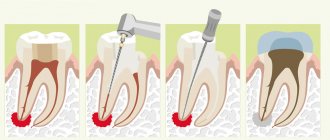
What symptoms indicate the appearance of a granuloma on the root of a tooth?
Over a long period of time, granuloma on the root of a tooth can develop asymptomatically, and this makes diagnosing the disease in the early stages of development extremely difficult. However, some signs may be present during the initial phases of granulomatosis.
Among them:
- The appearance of dental hypersensitivity to external irritants: hot/cold, sweet/sour foods;
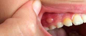
- Bursting sensations in the gum next to the tooth, on the root of which a granuloma has formed;
- A slight aching pain, most often occurring when biting;
- Change in color of the enamel of a diseased tooth.
If you observe similar signs in yourself, it’s time to contact your dentist to diagnose and begin treatment for granuloma! Our advanced dentistry clinic in Moscow, Firadent, uses modern diagnostic techniques and innovative equipment to help identify complex dental diseases in the early stages and carry out effective treatment!
Chronic apical granuloma of the tooth will sooner or later go into the acute phase of development, in which the disease acquires clear signs:
- Acute pain that intensifies when biting and chewing food;
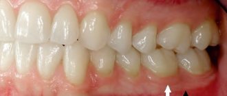
- Feels as if the tooth has become larger or is growing;
- Swelling of the gums and cheeks.
If you ignore these symptoms and do not consult a doctor to remove the dental granuloma, the intensity of the pain will increase, the swelling of the gums and swelling of the face will increase, purulent discharge may begin from the gums, a severe migraine, general weakness, and high temperature may appear. There is no need to try to endure such symptoms - seek professional dental help immediately!
How to treat tooth root granuloma in dentistry: all modern methods of treatment/removal of granulomas
What to do if you notice symptoms of granuloma? Of course, seek help from a dentist, because granulomatosis cannot be treated at home: neither antibiotics nor folk remedies will help. Treatment will begin with diagnostics, during which radiography or radiovisiography is performed. If you have a granuloma, the image will show a darkening at the root of the diseased tooth. Diagnostics will allow you to determine the shape of the granuloma (apical or periapical), assess the stage of development of the tumor, and choose the most effective treatment method for granulomatosis.
Treatment for granuloma can be:
- 1. Conservative (medication).
- 2. Surgical, involving the removal of tooth root granuloma.
We will consider all these methods and their features in detail below.
Clinical manifestations
Eosinophilic granuloma (EG), also known as Taratynov's disease, is a relatively benign variant of Langerhans' histiocytosis with the appearance of single foci in flat or tubular bones. The appearance of two or three or more lesions is much less common. Typically, Langerhans cell histiocytosis occurs in children and adolescents (up to 15–20 years), more often in boys (approximately 1.5:1). Frequency of occurrence: less than 1 person. per 100,000 population, which is 60–80% of all cases of LCH. In adults, eosinophilic granuloma, like hysteocytosis X in general, is much less common.
The most commonly affected bones are the skull bones, femurs, and less commonly, the pelvic bones, ribs, and vertebrae. There are also cases of the appearance of pathological foci in the thymus, skin, bladder, parathyroid glands, hypothalamus, lungs and gastrointestinal tract.
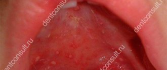
The clinical picture is determined by the location of the granuloma. An intraosseous formation may not cause any symptoms, but usually leads to the development of swelling, pain, and sometimes pathological fractures. When lesions appear in the jaw, tooth loss, damage to the mastoid process or temporal bone may occur with the development of otitis media. When the walls of the orbit are involved in the process, exophthalmos may develop. Sometimes polymorphic rashes are observed on the head, back, armpits, perianal and genital areas in the form of spots or plaques, small nodules or nodes with ulcerations. Common manifestations of Taratynov’s disease are common: increased fatigue, weakness.
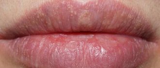
Figure 5. Photo of the manifestations of eosinophilic granuloma on the skin - typical elements of purpura, must be differentiated from seborrheic dermatitis
Types of granuloma
Depending on the cause of occurrence, granulomas are divided into:
- infectious (associated with bacteria, viruses);
- non-infectious (formed around foreign bodies);
- of unknown etiology (with Crohn's disease, sarcoidosis, etc.).
In addition, the following types of granulomas are distinguished::
- pyogenic – chronic formation caused by staphylococcal infection; localized on the face, legs, fingers and arms, often resulting from skin injuries;
- ring-shaped - a collection of small nodules arranged in the form of a ring; in most cases it is localized on the neck, hands, buttocks, knees and feet;
- tuberculosis - is a zone of necrosis, which is surrounded by epithelial cells and leukocytes; can be localized on the skin, lymph nodes, lungs, respiratory tract;
- purulent - most often located on the hands or face, mainly found in children; often these granulomas occur after wounds, but the exact cause of their appearance is unknown;
- scleroma - localized mainly on the mucous membrane of the upper respiratory tract, can lead to their stenosis (narrowing) or asphyxia;
- migrating subcutaneous granuloma - its location can vary, in most cases it is localized on the lower jaw and is a complication of osteomyelitis or chronic periodontitis;
- venereal - occurs against the background of sexually transmitted infections, for example, syphilis, chlamydia, is localized in the groin and genital area, occurs mainly in men;
- ligature (postoperative) – develops as a reaction to suture material after surgery.
Laboratory and instrumental diagnostics
A general blood test may show an increase in ESR, eosinophilia, leukocytosis, and a decrease in hemoglobin levels.
The use of various imaging methods (radiography, CT, MRI) allows us to identify foci of destruction up to 5 cm in size with clear boundaries without sclerotic changes, sometimes pathological fractures, flattening of the affected vertebrae (vertebra plana).
Pathomorphological examination is of decisive importance in the diagnosis of histiocytosis in adults and children. Microscopy reveals infiltrates of Langerhans cells (large oval cells with irregularly shaped nuclei), eosinophils, lymphocytes, and macrophages. Immunohistochemical analysis reveals the expression of CD1a, langerin, and S-100 proteins, characteristic of Langerhans cells. Electron microscopy allows one to see the Birbeck granules characteristic of Langerhans cells.
Differential diagnosis for EG is carried out with osteomyelitis, primary tumors or metastatic bone lesions, lymphoma, multiple myeloma, Papillon-Lefevre syndrome and bone cysts.
Figure 6. Birbeck granules, characteristic of Langerhans cells (electron microscopy data)
Symptoms of granuloma
Since most granulomas are not an independent disease, symptoms can be varied and depend on the background of the pathology they appeared against. Sometimes the first signs may go unnoticed.
Inflamed nodules first look like bright red spots. Next, a pink papule appears. In most cases, granuloma is not accompanied by discomfort. Often it can disappear and reappear on its own.
Nodules can appear on the skin, muscles, on the walls of blood vessels, bones, teeth, mucous membranes, and internal organs. Quite common is umbilical granuloma, which appears at the site of umbilical cord removal. It can also occur in adults due to wearing piercings.
Symptoms will vary depending on the location of the lesion.:
- if the skin is affected, itching, swelling, redness may occur, and peeling of the skin may also occur;
- if the liver is damaged, there may be no symptoms, sometimes dull pain occurs in the right hypochondrium, and in rare cases jaundice may appear;
- if the lungs are damaged, a cough and shortness of breath appear; with tuberculosis, sputum may be produced;
- when teeth are damaged, toothache, bleeding gums, and discharge of pus occur;
- in case of brain damage, symptoms depend on the location of the granulomas: paralysis, deterioration of vision, hearing, and smell may be observed.
Treatment and prognosis
In some cases, eosinophilic granuloma does not require treatment, does not manifest itself in any way and goes away on its own within a few years. In case of severe symptoms and/or pronounced bone defects, different approaches and their combinations are used: surgical removal of the lesion (curettage, excision) or its radiofrequency ablation; prescription of cytostatics or a combination of cytostatics and glucocorticosteroids (including those administered into the pathological focus); radiation therapy in the presence of large lesions or lesions that compress neighboring tissues and organs. Correction of concomitant pathologies is also carried out.
Anti-cytokine drugs, anti-CD1a monoclonal antibodies, and trans-retinoic acid drugs are considered promising treatment regimens for histiocytosis X.
EG generally has a favorable prognosis, up to the possibility of spontaneous recovery, however, when transitioning to other forms, the course of histiocytosis from Langerhans cells can be more malignant - this happens, for example, when the bone marrow is involved (a rather rare phenomenon). In some cases, Taratynov's disease requires surgical intervention. After the course of treatment, patients must be under clinical supervision of an oncologist for 3 years with monthly examinations and x-ray examinations every six months.
Diagnosis of granuloma
A large number of diseases can provoke the appearance of inflamed nodules. To determine the cause of their occurrence, the doctor examines the patient, studies complaints and medical history, then prescribes additional examinations. Their range may include :
- general blood analysis;
- blood chemistry;
- microscopic examination of smears from the genital organs (if granuloma venereum is suspected);
- biopsy of granuloma cells (examination of tissue under a microscope; allows you to determine the nature of inflammation);
- radiovisiography (computer x-ray; used to determine dental granuloma).
Clinical case
Based on Lam S. et al, Eosinophilic granuloma/Langerhans cell histiocytosis: Pediatric neurosurgery update. 2015
A 17-year-old young man was hospitalized due to an increasing scalp lesion over the past 6 weeks. The formation is painful on palpation and periodically bleeds due to ulceration, but no neurological deficit has been identified. CT and MRI revealed a large lesion in the frontal bone on the right, compressing the superior sagittal sinus. A total resection of the formation was performed, and the diagnosis of Langerhans cell histiocytosis of the skull bone was confirmed. At the outpatient stage, cytostatic therapy was carried out.
Figure 6. (a) CT examination without contrast - frontal scan (upper and middle part) and 3D reconstruction of the skull (lower part). (b) MRI scan. T1-weighted image in the coronal plane (top) and T2-weighted image in the sagittal plane
Sources
- Coppes-Zantinga A., Egeler RM The Langerhans cell histiocytosis X files revealed. Br J Haematol, 2002. - V. 116 - N. 1 - P. 3–9.
- Lam et al., Management of adult patients with Langerhans cell histiocytosis: recommendations from an expert panel on behalf of Euro-Histio-Net. Orphanet J Rare Dis, 2013 - V. 8. - N 72.
- Lam S., Reddy GD, Mayer R., Lin Y., Jea A. Eosinophilic granuloma/Langerhans cell histiocytosis: Pediatric neurosurgery update. Surg Neurol Int, 2015. - N. 6 (Suppl 17): S435 - S439.
- Langerhans' Cell Histiocytosis (Histiocytosis X). What is it? Harvard Medical School. Harvard Health Publishing. October, 2014. www.health.harvard.edu
- Sharma R., Singh R. et al. Langerhans cell histiocytosis (skeletal manifestations). Radiopaedia. https://radiopaedia.org/articles/langerhans-cell-histiocytosis-skeletal-manifestations-1
- Shea C. R, James W. D. et al. Langerhans Cell Histiocytosis. Medscape, 2022. https://emedicine.medscape.com/article/1100579‑overview
- Churilov L.P. Death on takeoff, or Who are you, Doctor Taratynov? Health is the basis of human potential: problems and ways to solve them, 2014. - T. 9 - No. 2 - P. 919–929.
- Yusupova L. A., Yunusova E. I., Garayeva Z. Sh., Mavlyutova G. I. Histiocytosis X. Practical Medicine, 2014 - T. 08 - No. 14.
Pyogenic granuloma as an interdisciplinary problem
Pyogenic granulomas can be considered as benign vascular tumors or as reactive vascular changes arising at sites of previous injury or inflammation [1].
Synonyms: botryomycomoma, telangiectatic granuloma, benign pedunculated granuloma, granuloma of pregnancy, lobular capillary hemangioma, eruptive angioma, etc.
The variety of listed synonyms indicates the absence of a common view on the etiopathogenesis of the disease and, as a result, causes significant difficulties in classification, diagnosis, treatment tactics and prognosis of this pathology.
The term “botryomycosis” was proposed by Boullinger, who in 1887 described lung lesions in horses, caused, according to the then assumption, by Botryomyces equina. Ten years later, in 1897, Poncet and Dor first presented patients with what they then believed to be equine botryomycosis. Further research proved the untenability of ideas about the mycotic nature of this disease, however, the term “botryomycoma” is still widely used today.
Different points of view are still expressed about the etiology of pyogenic granuloma. Previously, the disease was regarded as vegetative pyoderma; it is now considered a type of angiomas, developing as a reactive process at the site of microtrauma in the form of a vascular node with profiling capillaries, resembling granulation tissue. Since this formation is essentially neither infectious nor granulomatous, its most accurate name is “lobular capillary hemangioma” [2].
Most often, pyogenic granuloma is observed in young people and adolescents, as well as during pregnancy (in 5% of all pregnancies). Pyogenic granuloma has been reported in the gastrointestinal tract, larynx, nasal mucosa, conjunctiva and cornea [3].
Trauma (including microtrauma), pregnancy, infectious diseases or previous dermatosis are suggested as provoking factors. However, a history of trauma is noted in only 25% of cases.
Cases of the occurrence of multiple pyogenic granulomas on burn surfaces have been described after taking oral contraceptives, protease inhibitors and acne treatment with isotretinoin. It is known that pyogenic granulomas regress after pregnancy. In one study, an increased concentration of vascular endothelial growth factor was found in pyogenic granulomas during pregnancy; after childbirth, the content of this factor was practically not determined; apoptosis of endothelial cells and regression of the granuloma were noted [4].
Clinically, pyogenic granuloma is a soft or densely elastic, painless tumor-like formation on a stalk, 0.5–2.0 cm in diameter, dark red in color, with a smooth or lobulated surface. The stalk, surrounded by a “collar” of exfoliated epithelium, can be of varying length and sometimes gives the tumor a mushroom-like appearance. Appearing several weeks or months later at the site of injury (cut, burn, injection, abrasion, etc.), the granuloma quickly grows, darkens (may become cyanotic), and thickens. Its surface, initially moist, vascular (like a raspberry), erodes, becomes crusty, and bleeds easily with minimal trauma. Often a secondary infection occurs, ulcerations form (sometimes occupying the entire surface of the tumor), necrosis, and bloody-purulent discharge. Palpation is usually painless. More often it is presented by a solitary focus (multiple tumors are rare, but are sometimes observed after extensive burn injuries). Most often it is localized on the hands (especially on the fingers), feet, face, but can also be located on other areas of the skin - on the trunk, genitals, in the perianal area, along the edge of the eyelids and on the tongue. According to observations, in approximately half of cases, pyogenic granuloma is found at the site of an ingrown nail and is one of the most common complications of this pathology [5].
Giant granulomas, with a diameter of 3.0–5.0 cm or more, are traditionally considered a rare phenomenon, but in practice they are detected quite often. According to observations, they periodically occur in bedridden patients at the site of bedsores.
The course of pyogenic granuloma is characterized by rapid growth, the presence of a stabilization phase of the process, and complications in the form of a secondary infection with all the ensuing consequences. This tumor is not characterized by spontaneous regression, but it is not prone to dissemination and malignancy.
Differential diagnosis is usually not difficult. It is carried out with vegetative pyoderma, melanoma, Kaposi's sarcoma, angiosarcoma, glomus tumor, keratoacanthoma, spinous cell epithelioma.
To prevent bleeding and exclude malignancy, removal of the lesion is recommended [6].
Pyogenic granulomas are removed using tangential excision and electrodesiccation; in the latter case, the number of relapses is noticeably lower. During manipulation and excision, the lesions bleed heavily. Lidocaine with epinephrine must be used (with a ten-minute delay of manipulation - the effect of epinephrine), electrocoagulation is used for hemostasis. The focus of pyogenic granuloma, cut off with a scalpel blade, is sent for histological examination. Then curettage of the base of the lesion is performed, which helps stop bleeding and prevent relapse. Curettage and electrodesiccation of the base are carried out until bleeding stops.
There are reports of the effectiveness of cryo- and laser therapy.
We present a clinical case of a patient with pyogenic granuloma.
The patient, born in 1987, complained of a painless formation on the skin of the right hand, bleeding in the area of the rash. She was ill for about 2 weeks when the above complaints first appeared without a clear causal connection. Denies the fact of skin trauma. I did not contact a dermatovenerologist and did not treat myself. Education progressively increased in size.
Life history: tuberculosis, helminthic infestations, viral hepatitis, denies sexually transmitted diseases in the past. Current chronic diseases: denies. Operations, injuries: denies. Allergy history: not burdened. Currently, she is being seen by an obstetrician-gynecologist for pregnancy at 30 weeks.
Objectively: general condition is satisfactory. Position: active. Body temperature 36.7 °C. Upon examination, no pathology was found in the internal organs and systems.
Local status: the pathological skin process is limited in nature, localized on the skin of the palmar surface of the third finger of the right hand. It is represented by a bright red node up to 1.0 cm in diameter, along the periphery there is a rim of exfoliated epidermis, the phalanx of the finger is slightly swollen. On palpation, the formation is soft-elastic, heterogeneous, immobile, painless. Peripheral lymph nodes are not enlarged. Dermographism is mixed. There are no other pathological rashes on the skin or visible mucous membranes (Fig. 1).
During the examination: general blood test, general urinalysis, biochemical blood test - without pathology. Examination for hepatitis HBsAg - not detected. ELISA for antibodies to HIV - negative.
On dermoscopic examination, the formation was of a non-melanocytic nature; Large vascular lacunae of pink-red color and superficial ulceration are visualized.
Considering the bleeding of the formation, progressive increase in size, and state of pregnancy, the patient was referred to a surgeon for excision. Within a week, she was consulted by three surgeons from different health care facilities in Surgut, who doubted the correctness of the diagnosis. There were suggestions of “cancer”, “wart”, etc. A 2-week observation was recommended. During the observation period, the formation slightly increased in size. At the next visit, the surgeon performed outpatient excision of the pyogenic granuloma and treatment with antiseptic solutions (Fig. 2). When examined after 10 days, active epithelization was noted in the lesion and the absence of new rashes (Fig. 3).
Conclusion. Pyogenic granuloma, due to the lack of uniform approaches to treatment, is an interdisciplinary problem and presents certain difficulties for diagnosis among doctors of different specialties. It is advisable to conduct a preliminary dermoscopic examination for the purpose of differential diagnosis with malignant neoplasms of the skin and its appendages.
Literature
- Domanin A. A., Solovyova O. N. Calculation of the diagnostic significance of the morphological signs of pyogenic granuloma and capillary hemangioma. In the book: Treatment-diagnostic, morphofunctional and humanitarian aspects of medicine. Tver, 2011. pp. 57–59.
- Aladin A. S., Yaitsev S. V., Korolev V. N. A case of pyogenic granuloma of the anterior surface of the neck, simulating a malignant tumor (clinical observation) // Tumors of the head and neck. 2011. No. 2. pp. 49–54.
- Novoselov V. S., Gostroverkhova I. P., Novoselova N. V. Clinical cases from the practice of a dermatologist // Russian Medical Journal. 2008. No. 23. P. 1559.
- Skripkin Yu. K. Skin and venereal diseases. M.: Triada-Farm, 2005. 168 p.
- Hebif T.P. Skin diseases: diagnosis and treatment / Trans. from English V. P. Adaskevich; edited by A. A. Kubanova. 4th ed. M.: MEDpress-inform, 2016. 700 p.
- Bogatov V.V., Zemlyakova L.I. Use of a laser scalpel in the treatment of pyogenic granulomas of the maxillofacial region // Bulletin of the Smolensk Medical Academy. 2010. No. 2. pp. 30–32.
E. N. Efanova*, 1, Candidate of Medical Sciences Yu. E. Rusak*, Doctor of Medical Sciences, Professor E. A. Vasilyeva* I. N. Lakomova** R. R. Keldasova*
* BU VO KHMAO-Yugra Surgut State University, Surgut ** BU KHMAO-Yugra Surgut KKVD, Surgut
1 Contact information