Soft dental plaque is clearly visible to the eye, is easily collected with a probe, and actively absorbs dyes. Soft plaque may not be immediately visible. Therefore, to identify it, preliminary staining with contrast dyes is necessary. The use of various dyes makes it possible to detect the presence of dental plaque and the places where they accumulate the most.
One of the criteria for assessing oral hygiene is an indicator that informs about the size of the surface of the tooth crown covered with plaque. Since dental deposits are usually colorless, they are determined using dyes (Bismarck brown, basic fuchsin red solution, Lugol’s solution, fluorescent sodium solution, etc.)
The use of various dyes makes it possible to detect the presence of dental plaque and the places where they accumulate the most. These substances can be used both for individual control by the patient himself and to determine the level of oral hygiene by a doctor.
Dyes for personal use are usually either solutions for mouth rinsing or coloring tablets for dissolving or chewing. Based on the intensity and location of the staining, a person himself can adjust his teeth cleaning technique. This is also helped by the use of individual dental mirrors with or without lighting.
Dyes for medical use are usually solutions that are applied directly to the surfaces of teeth using swabs or soaked beads.
Means for indicating dental plaque are used:
— For the purpose of demonstrating plaque and hard deposits on the teeth.
— To assess the effectiveness of professional hygiene.
— To teach daily oral hygiene.
— To identify plaque in hard-to-clean places. What to choose to identify plaque in a given situation depends on the doctor.
Indicators of dental plaque include a number of substances. Erythrosine tablets and solutions stain dental plaque red. Their disadvantage is the simultaneous staining of the oral mucosa. After treatment with sodium fluorescein, dental plaque acquires a yellow glow when irradiated with a special light source, without staining the gums. Combined solutions have been developed that make it possible to determine the age of dental plaque. So, when treated with such a solution, an immature (up to 3 days) dental plaque turns red, and a mature one (over 3 days) turns blue. Preparations based on iodine, fuchsin, and Bismarck brown can be used as coloring agents. Examples of coloring substances include Dent tablets (Japan), Espo-Plak (Paro), Red-Cote liquid and tablets (Butler), Plaque test (Vivadent) - an indicator liquid for visual detection of dental plaque under halogen light. Coloring agents can be supplied as impregnated beads for use on dental surfaces
Negative or iodine-negative Schiller test
Often, only with the help of a test with Lugol's solution, it is possible to identify areas of pathologically altered epithelium, when upon examination they are visually practically indistinguishable from healthy ones in color and relief, since they do not rise above the surface of the surrounding tissues.
With a negative or iodine-negative Schiller test, the altered epithelium will be stained with Lugol's solution in different shades. The changed areas during the Schiller test have a lighter color, with clear edges - the so-called silent iodine-negative area (usually keratinized epithelium).
Pathologically changed cells are stained with Lugol's solution with different shades, depending on the type of damage and the degree of keratinization (keratinization) of the tissue. In this case, the iodine solution stains only the changed integument (epithelium) with varying intensities of canary yellow color.
Gingivitis: different causes - one outcome
In our age of the Internet, when a person notices symptoms of gingivitis, periodontitis or another problem that worries him, the first thing he does is not go to the doctor, but to a professional dental forum. But postponing a visit to the periodontist when bleeding and inflammation of the gums appears is not the wisest decision. You should not engage in self-diagnosis: entrust the health of your teeth and gums to professionals.
Gingivitis: my gums hurt
The process, which is characterized by gum inflammation, swelling, redness and bleeding, is called gingivitis and is one of the most common periodontal diseases in both children and adults. Only 3% of people can boast of absolutely healthy gums. How to get into such a small percentage of lucky ones? The answer is simple - follow the necessary measures to prevent gum inflammation, regularly visit the doctor and not let even seemingly harmless signs of an incipient disease take their course.
Gingivitis is the last of all diseases in periodontology in which the inflammatory process can still be stopped. Next comes periodontitis, a disease in which inflammation spreads to other periodontal tissues. From this point on, treatment is based only on bringing the disease into remission and attempts to relieve symptoms as much as possible at the time of relapses, as well as in the future when it is necessary to resort to tooth extraction. Therefore, gingivitis in children and adults requires increased attention in order to avoid serious periodontal problems.
What can gingivitis be like: classification
Gingivitis differs in the nature of its course:
- Acute gingivitis is a disease whose symptoms appear suddenly and progress quite quickly.
- Chronic gingivitis is a sluggish process, the symptoms of which increase gradually.
- Aggravated gingivitis (recurrent stage of a chronic process) is an increase in the symptoms of a chronic disease.
- Gingivitis in remission is the moment of complete relief of all symptoms.
The form is:
- catarrhal gingivitis, which is manifested by swelling and redness;
- ulcerative (ulcerative-necrotic) gingivitis, with necrotic (dead) areas of the gums;
- hypertrophic gingivitis, in which there is a significant increase in the volume of gum tissue and its bleeding;
- atrophic gingivitis, on the contrary, is characterized by a decrease in the volume of gingival tissue;
- desquamative (geographic) gingivitis, which is manifested by intense redness and abundant desquamation of the epithelium of the mucous membrane.
According to its distribution in the oral cavity, gingivitis can also be local (affects some areas of the teeth) and generalized (the process affects the gums of the entire jaw or both jaws). And according to severity - mild, moderate and severe gingivitis.
Etiology and pathogenesis of gingivitis: why, what and how
Most often, gingivitis develops as an independent disease, but sometimes the causes of its occurrence are acute and chronic diseases of the gastrointestinal tract, cardiovascular system, hematopoietic organs, infectious diseases, and changes in hormonal levels. In this case, gingivitis is one of the symptoms of the underlying pathology. The causes of gingivitis can be internal or external.
Internal include:
- tooth growth that injures the gums - the eruption of wisdom teeth;
- vitamin deficiency, hypovitaminosis (most often lack of vitamin C and zinc);
- weakened immune system;
- metabolic disease;
- allergic diseases;
- diabetes;
- stress, mental illness;
- anomalies and various deformations of the gums;
- diseases of the gastrointestinal tract.
External reasons are a number of factors:
- physical (injuries, burns);
- chemical (the influence of aggressive substances);
- medical (incorrectly applied fillings, incorrectly installed veneers, traumatic wearing of braces);
- bad habits (smoking, mouth breathing);
- biological (infectious process);
- hygienic (insufficiently thorough hygienic procedures).
Toxins from microorganisms that enter the oral cavity with food and water, as well as those that live there permanently, form dental plaque (plaques) due to insufficient hygiene measures. They are the most common cause of the inflammatory process.
Inflammation can develop differently depending on the cause. Chronic catarrhal gingivitis occurs most often due to unsatisfactory hygiene measures or as a result of gum injury or burns. Hypertrophic gingivitis is caused by crowded teeth, incorrectly installed fillings or dental crowns, as well as changes in hormonal levels, for example, during pregnancy (pregnant gingivitis) or puberty (adolescent or juvenile gingivitis). Necrotizing ulcerative gingivitis (Vincent gingivitis) is usually caused by an infectious process. It occurs due to the activation of two microorganisms (Vincent spirochete and spindle bacillus) against a background of weakened immunity, hypothermia, stress or malnutrition.
Forms of gingivitis: their symptoms and diagnostic methods
Signs of gingivitis directly depend on the nature of the disease and its form. Let's look at each form of gingivitis separately. So, complaints and visual inspection.
Catarrhal gingivitis
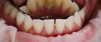
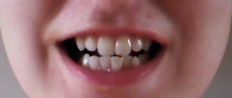
This form of the disease usually occurs without obvious pain. Its immediate symptom is bleeding gums when brushing teeth, eating solid foods and other mechanical effects on the dental system.
Ulcerative-necrotizing gingivitis
This is one of the most unpleasant forms of gingivitis, which is characterized by a feeling of itching of the gingival papillae, severe pain, copious flow of saliva, fever, inflammation of the lymph nodes and the formation of necrotic areas of the gums.
Hypertrophic gingivitis
Patients suffering from this form of gingivitis complain of severe pain, constant bleeding of the gums and a significant increase in the volume of the gums, which can partially cover the crowns of the teeth from the outside (not from the tongue). At the same time, the patient’s gum remains quite hard and under it, on the teeth, tartar forms, which creates favorable conditions for the proliferation of microorganisms. With hypertrophic gingivitis, teeth may move slightly.
Atrophic gingivitis
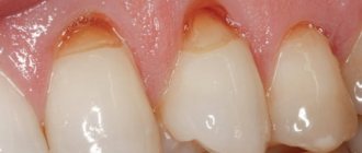
The last and most advanced stage of gingivitis, often leading to periodontitis, is atrophic gingivitis. With it, the gum tissue becomes thinner, decreases in size, the necks of the teeth, and sometimes their roots, are exposed. Teeth become more sensitive to temperature changes (cold or hot drinks, frosty air), to sour or sweet foods, to the mechanical impact of a toothbrush.
Desquamative (geographic) gingivitis
The symptoms of this form of gingivitis differ from others by pronounced red spots on the gums, desquamation of the upper layer of the epithelium, the appearance of blisters on the gums and the formation of mouth ulcers and erosions.
Diagnostic tests
Schiller-Pisarev test
This test is based on determining the level of glycogen in the gum. Its amount increases significantly during inflammation, while healthy gums do not contain glycogen. Lubricating the inflamed gums with Schiller-Pisarev solution gives a color change reaction from light brown to brown. This research method is used to make diagnoses of both periodontitis and gingivitis.
Assessment of oral hygiene level
A solution (2 g of potassium iodide, 1 g of crystalline iodine, 40 ml of distilled water) is applied to the outer surface of the six lower front teeth.
The assessment is carried out using a five-point system and each tooth is assessed separately:
- 5 points – complete staining of the entire tooth surface;
- 4 points – staining of ¾ of the tooth surface;
- 3 points – staining of half the tooth surface;
- 2 points - staining of a quarter of the tooth surface;
- 1 point - absence of any staining of the tooth surface.
Then the scores of all examined teeth are summed up and divided by their number (usually the test is carried out on six teeth). This is how the hygiene index is obtained.
As a result, the quality of hygiene is assessed:
- 1.1-1.5 points – good hygiene index;
- 1.6—2.0—satisfactory hygiene index;
- 2.1—2.5—unsatisfactory hygiene index;
- 2.6—3.4—poor hygiene index;
- 3.5-5.0 - very poor hygiene index.
Vacuum test according to Kulazhenko
Using a Kulazhenko vacuum apparatus, it is possible to determine the time of hematoma formation when a vacuum is applied to the gum area. Typically, the test is carried out in the incisor area by placing a tube of the device on the gum. The formation of a hematoma in 60 seconds indicates the normal condition of the gums, while the appearance of a hematoma in 29-30 seconds signals an inflammatory process.
Oxygen tension in gum tissue
The sensor of the device is applied to the gum, and the device records the level of tissue hypoxia. Reduced oxygen tension indicates a prolonged inflammatory process.
Differential diagnosis of gingivitis
It is based on the complaints presented to the patient, a visual examination of the patient, the results of functional tests and laboratory tests. The goal of differential diagnosis is to distinguish gingivitis from other periodontal diseases, such as periodontitis and periodontal disease.
The main feature that distinguishes gingivitis from other periodontal diseases is that the inflammatory process affects only the gum tissue, the remaining structures (muscle ligaments that hold the tooth in the jaw and bone tissue) remain unchanged.
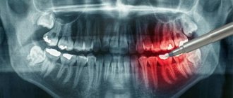
Along with this symptom, gingivitis is not characterized by periodontal pockets, exposure of the necks of teeth, or their mobility. And the x-ray shows no signs of bone resorption.
Identifying gingivitis in a timely manner, determining its form and prescribing the correct treatment is the task of a periodontist. But not to forget about prevention and regularly visit the dental clinic is the maximum program for the patient. This is the only way to avoid a more serious periodontal disease – periodontitis.
Schiller test value
Of great importance in the differential diagnosis of normal and atypical patterns is the color shade and the clarity of the zone boundaries.
The presence of clear boundaries indicates an unfavorable situation. Benign lesions usually have less sharp, blurred contours.
Thus, the test expands diagnostic capabilities and allows:
- preliminary assess the degree of damage, size, depth and number of pathologically changed areas;
- determine patient management tactics;
- evaluate the effectiveness of treatment measures,
- to clarify the features of changes in stratified squamous epithelium under various physiological conditions (pregnancy, menopause, contraception).