- When is root canal filling indicated?
- What are the purposes of temporary filling?
- What material should be used for temporary filling?
- How does filling occur?
- What does pain after filling mean?
- Types of fillings
- Types of pastes
- How much does temporary root canal filling cost?
The period for which a temporary filling is placed is determined by the attending physician.
In most cases, a drug that has a long shelf life is placed under the filling material. After this period, the doctor removes the temporary material. When medicinal pastes are used together with disinfectants and antiseptic medications, there is a significant increase in the effectiveness of treatment. In addition, there will be no cross-contamination of soft and hard tissues between visits to the doctor.
When is root canal filling indicated?
There are general indications for the procedure:
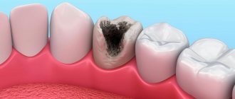
- The root canal is affected. This happens if the patient has a chronic form of pulpitis.
- During the purulent stage of pulpitis, regardless of whether the tooth cavity is closed/open.
- If intraradicular soft tissues are excessively affected.
After mechanical processing is completed, a funnel-shaped channel is formed. It has a minimum diameter at the apex, and a minimum at the mouth. Also, the balance between the “diameter and wall thickness” of the channel is observed.
Preparation for the procedure
Proper preparation is the most important aspect of endodontic treatment. If you do not provide for all possible nuances, the effectiveness of the procedures may be reduced to zero. Before filling directly, it is necessary, first of all, to thoroughly clean the root canal of any plaque that has formed, and also remove all damaged tissue.
In general, all preparatory actions are quite standard. They are carried out in stages and have the following action plan:
- carious lesions are eliminated, as well as dead tissue (specialized drills are used for this);
- pulp is removed from open canals;
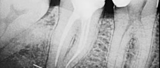
- An x-ray is taken to determine the specific shape and length of the canal;
- The root canal is expanded to the required size using various attachments and other tools.
Please note that strict adherence to all of the above points is the key to effective treatment. If you neglect at least one of these stages, with a high degree of probability the current situation will only worsen.
What material should be used for temporary filling?
There are special requirements for the material used for filling:
- Creating complete tightness, which is achieved by penetrating even the smallest cracks. This helps create a kind of barrier that prevents the development of bacteria. If microorganisms develop a small cavity into which they can penetrate, the inflammatory process will re-develop.
- The materials should be clearly visible on x-ray. Thanks to this, the doctor is convinced that high-quality work has been done.
- High aesthetics. The material should not be visible through tooth enamel.
- Saliva, hard or soft tissues of the oral cavity should not provoke an allergic reaction by contacting the filling mass.
- Materials for temporary filling of root canals should be easily removed from the roots. After the substance hardens, a slight shrinkage effect may appear, but no cavities are formed.
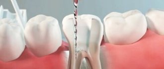
How does filling occur?
Before temporary filling of the canals begins, the patient is sent for an X-ray examination. Using the image, the extent and depth of the lesion is determined. Then a technique is selected and the procedure begins. It consists of the following steps:
1. Administration of anesthesia. Local anesthesia is used. Many patients have a fear of the sound of the drill, even if there is no pain. These patients are given intravenous sedation.
2. Clean the tissues affected by caries (enamel, dentin).
3. The dental nerve is removed.
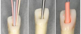
4. Probe the root passage. Its depth is determined.
5. The width of the passage is expanded and drilled.
6. Wash with an antiseptic solution and dry.
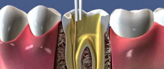
7. If necessary, a pin is inserted.
8. Material is placed into the cavity, taking into account the developed technique.
9. Carrying out a control x-ray examination.
General overview
The composition used affects the periapical structure. The reaction of irritated tissues to the formation of a necrotic layer is the formation of a connective structure, which subsequently undergoes mineralization. The alkaline pH level is sufficient to neutralize the lactic acid of osteoclasts, so the mineral structure of dentin and bone tissue remains protected.
Calcium hydroxide, used as part of endodontic therapy, helps solve a number of functional problems, such as:
- Reducing the number of pathogenic bacteria located in the canal cavity;
- Maximum possible extraction of necrotic tissue;
- Reducing the volume of concentration of bactericidal endotoxins.
Using only mechanical instruments to treat the cavity, despite the use of an irrigator, does not provide a sufficient disinfectant effect, since not only pathogens, but also dead tissue particles remain in the canal. Calcium hydroxide, provided by the temporary filling protocol, has a positive effect on the general condition of the affected root, and creates conditions for full treatment.
What does pain after filling mean?
The presence of discomfort for three to five days after the procedure is normal. The reason for their appearance is irritated gum tissue and pulp. The patient may complain of a feeling of fullness inside the treated tooth. This is also normal.
To avoid negative consequences, you should contact only an experienced and qualified specialist. Otherwise, the following consequences may arise due to mistakes made:
- A through hole is formed in the root canal.
- Secondary inflammation develops under the filling material.
- Flux and fistula develop.
- A cyst or granuloma forms.
- Allergic reaction to the material.
By choosing a clinic that has a good reputation, you can not be afraid of the above problems.
Types of fillings
There are 2 types - temporary and permanent root canal filling.
The first method uses a non-hardening paste, which is distinguished by the presence of medicinal properties. Getting a good result occurs due to calcium hydroxide. The use of temporary filling is indicated for advanced pulpitis, when the inflammatory process has spread quite deeply. A special material is placed into the cavity and covered with a filling. Installation of a permanent filling may take several days or even months.
The second method (permanent filling) is carried out for more than a dozen years. The procedure is allowed immediately during the first visit to the doctor, if there is no inflammatory process, or after treatment with temporary material has ended.
Treatment regimen
- Diagnostics Before treatment, an x-ray or computed tomography must be done to determine the number of channels, their location and shape.
- Anesthesia The doctor selects an anesthetic and, if necessary, conducts sensitivity tests to various drugs.
- Installation of a rubber dam An obligatory aspect of modern treatment is the isolation of the tooth from oral fluid using a special latex scarf (rubber dam).
- Instrumental and medicinal canal treatment The essence of root canal treatment is high-quality disinfection of the tooth cavity. To do this, the doctor expands the canal with instruments, gives it the desired shape and rinses it with disinfectant solutions to destroy the infection.
- Preparing the canals The doctor measures the length of the canals using an apex locator, after which they are mechanically expanded and smoothed out the walls.
- Filling canals Cement, a pin, or a combination of both are injected into the prepared cavities, depending on the chosen treatment method.
- Control image The doctor evaluates the quality of canal filling, after which he performs tooth filling and other necessary procedures. In addition, the patient receives recommendations for further oral care.
Types of pastes
Paste containing an antibiotic and a corticosteroid
In most cases, such a paste contains 2-3 broad-spectrum antibacterial drugs and a corticosteroid. Basically, it is dexamethasone. The paste contains it in a certain amount, therefore, while reducing inflammation and allergic reactions, there is no disruption of the periodontal protective reactions. The third component is a radiopaque filler. Thanks to him, it is possible to objectively assess how well the channels have been filled. This paste has a strong, but not long-lasting effect. The duration of their presence in the dental canals varies from three to seven days.
Paste containing metronidazole
This component helps to effectively suppress pathogenic microflora in the root canals, stop kagabolic tissue destruction, and block the inflammation process. The patient will not have an allergic reaction to such a paste, and there is also no need to be afraid of addiction.
A paste with this component is used in case of excessive infection of the canals. In addition, such a remedy can cure the acute stage of periodontitis. As a result, re-infection can be easily prevented. Also, the disease progresses much easier.
Paste based on a long-lasting antiseptic mixture
This type of paste consists of antiseptic components that have a strong effect - camphor, thymol, cresol, menthol, etc. This is a radiopaque paste, it does not harden, and gradually dissolves in the root canal. Indications for the use of such a paste are the presence of pulpitis, periodontitis, and problems with baby teeth.
Calcium hydroxide paste
A highly alkaline reaction occurs; when the canal is filled with this substance, various bacteria are destroyed and necrotic tissue is destroyed. In addition, stimulation of ostogenesis, dentinogenesis and cementogenesis begins.
The use of a non-hardening paste, which includes calcium hydroxide, is necessary for severe periodontitis, cystogranuloma. This mass is placed in the channel; a special channel filler is used for this purpose.
After six weeks, the paste is replaced. A new portion is placed into the root canals. After this, the paste is changed every eight weeks until the desired result is achieved. If the pain and inflammation have disappeared, and exudation has stopped, the doctor cleans the canals and fills them with permanent filling material.
A Look at Some Popular Root Canal Obturation Materials
Malanin Igor Valentinovich Doctor of Medical Sciences, Professor, Academician of the Russian Academy of Economics, President of the European Academy of Dentistry, Head of the Kuban Scientific School of Dentistry
Endodontics today continues to strive for high technology and new materials. As more and more researchers have proposed solutions to common clinical situations, the complexity of the arsenal of materials for root canal obturation has increased. The high level of manual skills required in endodontics inevitably determines the improvement of personal skills. Hence, it is quite obvious that numerous publications related to a specific material or method have appeared.
Most of the currently used root canal obturation materials have been used successfully for several decades. The focus has typically been on clinical data and research aimed at improving or modifying established clinical practices. This often resulted in improvements to known techniques rather than the development of new materials.
In addition to personal, subjective influences, the literature on materials for root canal obturation reflects the prevailing concepts in endodontic practice that are popular at a given time. This occurs despite the almost universal belief that the core of clinical endodontic practice is the cleaning, shaping and filling of the root canal system. For example, when experts were absolutely sure that pulpless teeth were “foci of infection,” they placed their main emphasis on instrumental techniques aimed at “disinfecting” the root canal and on filling materials with a strong and long-lasting antiseptic effect. Later, endodontic techniques were gradually reoriented to reduce traumatic instrumentation damage to the periodontal ligament and to use materials that were better tolerated by the pulp and periapical tissues. Until recently, materials for root canal obturation were not studied as such outside of clinical techniques.
What principles guides a modern endodontist when choosing a material for obturation of root canals? Does the doctor delve into the composition of the material he uses, or does he fill “like everyone else”? As a rule, “experienced” doctors have been using the same material for decades.
Modern, newly created material for obturation of root canals has a short life. Many of the technologies and treatments that students learn today will be modified or replaced by others by the time these students become doctors. In order to keep up with the times, the dentist must have the ability to evaluate the potential of each new material and treatment method.
It is well known that the last stage of endodontic treatment is the complete, dense and hermetically sealed filling of the root canal system and all hard-to-reach areas with non-irritating materials. For successful treatment, three-dimensional filling of the entire space of the canal, the apical hole in the area of the dentinal-cement junction and additional canals is necessary with an inert, biologically compatible material that has spatial stability. Amputation methods for the treatment of pulpitis, used until recently in domestic dentistry, are considered a gross mistake in modern dental practice.
Root canal materials come into contact with biological tissue that is not protected by an epithelial layer, so their biocompatibility is of particular importance. It is generally accepted that a biologically acceptable material must be inert. In practice this cannot always be achieved. Therefore, material creators strive to achieve a favorable interaction between the material and the biological environment in which it is located, and which would not have a negative impact on the material itself. It is important that the material does not cause an inflammatory reaction in the tissue, as this can cause irritation, pain and necrotic changes.
A constant problem in endodontic treatment is the possibility of recurrent infection at the apex of the tooth due to the presence of microorganisms there. This dictates another requirement for materials for filling root canals - to have an antimicrobial effect.
In modern dentistry, it is quite difficult to combine these two material requirements, since this implies the need to take into account a high degree of selectivity of the biological response. After all, it is well known that a material with an antimicrobial effect causes an inflammatory reaction in adjacent tissues, and those materials that do not cause it have the best bacteriostatic properties. If we agree that complete sealing of the root canal cannot be achieved, the materials used must have sufficient antimicrobial activity to prevent the infiltration of microbes into the canal space and their proliferation. At the same time, the antimicrobial properties of the material should not be achieved at the expense of its biocompatibility.
Gutta-percha is a biocompatible material with very low cytotoxicity, so only the cements used with it will determine the tissue reaction.
Gutta-percha points are used in combination with cement, which is necessary to fill the spaces between the point and the root canal wall, thus preventing the penetration of microorganisms. It also lubricates the pins as they compact, filling channel irregularities and side channels.
The use of cements to seal the root canal without obturating pins is not recommended. When cements are introduced into the canal in large quantities, they undergo more intense dissolution and exhibit excessive shrinkage during curing. In addition, it is quite difficult to determine adequate filling of the canal, and there is a danger of cement leaking beyond the root apex into the surrounding tissue.
Until recently, it was generally accepted that filling a canal with cement could not guarantee against permeability of tooth tissue, and therefore the main attention was paid to imparting antimicrobial properties to these materials.
In clinical practice, a large number of materials are used to fill root canals, including:
- Zinc oxide eugenol cements (eg Tubliseal, Kerr);
- Polymer cements (AH Plus, Dentsplay; Diaket, ESPE);
- Cements containing calcium hydroxide (Apexit, Ivoclar; Sealapex, Kerr);
- Glass ionomer cements (Ketak Endo, ESPE; Endion, Voko);
- Polydimethylsiloxanes (RCA RoekoSeal, Roeko).
In this article, the author did not intend to describe all known materials for permanent root canal filling. But I would like to focus on some of them, due to their prevalence in Russia and specifically in the Krasnodar Territory.
Endomethasone - unfortunately, this material is by far the most purchased and popular endodontic material in Russia.
Endomethasone is a material based on zinc oxide eugenol paste; contains corticosteroids (hydrocortisone and dexamethasone), antiseptics, diiodothymol and paraformaldehyde, as well as radiopaque filler.
Antiseptics provide sterilization of organic residues in microchannels, deltoid branches, and affect the microflora of the periapical lesion during periodontitis. As the paste hardens, the effect of these substances weakens and then stops. If endomethasone is excreted beyond the apex, then eugenol diffuses quite quickly into the bloodstream, and then the remaining components of the paste gradually dissolve (first behind the apical foramen, and then in the canal).
As for corticosteroids, in addition to their positive effects, they also have a number of negative properties. For example, they weaken the protective mechanisms of the periapical area, in particular due to the suppression of phagocytosis, resulting in the proliferation of microorganisms; Their side effects cannot be ruled out either.
Included in the composition of endomethasone to reduce complications after filling, there are also products containing formaldehyde. The resorcinol formaldehyde method, proposed at the beginning of the last century and very widely used for a long time, was subjected to careful analysis in the 80s and early 90s for its toxic effect, of which quite a lot of examples have accumulated.
Electron microscopic research has demonstrated that formaldehyde denatures pulp proteins and, deposited in crystalline form on the surface of the denatured alcohol, binds tightly to it. If the proteinaceous material in the root canals is sufficient to bind formaldehyde, its systemic effects are negligible. If the pulp is partially or completely removed, the antiseptic can enter the periodontium, causing local and general adverse effects.
Many negative effects are associated with the use of paraformaldehyde and corticosteroids. When formaldehyde comes into contact with living tissues, it spreads throughout the body. Systemic studies found labeled paraformaldehyde in the blood, regional lymph nodes, kidneys and liver after pulpectomy in dogs using labeled 14-degree formocresol. In addition, it is well known that formaldehyde has mutagenic and carcinogenic properties. In this regard, the question arises about the possibility of contact with living tissues.
The popularity of this material among some doctors is explained by the fact that the addition of corticosteroid drugs and paraformaldehyde to endomethasone can significantly reduce the risk of developing painful reactions from the periodontium after endodontic treatment, even with accidental (permanent) removal of the material beyond the apex.
If the material is not brought to the apex or the canal is processed poorly, endomethasone is good for so-called chemical pulpotomy. In the first case, the antiseptic effect is leading, in the second, it is also possible to cause aseptic necrosis and mummification of the pulp without complete mechanical extraction. In other words, this drug is the drug of choice for doctors who have insufficient manual skills.
Without listing the negative properties of endomethasone and similar drugs, I would like to add that not only the International Dental Association and the American Dental Association, but also many Russian educational centers do not recommend (prohibit) filling root canals with pastes, since the latter do not provide reliable obturation.
It is also inappropriate to use cements containing calcium hydroxide (Apexit, Ivoclar; Sealapex, Kerr) for permanent filling. This is due to the fact that calcium hydroxide released from the material reduces its spatial volume, which is unacceptable for permanent filling. Doctors also forget that calcium hydroxide acts for a short time and is used for temporary filling.
AN-26, AN Plus
According to the author, a good alternative to the above materials is AN-26, which is widely used not only in the Krasnodar region, but also in world endodontic practice. It was first reported around 1957. It is an epoxy resin with poor solubility. It consists of silver powder (10%), bismuth trioxide (60%), titanium dioxide (5%) and hexamethylene tetramine (25%), which are mixed to the consistency of a thick paste with a liquid - bisphenol diglycidyl ether (100%). It has good adhesive properties, antibacterial activity, low toxicity and is well tolerated by periapical tissues.
AN 26 is a thin-flowing, slowly curing material. If it cures in contact with tissue fluid, it releases small amounts of formaldehyde. Hardening time is approximately 34 hours. With some filling techniques, this is considered an advantage, since it provides time for filling correction after X-ray control. Subsequently, a modification of this filling material was presented - AN Plus, which is a two-paste system that excludes the content of hexamethyl-tetramine, which is responsible for the release of formaldehyde.
AN-26 contains silver powder, therefore, in order to avoid discoloration of the tooth, all remnants of root cement must be removed to the level of the gum edge. In this regard, AN-26 and AN Plus produced by modern industry do not contain silver.
The mild cytotoxic reaction to freshly prepared AN26 may be due to the release of formaldehyde, which is formed as a by-product of the polymerization process. Because AH26 takes some time to polymerize, patients may experience some degree of sensitivity that may be associated with its use. AH Plus has been shown to release only a small amount of formaldehyde (3.9 mg/kg) compared to AH26 (1347 mg/kg). However, AH26 has cytotoxicity, although this is significantly reduced after curing of the material.
Endion
In their daily work, many doctors prefer the use of glass ionomer cements for permanent filling of root canals due to their biocompatibility with tooth and periodontal tissues. Clinical studies of the material, Endion (Voko), conducted by the author, showed good long-term results (over 7 years), which were compared with data from studies of other filling materials. Studies have shown high biocompatibility, good adhesion to dentin and low shrinkage.
The disadvantage of glass ionomer cements is the difficulty of removing them from root canals. Although, according to the author, it is relatively easily removed from the root canal using ultrasound.
Also, the “dislike” of many doctors for this material is explained by the fact that due to the short hardening time, high-quality lateral condensation is difficult. When it goes beyond the apex, severe pain occurs. However, with improved manual skills, these problems can be easily eliminated.
Epiphany
In modern endodontic practice, gutta-percha is recognized as a weak link. Although the requirements for optimal instruments for root canal preparation are generally known and achievable, it is impossible to hermetically fill the root canal with gutta-percha. In fact, coronal restoration, not gutta-percha, is one of the cornerstones of success in endodontic treatment. Most materials used for coronary repair would be much more successful if they could be efficiently placed into a long, narrow canal and just as efficiently removed if complications arise after treatment.
Research has shown that the Epiphany obturator system combined with Resilon is more resistant to microleakage (6 times more) than a gutta-percha obturator system. The Epiphany system, combined with Resilon obturation material, strengthens the root by more than 20% compared to standard obturation techniques.
Advantages of the Epiphany obturator system: seals better; strengthens the root; easy to remove; provides immediate coronary sealing;
The main component of the Epiphany obturation system is Resilon, a root canal obturation material with a soft resin thermoplastic synthetic polymer backing. It contains bioactive glass and radio-resistant fillers in a resin matrix. The material is similar to gutta-percha in handling, physical properties and radiographs. It has the same properties for successful unfilling and re-treatment, softens under the influence of high temperatures, dissolves in solvents like chloroform, remains thermoplastic at low temperatures, and is biocompatible. Like gutta-percha, there are main cones in all ISO sizes, as well as additional cones in a variety of sizes available.
Resilon is based on a resin that, in combination with Epiphany dual-cure sealer and Epiphany primer, is bonded into the root canal in the form of a monoblock. The emerging monoblock also helps to strengthen the obstructed root - by filling the root canal, coronal sealing occurs, thereby eliminating the possibility of pathogenic bacteria penetrating into the apex. By replacing gutta-percha and the usual filler with materials from the Epiphany obturation system, there is no need to change the canal obturation technique. The fundamental point is the complete cleansing and formation of the channel.
The author, having worked with Epiphany material for a rather short time, nevertheless made an attempt to reflect his impressions of this material:
Because Epiphany obturation can be light cured immediately after filling the canal, an immediate coronal seal will occur. This will save the patient from having to visit again.
Epiphani can be used with any obturation technique and has high radiopacity.
If it is necessary to remove Epiphany from the canals, there are no difficulties; it is easily removed, no more difficult than gutta-percha with zinc oxide sealant.
Today, Epiphany represents an alternative to gutta-percha-based obturation systems, due to the fact that it has all the advantages of gutta-percha, but at the same time it is free from the disadvantages of gutta-percha.
Despite the introduction into practice of a large number of materials for filling root canals, many endodontists, unfortunately, prefer to use materials based on zinc oxide eugenol paste for these purposes.
Today, the success of endodontic treatment is a reality. Many of our happy patients, having gotten rid of pain, will agree with this. However, incorrectly performed techniques cannot be considered successful solely on the basis of the patient’s lack of obvious symptoms.
We are our own harshest critics and set too strict criteria for success. For oncological diseases, doctors consider survival to be 5 years a success, and for femoral prosthetics - 3-5 years. Are these numbers arbitrary? Do they have a real basis? Do doctors consider them reasonable if most of their patients fit within this 3-5 year period? Should we, like this, strive for short-term success or should long-term failure be anticipated in dentistry?
We must not deceive ourselves. Failures do happen and will continue to occur, despite the great efforts of doctors and the constant improvement of techniques. Our goals may be noble and lofty, but we cannot always achieve them, and this is often due to the fact that we are dealing with a human body that does not always behave as it is written in books.