Detailed description of the study
Salivary gland cancer is a malignant tumor of the salivary gland formed from epithelial cells. Tumors of the parotid salivary glands (PST) are a rare type of neoplasm. They mainly develop between the ages of 20 and 40. More common in men than in women. Tumors are usually located in the soft tissues of the face and often cause serious cosmetic defects, causing paresis of the facial nerve.
The main risk factors for developing salivary gland cancer are:
- Chronic inflammatory diseases of the oral cavity;
- Hormonal imbalances;
- Genetic disorders;
- Smoking;
- Nutritional factors (low vitamin content, high fat content in food).
Most often the tissues of the parotid salivary gland are affected - 80%, less often the tumor develops in the tissues of the submandibular salivary gland - 15%, sublingual or minor salivary gland.
The following options for the development of pathology are distinguished:
- Primary cancer (from the tissues of the gland itself);
- Secondary cancer (metastasis or germination from neighboring organs).
According to histological parameters, tumors of the salivary glands represent the most heterogeneous group of human neoplasms. More than half of them are benign formations.
Benign neoplasms of the parotid salivary glands are detected in more than 30% of patients who complain, malignant ones - in 65%. Among the morphological types of benign tumors of the parotid salivary gland, the most commonly diagnosed are pleomorphic adenoma (a benign formation that forms in the glandular epithelium), adenolymphoma, lipoma and a very rare adenoid cystic tumor (formed by epithelial cells lining the lumen of the gland).
Malignant tumor formations are most often represented by: mucoepidermoid cancer (formed by cells secreting mucus and epithelium) and adenocarcinoma. Acinic cell carcinoma also occurs, which mainly affects the tissue of the parotid salivary gland.
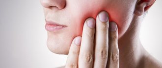
Usually, during examination of patients, palpation reveals a dense, painless formation in the area of the salivary glands, which, according to patients, grows generally very slowly. In approximately a third of cases, pain may be present.
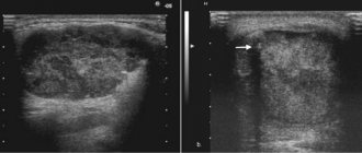
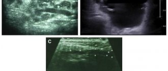
The “gold standard” for diagnosing tumors of the salivary glands is aspiration puncture of the tumor followed by histological examination of the obtained material, carried out before surgery or intraoperatively. The diagnostic value of this method is up to 90%, because the tissue of the affected area is studied directly under a microscope.
Aspiration puncture (biopsy) is performed using a special needle under ultrasound control. The study allows you to confirm or clarify the diagnosis, track the growth dynamics of the tumor and analyze the changes that occurred during tumor therapy.
During histological examination, three indicators are assessed:
- Tumor size and extent of its spread (T);
- The presence of damage to regional lymph nodes by metastases (N);
- Determination of metastases in other organs and tissues (M).
Based on the degree of malignancy, there are three types of tumors:
- High differentiation, when the cells are similar to glandular epithelial cells, such formations grow slowly and often have a favorable prognosis;
- Low differentiation, when cells are very different from normal, is characterized by rapid growth and early metastases;
- Medium differentiation.
Types and functions of salivary glands
Salivary glands are paired organs that differ in location:
- large parotid - located on the side of the lower jaw, under the left and right ears. They produce saliva containing high concentrations of potassium and sodium chlorides;
- large submandibular - adjacent to the lower jaw, have an excretory duct in the area of the lingual frenulum, produce saliva with a low acid index;
- large sublingual - located in the oral cavity under the mucous membrane, secrete alkaline saliva containing protein saturated with mucin (high molecular weight glycoproteins).
The minor salivary glands are located on the mucous membrane of the cheeks, lips, and tongue.
The role of the salivary glands is to secrete a fluid that contains special enzymes and is involved in the digestive process. They also perform the following functions:
- exocrine - production of proteins, fluids;
- endocrine - produce biologically active substances;
- excretory - participation in the removal of metabolic products from the body.
References
- Malignant tumors of the salivary glands, Clinical guidelines. Association of Oncologists of Russia, 2022. - 55 p.
- Balkanov, A.S., Bychenkova, O.A., Sipkin, A.M. and others. Combined treatment of parotid salivary gland cancer. Almanac of Clinical Medicine, 2022. - No. 4. - P. 309-313.
- Polyakov, V.G., Shishkov, R.V., Ermilova, V.D. and others. Children's Oncology, 2004. - No. 1. - P. 45-47.
- Carlson, E., Schlieve, T. Salivary Gland Malignancies. Oral and maxillofacial surgery clinics of North America, 2022. - Vol. 31(1). — P. 125-144.
Bougienage during examination of the salivary gland duct
Examination of patients with diseases of the salivary glands requires certain skills from the dentist, since often the first symptoms of this disease are inflammatory processes in the oral cavity.
After collecting anamnesis, visual and palpation examination, the following may be prescribed:
- sialometry using the Andreeva method - after making a bougienage into the duct of the salivary gland, a cannula (special tube) is inserted through which saliva is collected for quantitative and qualitative research;
- sialography (contrast x-ray diagnostics) - 1 - 2 ml of iodolipol or other iodine-containing substance, heated to a temperature of 37 - 40 degrees, is injected into the ducts of the salivary glands using a blunt needle. The mouth of the duct is first bougiened with a conical probe. An x-ray is taken immediately;
- pantomosialography (contrast x-ray diagnostics of several glands at once) - this procedure helps to determine the ongoing hidden inflammatory process of paired salivary glands, narrowing or widening of the ducts;
- digital (digital) subtraction sialography - performed with the introduction of a contrast agent.
Treatment
Removal of a salivary gland adenoma is a simple surgical operation; the prognosis for treatment in most cases is favorable. The only difficulty that can be encountered during treatment is damage to the facial nerve. However, a solution has been found for this problem.
To gain access to the tumor, the surgeon dissects the facial nerve by carefully lifting it upward. Only after this the tumor is removed. Removal of the node is carried out within a few minutes in the maxillofacial departments of dental clinics. If the results of histological analysis of the tissues of the removed tumor confirm its benign nature, no additional treatment will be required.
Complications
Despite the fact that treatment of salivary gland adenoma does not pose a serious problem, it is important to remember that untimely removal of the tumor can lead to a number of complications. The most serious of them is the transformation of a benign neoplasm into a malignant one. Unlike malignant tumors, salivary gland adenoma can exist asymptomatically, only in some cases causing facial asymmetry. Therefore, if you detect the slightest swelling, you must immediately consult a doctor. Timely removal of the tumor will prevent many very unpleasant problems.
The most common postoperative complications:
- the risk of developing Frey's syndrome (occurs when autonomic nerve fibers are damaged, accompanied by redness and sweating of the operated area of the face);
- a feeling of dry mouth when the gland is completely removed.
Observation and regular consultations with a doctor can reduce all postoperative complications to a minimum.
ALL-RUSSIAN CENTER FOR EYE AND PLASTIC SURGERY
On the pages of this site you can get acquainted with the scientific and medical center of federal significance, located in a picturesque corner of the city of Ufa. The basis for the creation of the Center was the development in Ufa of an innovative technology for tissue transplantation, protected by the Alloplant trademark. This is one of the rare cases when a fundamentally new treatment method has led to the opening of a federal scientific institution. The Center received All-Russian status in 1990. Since 1983, our team has been known as the Republican Laboratory of Tissue Conservation. The first Alloplant biomaterial transplantation operation was performed in 1973.
The All-Russian Center for Eye and Plastic Surgery today is a well-known scientific institute of regenerative surgery in the country and abroad. As is known, methods of regenerative surgery are being developed in various scientific and medical centers on the basis of cell, tissue and organ transplantation. Our institution creates and implements methods for the regenerative restoration of anatomical structures based on tissue transplantation. Donor tissues subjected to deep physical and chemical processing are commonly called biomaterials. Tissue transplantation fully meets the requirements of Russian and international legislation, is much cheaper than cellular technologies in terms of costs and can be implemented in both multidisciplinary and specialized medical institutions.
Our Center has all the necessary structural units in the field of transplantology from experimental development to clinical implementation.
It includes a scientific department of morphologists with laboratories of electron and laser microscopy, immunohistochemistry; a multidisciplinary tissue bank with a tissue conservation laboratory, a radiation sterilization unit for biomaterials and a laser modeling complex. The clinical divisions of the Center include specialized ophthalmic surgery departments, associated services of otorhinolaryngology and plastic surgery. Services for neurophysiological and psychological rehabilitation of patients are separated into a separate complex.
It is necessary to dwell separately on the systemic effect of transplants, first noticed by Academician V.P. Filatov and called biostimulation. Our studies have shown that Alloplant biomaterials, acting through the immune, nervous and endocrine systems, make it possible to rehabilitate patients with severe damage to the musculoskeletal system, peripheral and central nervous system, etc. For the clinical implementation of this area, a special department of biophysical methods of diagnosis and treatment “Aura” has been created.
All presented laboratories and departments form a single scientific, medical and production complex within the Center, within which the treatment and rehabilitation technologies created here are constantly being improved.
More detailed information about each service can be found on the corresponding pages of the site.
To quickly find the necessary sections of the site, use the Navigator - a visual menu. it is available on all pages of the site - just click on the Navigator icon.
An exceptional property of the Alloplant biomaterials we created was the ability to stimulate the regeneration of various tissues and anatomical structures. The development of the described biomaterials required solving a whole complex of scientific, organizational, technological and production problems. These include the technology of physical and chemical processing of donor tissues, which makes it possible to reduce the immunogenicity of the final biomaterial and achieve its decontamination while maintaining bioplastic properties. The technological equipment of our production was created through integration with the Russian Federal Nuclear Center (Sarov). Today we actively cooperate with this wonderful and highly respected scientific institution, constantly improving methods of selective radiation sterilization of biomaterials and their laser modeling. By creating biomaterials of various biochemical composition, spatial structure and biomechanical properties, the Center’s team solved many applied problems of regenerative medicine.
In particular, it was possible to achieve regeneration, both experimentally and in the clinic, of various types of epithelial cover, connective structures, blood and lymphatic vessels, peripheral nerves, optimize osteogenesis, etc.
As a result of many years of scientific and innovative activity of our Center, the technology of regenerative medicine based on Alloplant biomaterials was created and introduced into industrial and clinical practice.
Over 95 types of Alloplant biomaterials are used in clinical practice in more than 600 clinics in all regions of the Russian Federation and CIS countries.
The center cooperates widely with a number of scientific and medical institutions and universities in Russia, which ensures the introduction of biomaterials into various fields of medicine. One of the important activities of the team is training doctors in Alloplant transplantation technologies under postgraduate professional education programs.
With best wishes, Chief Scientific Consultant of the All-Russian Center for Eye and Plastic Surgery, Honored Doctor of the Russian Federation, Professor Ernst Muldashev