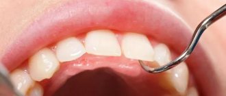
Biometrics in the study of the jaw is of decisive importance, as it allows one to diagnose various malocclusions, the shape and size of the dentition, as well as other deviations from the physiological norm. It is not possible to conduct biometric studies directly in the oral cavity, and it is for this reason that such studies are often carried out on diagnostic models, which can be done in a dental office.
Each such model not only performs a diagnostic function, but also serves as a subsequent control model when applying various treatment methods in orthodontics. Constructive occlusion in orthodontics is
a bite that the dentist tries to recreate for each patient individually. This bite has individual biometric indicators and is recreated, as a rule, using wax templates. Any model must have the following markings:
- Patient's name;
- date of the study;
- patient's age;
- medical history number.
This approach eliminates the possibility of medical error during the study and helps to record its results. The size of individual teeth or the dentition as a whole is important when studying diagnostic models. This helps solve a wide variety of problems facing specialists to restore the patient’s anatomically correct bite and ensure proper functional load.
Biometric methods for studying diagnostic models of jaws
All patients admitted for orthodontic treatment underwent a study of diagnostic models of the jaws. Diagnostic models were cast from casts obtained using a well-known technique from silicone or alginate materials. The models were cast from ordinary or high-strength plaster. Diagnostic models were measured to make a diagnosis and determine the causes of retention.
Teeth measurements.
Using a special caliper, the mesiodistal dimensions of the crown parts of the teeth were measured. The measurements were carried out in the region of the equator. In all patients, the Tonn index was determined, which reflects the relationship between the sizes of the teeth of the upper and lower jaw:
Sum of width of 4 upper incisors
Index Ton ———————————————— = 1.33
Sum of width of 4 lower incisors
If the ratio is greater than 1.45, it means the patient has individual macrodentia of the upper teeth, which may cause tooth retention. A ratio value less than 1.25 indicates individual macrodentia of the lower teeth.
Measurements of the longitudinal length of the dentition were carried out using the Nance method using a ligature wire, which was placed from the distal surface of the first molar to the distal surface of the first molar of the opposite side, giving the wire the shape of the dentition. In the area of the lateral teeth, the wire was placed in the middle of the chewing surface, and on the front teeth - along their cutting edges. The longitudinal length of the dentition is normally equal to the sum of the mesiodistal dimensions of 12 teeth.
Measuring the dentition.
The transverse dimensions of the dental arches were determined using the Pon method with the Linder-Hart correction, which is based on the relationship between the sum of the mesiodistal dimensions of the 4 upper incisors and the distance between the first premolars and the first molars on the upper and lower jaws. For this purpose, Pon proposed measurement points that normally coincide when the upper and lower jaws are closed. In the area of the first premolars, the width of the dentition, according to Pont, is measured:
- on the upper jaw - between the points in the middle of the intertubercular fissure;
- on the lower jaw - between the distal contact points on the slope of the buccal cusps.
In the area of the first permanent molars, the width of the dentition is measured:
- on the upper jaw - between the points in the anterior recesses of the longitudinal fissure;
- on the lower jaw - between the distal buccal cusps.
Pon derived the premolar and molar indices.
Premolar index=
(Sum of transverse dimensions of 4 upper incisors/distance between premolars) x 100 = 80.
Molar index=
(Sum of transverse dimensions of 4 upper incisors/distance between molars) x 100 = 64.
The obtained measurements are compared with tabular data.
The apical base is a conditional line passing at the level of the apexes of the roots of the teeth in the upper and lower jaws. In the vestibule of the oral cavity, it is projected onto the transitional fold. The width of the apical base is measured using the House method modified by N.G. Snagina.
The width of the apical base of the upper jaw is determined on the plaster model along a straight line between the deepest points in the fossa canina area, and on the model of the lower jaw, the measurement is carried out between the same teeth, departing from the level of the gingival margin by 8 mm. Normally, the width of the apical base of the upper jaw is 44%, the lower - 40% of the sum of the mesiodistal dimensions of the 12 permanent teeth of each jaw.
The discrepancy between the width of the crowns and the size of the apical base can cause dental anomalies, including retention of individual teeth.
To determine the relationship of the dentition in the sagittal direction, Engle's classification was used.
It is based on the type of closure of the first molars. The first class is characterized by normal closure of the molars in the sagittal plane. The mesiobuccal cusp of the first molar of the maxilla is located in the intercuspal fissure of the first molar of the mandible. In this case, all changes occur in front of the molars. There may be a crowded position of the incisors, a violation of their closure.
The second class is characterized by a violation of molar closure, in which the intercuspal fissure of the first molar of the lower jaw is located behind the mesiobuccal cusp of the first molar of the upper jaw. This class is divided into two subclasses: the first subclass - the upper incisors are inclined in the labial direction (protrusion); second subclass - the upper incisors are inclined palatally (retrusion).
The third class is characterized by a violation of the closure of the first molars, in which the intercuspal fissure of the first molar of the lower jaw is located in front of the mesiobuccal cusp of the first molar of the upper jaw.
The vertical gap between the incisors was measured using the method of Khoroshilkina F.Ya (1982) [80].
— I degree (up to 5 mm);
— II degree (from 5 to 9 mm);
— III (more than 9mm).
The vertical gap between the incisors determines the severity of the anomaly.
Measurement of dentoalveolar height on diagnostic jaw models was carried out according to the method of A.P. Romanovskaya (1988) [56].
We measure the central incisors vertically in the center from the cutting edge to the transitional fold. We measure the first premolars in the center of the buccal cusp from the occlusal surface to the transitional fold. The first molars are located in the center of the mesiobuccal cusp from the occlusal surface to the transitional fold.
By measuring the patient's models and comparing them with the derived norm, it is possible to reliably determine the degree of dentoalveolar lengthening or dentoalveolar shortening.
Table 2.2.3.1
Average values of dentoalveolar height for orthognathic occlusion according to Romanovskaya A.P.
| Dentoalveolar height in the area of the anterior and lateral teeth of the upper and lower jaw | ||||||
| in the area of 11-21 teeth | in the area of 14-24 teeth | in the area of 16-26 teeth | in the area of 31-41 teeth | in the area of 34-44 teeth | in the area of 36-46 teeth | |
| Orthognathic bite, (normal, in mm.) | 15.0±0.08 | 14.9±0.08 | 13.5±0.08 | 14.7±0.08 | 14.7±0.08 | 13.5±0.07 |
Using measurements of diagnostic models, the probable causes of vertical incisal disocclusion were determined. After a thorough examination, a treatment plan was drawn up. We also planned the designs of the main and auxiliary orthodontic equipment necessary to achieve the goals.
Purpose
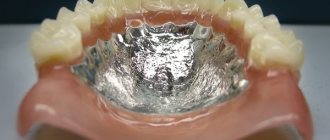
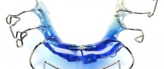
Doctors recreate a constructive bite to restore the correct position and shape of teeth using orthodontic treatment or various types of restorations. In order to correct the position of the jaws, patients are fitted with devices of various design features and functional significance: bite rings, strips, single-jaw plates, double-jaw devices and fixing mouthguards.
Methodology for measuring models according to Pon
To determine the pathology of the dentition in the transversal plane, the simplest and most common method for studying models is the Pon method. The method is based on a certain relationship between the transverse dimensions of the crowns of the four upper incisors and the width of the dentition in the area of premolars and molars. These data were obtained from numerous measurements of properly formed bites.
As a result, the indices were calculated: premolar (72-82, average 80) and molar (60-65, average 64).
Sum of transverse dimensions of 4 incisors x 100%
Premolar index = ————————————————————————— = 80
Distance between premolars
Sum of transverse dimensions of 4 incisors x 100%
Molar index =—————————————————————————— = 64
Distance between molars
To determine the average individual norm for the width of the dental arches in the area of premolars and molars, Pon compiled a table (Table No. 3) taking into account the width of the four upper incisors.
For the lower jaw, the sum of the transverse dimensions of 4 incisors and the corresponding distance between the premolars and molars are determined from the data of the upper jaw.
After determining the sum of the transverse dimensions of 4 incisors and the distance between the premolars and molars, they are compared using the table with the width of the dental arches that the patient has.
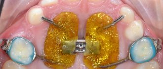
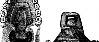
The width of the patient's dental arches is measured at Pon's points. For the upper jaw, this is the middle of the intercuspal fissure of the first premolars and the anterior depression of the intercuspal fissure of the first molars (Figure No. 7); for the lower jaw - this is the most distal point of the slope of the buccal cusp of the first premolar (the point between the premolars) and the top of the anterior buccal cusp of the first lower molar (four-cusp tooth) or the top of the middle cusp (five-cusp tooth); (Figure No. 8).
What is being measured
For calculations in orthodontics, high-precision measurements are a prerequisite. To accomplish this, specialists have special calipers, compasses and protractors that allow them to estimate the distances and angles between the structures of the dentofacial apparatus.
The following indicators must be recorded:
- dimensions of the coronal part protruding above the mucous layer (height and width);
- length and width of dentition;
- the relationship of the upper and lower arc in three planes;
- length of the anterior segment of the dental arch;
- transversal and sagittal dimensions of the dentition in children during the period of temporary occlusion;
- height of the apical base of the jaw;
- symmetry and relationship of the anterior and lateral segments of both arches.
The list of indicators to be measured may be changed and supplemented depending on which calculation method is intended to be used for a given patient.
Pont index and its modifications
With normal occlusion, the measuring points of the lower jaw coincide with the corresponding points of the upper jaw. These indicators are applicable for permanent dentition.
In a mixed dentition in the absence of premolars, the distance between the distal fossae of the first primary molars on the upper jaw or the disto-buccal cusps on the lower jaw is measured. In cases where the upper jaw does not yet have all the incisors, the width of the dental arch can be determined by the sum of the transverse dimensions of the lower incisors.
Ton revealed the relationship between the total width of the 4 upper and lower incisors.
The width of the 4 lower incisors, multiplied by 4 and divided by 3, gives the sum of the 4 upper incisors, based on this the Tone index was determined to be 1.35. Linder and Hart checked Pon's method and made some adjustments according to their data: the premolar index is 85, and the molar index is 65. Table No. 3.
More accurately, the narrowing of the teeth can be determined by the percentage ratio of the width of the dental arch to the sum of the mesiodistal dimensions of 12 teeth (the method was proposed by N.G. Snagina). The measurement points on the upper and lower jaws coincide with Pon's points.
There are three degrees of narrowing of the dentition.
Narrowing of the dentition of the 1st degree is characterized by a decrease in the width of the dental arch in the area of premolars and molars in the range from 1 to 4 mm.
Narrowing of the dentition of the 2nd degree is characterized by a decrease in the width of the dental arch in the area of premolars and molars to 6 mm.
Narrowing of the dentition of the 3rd degree is characterized by a decrease in the width of the dental arch in the area of premolars and molars by 6 mm or more.
Fig No. 7 Fig No. 8
What disorders are diagnosed?
Using calculations in orthodontics, the following deviations from the norm can be identified:
- individual discrepancy between the size of the teeth in relation to the jaw;
- narrowing or shortening of the dentition;
- tight position of teeth;
- abnormal condition of the apical bases;
- any malocclusion;
- disproportions of the anterior or lateral segments;
- crowding or enlargement of interdental spaces;
- degree of crown rotation, etc.
Calculations based on biometric models are applicable in the diagnosis of most orthodontic pathologies. This is due to the simplicity and accessibility of the method, as well as its high information content. The main advantage is that these techniques are feasible without expensive equipment, and the data can be obtained fairly quickly.
Molar Index
All patients admitted for orthodontic treatment underwent a study of diagnostic models of the jaws. Diagnostic models were cast from casts obtained using a well-known technique from silicone or alginate materials. The models were cast from ordinary or high-strength plaster.
Diagnostic models were measured to make a diagnosis and determine the causes of retention.
Teeth measurements. Using a special caliper, the mesiodistal dimensions of the crown parts of the teeth were measured.
The measurements were carried out in the region of the equator.
In all patients, the Tonn index was determined, which reflects the relationship between the sizes of the teeth of the upper and lower jaw:
Sum of width of 4 upper incisors
Index Ton ———————————————— = 1.33
Sum of width of 4 lower incisors
If the ratio is greater than 1.45, it means the patient has individual macrodentia of the upper teeth, which may cause tooth retention.
A ratio value less than 1.25 indicates individual macrodentia of the lower teeth.
Measurements of the longitudinal length of the dentition were carried out using the Nance method using a ligature wire, which was placed from the distal surface of the first molar to the distal surface of the first molar of the opposite side, giving the wire the shape of the dentition. In the area of the lateral teeth, the wire was placed in the middle of the chewing surface, and on the front teeth - along their cutting edges.
The longitudinal length of the dentition is normally equal to the sum of the mesiodistal dimensions of 12 teeth.
Measuring the dentition. The transverse dimensions of the dental arches were determined using the Pon method with the Linder-Hart correction, which is based on the relationship between the sum of the mesiodistal dimensions of the 4 upper incisors and the distance between the first premolars and the first molars on the upper and lower jaws.
For this purpose, Pon proposed measurement points that normally coincide when the upper and lower jaws are closed. In the area of the first premolars, the width of the dentition, according to Pont, is measured:
- on the upper jaw - between the points in the middle of the intertubercular fissure;
- on the lower jaw - between the distal contact points on the slope of the buccal cusps.
In the area of the first permanent molars, the width of the dentition is measured:
- on the upper jaw - between the points in the anterior recesses of the longitudinal fissure;
- on the lower jaw - between the distal buccal cusps.
Pon derived the premolar and molar indices.
Premolar index=
(Sum of transverse dimensions of 4 upper incisors/distance between premolars) x 100 = 80.
Molar index=
(Sum of transverse dimensions of 4 upper incisors/distance between molars) x 100 = 64.
The obtained measurements are compared with tabular data.
The apical base is a conditional line passing at the level of the apexes of the roots of the teeth in the upper and lower jaws.
In the vestibule of the oral cavity, it is projected onto the transitional fold. The width of the apical base is measured using the House method modified by N.G. Snagina.
The width of the apical base of the upper jaw is determined on the plaster model along a straight line between the deepest points in the fossa canina area, and on the model of the lower jaw, the measurement is carried out between the same teeth, departing from the level of the gingival margin by 8 mm.
Normally, the width of the apical base of the upper jaw is 44%, the lower - 40% of the sum of the mesiodistal dimensions of the 12 permanent teeth of each jaw.
The discrepancy between the width of the crowns and the size of the apical base can cause dental anomalies, including retention of individual teeth.
To determine the relationship of the dentition in the sagittal direction, Engle's classification was used.
It is based on the type of closure of the first molars. The first class is characterized by normal closure of the molars in the sagittal plane. The mesiobuccal cusp of the first molar of the maxilla is located in the intercuspal fissure of the first molar of the mandible. In this case, all changes occur in front of the molars. There may be a crowded position of the incisors, a violation of their closure.
The second class is characterized by a violation of molar closure, in which the intercuspal fissure of the first molar of the lower jaw is located behind the mesiobuccal cusp of the first molar of the upper jaw.
This class is divided into two subclasses: the first subclass - the upper incisors are inclined in the labial direction (protrusion); second subclass - the upper incisors are inclined palatally (retrusion).
The third class is characterized by a violation of the closure of the first molars, in which the intercuspal fissure of the first molar of the lower jaw is located in front of the mesiobuccal cusp of the first molar of the upper jaw.
The measurement of the vertical gap between the incisors was carried out according to the method of Khoroshilkina F.Ya (1982).
— I degree (up to 5 mm);
— II degree (from 5 to 9 mm);
— III (more than 9mm).
The vertical gap between the incisors determines the severity of the anomaly.
Measurement of dentoalveolar height on diagnostic jaw models was carried out according to the method of A.P. Romanovskaya (1988).
We measure the central incisors vertically in the center from the cutting edge to the transitional fold. We measure the first premolars in the center of the buccal cusp from the occlusal surface to the transitional fold. The first molars are located in the center of the mesiobuccal cusp from the occlusal surface to the transitional fold.
By measuring the patient's models and comparing them with the derived norm, it is possible to reliably determine the degree of dentoalveolar lengthening or dentoalveolar shortening.
Table 2.2.3.1
Average values of dentoalveolar height for orthognathic occlusion according to Romanovskaya A.P.
| Dentoalveolar height in the area of the anterior and lateral teeth of the upper and lower jaw | ||||||
| in the area of 11-21 teeth | in the area of 14-24 teeth | in the area of 16-26 teeth | in the area of 31-41 teeth | in the area of 34-44 teeth | in the area of 36-46 teeth | |
| Orthognathic bite, (normal, in mm.) | 15.0±0.08 | 14.9±0.08 | 13.5±0.08 | 14.7±0.08 | 14.7±0.08 | 13.5±0.07 |
Using measurements of diagnostic models, the probable causes of vertical incisal disocclusion were determined.
After a thorough examination, a treatment plan was drawn up. We also planned the designs of the main and auxiliary orthodontic equipment necessary to achieve the goals.