The lower jaw (mandibula) is unpaired, horseshoe-shaped, the only movable bone of the skull. It consists of two symmetrical halves, completely fused by the end of the 1st year of life. Each half has a body and a branch. At the junction of both halves in old age, a dense bony protrusion forms.
In the body (corpus mandibulae) there is a base (basis) and an alveolar part (pars alveolaris) . The body of the jaw is curved, its outer surface is convex and its inner surface is concave. At the base of the body the surfaces merge into one another; in the alveolar part they are separated by alveoli. The right and left halves of the body converge at an angle that is individually different, forming a basal arch. The shape of the basal arch is one of the main features characterizing the shape of the lower jaw. To characterize the basal arch, the latitudinal-longitudinal index is used (the ratio of the distance between the angles of the lower jaw to the distance from the middle of the chin to the middle of the line connecting the angles of the lower jaw). There are jaws with a short and wide basal arch (index 153-175), with a long and narrow one (index 116-132) and with an intermediate shape. The height of the jaw body is greatest in the area of the incisors, the smallest is at the level of the 8th tooth. The thickness of the jaw body is greatest in the region of the molars, and the smallest in the region of the premolars. The cross-sectional shape of the jaw body is not the same in different areas, which is determined by the number and position of the roots of the teeth. In the area of the front teeth it approaches triangular with the base facing downwards. In areas of the body corresponding to large molars, it is close in shape to a triangle with the base facing upward (Fig. 1-12).
Structure of the lower jaw
A - top view : 1 - head of the lower jaw; 2 - pterygoid fossa; 3 - coronoid process; 4 - mandibular pocket; 5 - molars; 6 - body of the lower jaw; 7 - premolars; 8 - fang; 9 - incisors; 10 - mental tubercle; 11 - chin protuberance; 12 - interalveolar septa; 13 - dental alveoli; 14 - chin hole; 15 - interroot septa; 16 - angle of the lower jaw; 17 - outer wall of the alveoli; 18 - oblique line; 19 — inner wall of the alveoli; 20 - retromolar fossa; 21 - buccal ridge; 22 — notch of the lower jaw; 23 - tongue of the lower jaw; 24 - neck of the lower jaw. ; B - rear view : 1 - incisors; 2 - fang; 3 - premolars; 4 - molars; 5 - coronoid process; 6 - condylar process; 7 - tongue of the lower jaw; 8 - mylohyoid groove; 9 - mylohyoid line; 10 - submandibular fossa; 11 - pterygoid tuberosity; 12 - digastric fossa; 13 - mental spine; 14 - sublingual fossa; 15 - angle of the lower jaw; 16 - canal of the lower jaw; 17 - neck of the lower jaw.
The structure of the lower jaw . B — internal view : 1 — buccal ridge; 2 - temporal crest; 3 - notch of the lower jaw; 4 - head of the lower jaw; 5 - neck of the lower jaw; 6 - tongue of the lower jaw; 7 - opening of the lower jaw; 8 - mylohyoid groove; 9 - mandibular ridge; 10 - pterygoid tuberosity; 11 - mylohyoid line; 12 - angle of the lower jaw; 13 - submandibular fossa; 14 - sublingual fossa; 15 - digastric fossa; 16 - compact substance of the lower jaw; 17 - spongy substance of the lower jaw; 18 — incisors; 19 - fang; 20 - premolars; 21 - molars
In the middle of the outer surface of the jaw body there is a chin protuberance (protuberantia mentalis), which is a characteristic feature of modern humans and determines the formation of the chin. The angle of the chin relative to the horizontal plane in modern humans ranges from 46 to 85°. On both sides of the mental protuberance, closer to the base of the jaw, there are mental tubercles (tubercula mentalia). Outside of them is the mental foramen (foramen mentale), which is the outlet of the mandibular canal. The vessels and nerves of the same name exit through the mental foramen. Most often, this hole is located at the level of the 5th tooth, but it can move anteriorly to the 4th tooth, and posteriorly to the space between the 5th and 6th teeth. The dimensions of the mental foramen range from 1.5 to 5 mm, its shape is oval or round, sometimes it is double. The mental foramen is removed from the base of the jaw by 10-19 mm; on toothless jaws of adults with an atrophied alveolar part, it is closer to the upper edge of the jaw.
In the lateral parts of the body of the lower jaw there is an obliquely located ridge - an oblique line (linea obliqua), the anterior end of which corresponds to the level of the 5-6th tooth, and the rear end, without sharp boundaries, passes to the anterior edge of the ramus of the lower jaw. On the inner surface of the jaw body , near the midline, there is a bone spine, sometimes double, called the mental spine (spina mentalis). This place is the beginning of the geniohyoid and genioglossus muscles. Below and lateral to the mental spine is the digastric fossa (fossa digastrica), in which the digastric muscle begins. Above the digastric fossa there is a flat depression - the sublingual fossa (fovea sublingualis) - a trace from the adjacent sublingual salivary gland. Further posteriorly, the mylohyoid line (linea mylohyoidea) is visible, on which the superior pharyngeal constrictor and mylohyoid muscle begin. The maxillary-hyoid line runs between the digastric and sublingual fossae at the level of the 5-6th tooth and ends on the inner surface of the jaw branch. Under the maxillary-hyoid line at the level of the 5-7th tooth there is a submandibular fossa (fovea submandibularis) - a trace from the submandibular salivary gland located in this place. The alveolar part of the jaw body contains 8 dental alveoli on each side. The alveoli are separated from each other by interalveolar septa (septa interalveolaria). The walls of the alveoli facing the lips and cheeks are called vestibular, and the walls facing the tongue are called lingual. On the surface of the body, the alveoli correspond to alveolar elevations (juga alveolaria), which are especially well expressed at the level of the canine and 1st premolar. Between the alveoli of the incisors and the mental protuberance there is a subincisal depression (impressio subincisiva). The shape, depth and width of the alveoli, the thickness of their walls are different for teeth of different groups. The alveoli of the incisors (especially the central ones) are compressed from the sides, their bottom is shifted to the vestibular compact plate, therefore the thickness of the lingual wall of the alveoli is greater than the vestibular one. The alveoli of the canine and especially the premolars are rounded, the lingual wall is thicker than the vestibular one. The deepest alveoli of the canine and 2nd premolar. The thickness of their walls is greater than the alveoli of the incisors. The alveoli of molars are distinguished by the presence of interradicular septa. In the alveoli of the first two molars there is one septum separating the anterior and posterior chambers for the corresponding roots. The alveolus of the 3rd molar is varied in shape and number of septa, which is associated with the variability of the shape of this tooth. Most often the alveolus is conical, without septa, but may have one and sometimes two septa. The walls of the alveoli of the molars are thickened due to the oblique and mylohyoid lines. This strengthens the lower molars and protects them from loosening in the buccolingual direction during transversal lateral chewing movements.
The area located behind the 3rd molar is triangular in shape and is called the posterior molar fossa (fovea retromolaris). Laterally from this fossa, on the outer plate of the alveolar part, there is a mandibular pocket (recessus mandibulae), which stretches from the 2-3rd molar to the coronoid process (Fig. 1-13).
Rice. 1-13. The structure of the lower jaw, the outer surface (diagram according to V.P. Vorobyov ), part of the dense bone substance of the outer plate has been removed: 1 - condylar process; 2 - coronoid process; 3 - opening of the lower jaw; 4 - tongue of the lower jaw; 5 - buccal ridge; 6 - retromolar fossa; 7 - incisors; 8 - alveolar elevations; 9 - chin eminence; 10 - fang; 11 - premolars; 12 — roots of teeth; 13 - canal of the lower jaw; 14 - angle of the lower jaw; 15 - chewing tuberosity; 16 — notch of the lower jaw; 17 — tongue of the lower jaw (external view); 18 - molars
The structure of the alveoli of the lower jaw is similar to the structure of the alveoli of the upper jaw. The wall of the upper third consists of two layers: solid and compact plates (inner and outer). In the area of the bottom and lower third of the alveoli, under the hard plate there is a spongy substance. In the spongy substance of the body of the lower jaw there is a canal of the lower jaw (canalis mandibulae), through which vessels and nerves pass. The canal begins with the opening of the lower jaw (foramen mandibulae), on the inner surface of the branch and ends with the mental opening on the outer surface of the body. The canal has an arcuate direction with a convexity facing down and forward, lies closest to the bottom of the alveoli of the 2-3rd molar and passes between the chambers for their roots. Small tubules extend from the canal, through which vessels and nerves pass to the roots of the teeth; they open at the bottom of the alveoli. Medially from the mental foramen, the mandibular canal continues in the form of a small tubule to the midline and along this length gives off lateral branches to the bottom of the alveoli of the anterior teeth.
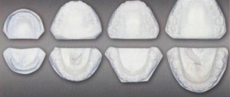
The branch of the lower jaw (ramus mandibulae) has outer and inner surfaces, anterior and posterior edges, which pass into the coronoid process (processus coronoideus) and the condylar process (processus condylaris), respectively. These processes are separated by the notch of the lower jaw (incisura mandibulae). The coronoid process serves to attach the temporal muscle, and the condylar process serves to form the temporomandibular joint (TMJ). The shape of the mandibular ramus varies individually (Fig. 1-14).
Rice. 1-14. Extreme forms of the basal arch of the lower jaw , ventral view: A - wide and short; B - narrow and long
The condylar process has a head (caput mandibulae) with an articular surface for connection with the mandibular fossa of the temporal bone and a neck (collum mandibulae). On the anteromedial surface of the neck of the condylar process there is a pterygoid fossa (fovea pterygoidea) - the attachment site of the external pterygoid muscle. The head of the articular process is flattened and occupies a position in which the axes drawn through the largest size of both heads intersect at the foramen magnum at an angle of 120-178°, open anteriorly. The shape and position of the head are individually different and depend on the operating conditions of the TMJ and the condition of its components. Deviations leading to changes in the volume and direction of movement in the joint change the shape and position of the articular heads. The anterior edge of the ramus of the lower jaw laterally passes into an oblique line on the outer surface of the jaw body, and medially reaches the posterior alveoli, thus limiting the retromolar fossa. The medial part of the ridge, formed at the site of the transition of the anterior edge into the walls of the posterior alveoli, stands out under the name of the buccal crest (crista buccinatoria), from which the buccal muscle begins.
The posterior edge of the branch passes into the base of the jaw, forming an angle (angulus mandibulae), the value of which ranges from 110 to 145° (usually 122-133°) and changes throughout life. In newborns it is close to 150°, decreases in adults with preserved teeth and maximum chewing load, and increases again in old people with complete loss of teeth (Fig. 1-15). The outer surface of the branch contains a masticatory tuberosity (tuberositas masseterica), which occupies most of the branch and the angle of the jaw and is the site of attachment of the masticatory muscle. On the inner surface of the branch in the area of the angle and adjacent sections there is a pterygoid tuberosity (tuberositas pterygoidea) - the place of attachment of the medial pterygoid muscle. On the same surface, in the middle, there is an opening for the lower jaw (foramen mandibulae), which is covered in front and above by an inconsistently pronounced bony protrusion - the tongue (lingula mandibulae). Above and anterior to the uvula is the mandibular ridge (torus mandibularis) - the place of attachment of two ligaments: the maxillary-pterygoid and the maxillary-sphenoid. The branches of the lower jaw are usually turned outward, so that the distance between the condylar processes of the right and left branches is greater than the distance between the outer points of the angles of the jaw. Can be distinguished as extreme forms of the jaw with maximally and minimally deployed branches. The degree of divergence of the branches depends on the shape of the upper half of the face. With a wide upper half of the face, the branches of the lower jaw are less developed than with a narrow upper half of the face. The smallest width of the branch, which usually falls in the middle of its height, ranges from 23 to 40 mm (usually 29-34 mm). The width and depth of the jaw notch are also individually different: the width of the notch is from 26 to 43 mm (usually 32-37 mm), the depth is from 7 to 21 mm (usually 12-16 mm). People with a wide upper half of the face usually have jaws with the greatest width of the notch and vice versa.
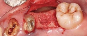
Permanent fixation of fragments
Fragments of the lower jaw can be fixed using conservative and surgical methods.
Permanent fixation can be carried out using: steel standard tape splints (Vasiliev splints), Tigerstedt dental wire splints, dental dentogingival and supragingival plastic splints, which are made in dental laboratories (they are rarely used nowadays).
In recent years, some clinics have begun to use orthodontic screws, which are inserted through the mucosa into the alveolar processes of the jaws, 3 pieces on each side, after which a rubber rod is put on the heads of the screws, providing intermaxillary fixation. This avoids the time-consuming procedure of double-jaw splinting. On the other hand, when inserting screws, the roots of the teeth can be damaged; in addition, there are often situations when the screw, under the action of a rubber rod, becomes mobile, which leads to inadequate intermaxillary fixation and removal of the screw.
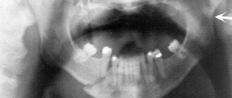
If conservative treatment methods do not produce an effect, osteosynthesis is used - this is a surgical method that, with the help of devices, allows you to fix the fragments and eliminate their mobility. Osteosynthesis is performed if the patient has comminuted fractures of the lower jaw, or the displacement of the fragments is so pronounced that they do not allow closed reduction. Osteosynthesis is also indicated in the absence or insufficient number of teeth in the patient.
Approximately 30% of patients with mandibular fractures require surgical treatment. Currently, osteosynthesis is one of the leading methods of surgical care, and is used much more often than in the past.
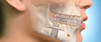
This is due both to an increase in the arsenal of technical means for performing surgery (the presence of physiodispensers, titanium miniplates), which leads to improved surgical techniques, greater predictability of results, and to the requests of patients, which are aimed at a more comfortable course of the post-traumatic period, reducing the time of intermaxillary fixation, and in some cases, complete abandonment of it.
Methods for treating fractures using surgical techniques make it possible to compare and fix mobile bone fragments in a normal anatomical position. This helps reduce treatment time and achieve early restoration of lower jaw function. However, according to various data from Russian and foreign clinics, there is also a negative aspect of using osteosynthesis - about 30% of patients experience complications after using this technique. This is due to the use of materials to hold fragments: steel, titanium, etc. Even the most bioinert alloys are not ideal. Being in bone tissue, they undergo corrosion and cause galvanosis, which negatively affects the recovery process and can cause purulent-inflammatory complications and pain reactions.
Recommendations
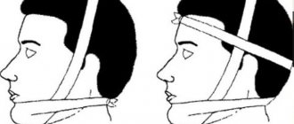
Treatment of mandibular fractures involves long-term immobilization of the jaws, which becomes a significant psychological problem for the patient. The success of treatment directly depends on how closely both the doctor and the patient act together and take the process seriously.
For the entire period of healing of the fracture, the patient is prescribed a diet (maxillary table No. 1 and No. 2); food is allowed to be taken only in a creamy consistency (well boiled and passed through a blender). In most cases, opening the mouth is impossible, because... The patient has an intermaxillary rubber traction device installed. Food is supplied through tubes, tubes and sippy cups.
From the moment of double-jaw splinting or osteosynthesis, the splints are left in place for an average of 21–30 days. If the doctor has confidence in the favorable course of the recovery process, then it is possible to reduce the period of wearing the intermaxillary rubber traction.
Even after removing the splints, the patient cannot fully open his mouth for 1-2 weeks. To restore the function of the chewing and facial muscles, he is prescribed myogymnastics.
Metal Kirschner spokes
This type of wire was first used to treat fractures of the lower jaw in 1933. Intraosseous insertion of these wires can be carried out both percutaneously (without incisions) and with soft tissue incisions.
In 1975 V.V. Donskoy used an original technique, with which he inserted a wire into the branch of the lower jaw through the mucosa without an incision, then carried out a reposition, and fixed it like a splint to the teeth or to a splint. Later, in 1988, Deryabin E.I. and Osipov V.Yu., and Yu.G. Kononenko and G.P. Ruzin proposed modifications of this method in 1991. Today there are many techniques where the wire suture is combined with knitting needles, staples, surrounding wire ligatures, etc.
Bone or wire suture
In 1825, surgeon Rogers from Dublin performed the world's first operation using silver wire. With its help, he connected the fragments of the lower jaw. Later, in 1863, Russian surgeon Yu.K. Szymanowski successfully used a bone suture. From this point on, the bone suture was successfully used for osteosynthesis for many years. The main material was initially stainless steel, later it was replaced by titanium, nichrome, tantalum, etc.
There are various modifications of the bone suture (loop-shaped, cruciform, figure-of-eight, trapezoidal, double, etc.). The choice depends on the nature and location of the fracture.
The application of a bone suture occurs according to certain rules. It is important to make holes for the material in those areas where damage to the mandibular canal and tooth roots is excluded, and no closer than 1.0 cm from the fracture line. Ideally, the suture should cross the fracture line in the middle of the distance between the edge of the mandible and the base of the alveolar process.