- Causes and risk factors
- Localization
- Metastases
- Symptoms of Oral Cancer
- Stages of the disease
- Treatment
- Prognosis and survival
- Prevention
Malignant tumors of the oral cavity account for about 6% of all cancers in general. Based on the histological structure (type of tumor-forming cells), the following types are distinguished:
- Tumor of epithelial cells - cancer
- Tumor of connective tissue cells - sarcoma
- Melanoma
Each type includes several varieties.
Several groups of precancerous diseases are also distinguished separately. Precancerous diseases of the oral cavity are divided into:
- Obligate - with a high incidence of malignancy. These include Bowen's disease, warty precancer, limited hyperkeratosis, Manganotti cheilitis.
- Optional - with a lower incidence of malignancy. This group includes the verrucous form of leukoplakia, papillomatosis, erosive-ulcerative and hyperkeratotic forms of systemic lupus erythematosus and lichen planus, post-radiation stomatitis and post-radiation cheilitis, keratoacanthoma.
Among malignant tumors of the oral cavity, cancer is the most common.
Oral cancer, in turn, is divided as follows.
- Intraepithelial carcinoma (Carcinoma in situ, cancer in situ) is characterized by the absence of invasion into the basement membrane, despite the malignancy of the neoplasm.
- Squamous cell carcinoma is the most common.
Causes and risk factors
The causes of the development of malignant neoplasms of the oral cavity can be divided into local and general risk factors.
Common factors include age, a history of various hazards (exposure to radiation, etc.), and hereditary predisposition.
Local factors are local factors affecting the oral cavity. These include chewing nasvay (tobacco- and drug-containing mixtures), smoking, the habit of drinking scalding hot drinks, chronic traumatization of the mucous membrane (tooth fragments, deformed dentures), as well as the presence of precancerous diseases. A separate risk factor for detecting the disease in later stages is the lack of annual dental examinations.
It is the neglect of preventive visits to the doctor that prevents the diagnosis of cancer at early, treatable stages, detection and treatment.
Localization
Oral cancer is usually classified by location. This is due to the fact that the area under discussion includes a large number of anatomical formations with significant diversity.
When choosing treatment tactics and the type of operation, the position of the tumor in the mouth plays a significant role. Different parts of the oral cavity are innervated differently, have different blood supply, and have different functional significance, so the prospects for treating absolutely identical tumors located in different places can differ significantly.
Based on location, oral cancer is usually divided into:
- Cheek cancer
- Floor of mouth cancer
- Tongue cancer
- Cancer in the alveolar ridge area
- Palate cancer
- Gum cancer
Cancer of the buccal mucosa
Cancer of the buccal mucosa ranks second in frequency (after cancer of the tongue) in the structure of oral cancer. Local factors, chemical and physical agents that cause chronic trauma to the mucous membrane have a significant influence on the increase in risk. To a greater extent than with cancer of other areas, such a predisposing factor as chronic traumatization by dentures and sharp edges of damaged teeth is relevant.
Floor of mouth cancer
This type of tumor accounts for 10-15% of all oral cancers. The floor of the mouth is formed by the structures between the tongue and the hyoid bone. The mucous membrane lining the bottom of the mouth has a developed submucosa, consisting of loose connective tissue and fiber. This area is richly supplied with blood. All this creates favorable conditions for tumor growth, spread and metastasis.
Tongue cancer
Tongue cancer is the most common type of oral cancer. The tongue is a mobile organ with a large number of nerve endings (receptors). Thanks to this, patients, as a rule, pay attention to the tumor that has arisen and have the opportunity to seek help in a timely manner. A developed network of blood and lymphatic vessels contributes to early tumor metastasis, primarily to peripheral lymph nodes.
Cancer in the alveolar ridge area
Cancer in this area develops either from mucosal cells or from the epithelial islets of Malasse. Epithelial islands of Malasse are the remains of epithelial cells in the thickness of the periodontium. Normally, these cells do not manifest themselves in any way, but under unfavorable conditions they can become a source of tumor. A distinctive feature of these tumors is the relatively early onset of symptoms, the teeth in the tumor growth area are exposed to it, and the patient begins to complain of pain.
Palate cancer
Palate cancer is rare. The hard and soft palates are separated, therefore the histological types of tumors of the soft and hard palate are different. Cylindromas and adenocarcinomas are more typical of the hard palate; the soft palate is more susceptible to squamous cell carcinoma.
Squamous cell carcinoma (SC) accounts for more than 90% of all malignant tumors of the oral mucosa (MOT) [1], this localization accounts for 2% of newly diagnosed malignant tumors per year [2]. PR ORS is considered a serious problem in some countries due to its incidence and high mortality rate [3].
The incidence of cancer of the lip, oral cavity, and oropharynx is rapidly increasing worldwide. The GLOBOCAN (Global Cancer Statistics) report shows that PR SOR is in 11th place in terms of frequency of occurrence and has a worse prognosis compared to other cancers [4]. If the oropharynx region is taken into account, then this malignant tumor is in 6th place in terms of frequency of occurrence in the world [1].
On average, 4.0 cases are detected per 100 thousand people per year and the mortality rate is 1.9 cases. A high incidence rate is observed in South Asia (India, Pakistan, Sri Lanka, Taiwan) with an incidence of more than 10 cases per 100 thousand population per year. The disease is also frequently detected in Eastern and Western Europe (Hungary, Slovakia, Slovenia and France), Latin America and the Caribbean (Brazil, Uruguay and Puerto Rico), and Melanesia (Papua New Guinea). PR SOR is more common in men (5.5 cases per 100 thousand people) than in women (2.5 cases per 100 thousand people). However, the opposite situation is observed in the ratio between men and women in India (1:2) and Thailand (1:1.56). Mostly, PR SOR occurs in people aged 50 to 70 years [1]. Despite the fact that in most cases this pathology is diagnosed in elderly men, there is evidence that recently more and more young non-smoking women are susceptible to this disease [5, 6].
Smoking is the most important cause of PR SOR [1]. It is known that PR OR is registered 5–9 times more often in smokers compared to non-smokers and 17 times more often in those who smoke 80 cigarettes or more per day [4].
Alcohol abuse enhances the negative effects of nicotine [1]. Those who drink more than 100 g of alcohol per day have a 30-fold higher risk of developing OP, and if they drink alcohol less frequently, the risk is 3-9 times higher [4]. Smokeless tobacco is also widely used throughout the world as chewing or snuff. In this form, it also leads to the development of PR SOR, however, some studies (the effect of Swedish snuff) have shown that there is no risk of malignancy. Betel nut and/or tobacco are often mixed with other substances: slaked lime, betel buds, sweeteners, spices. Betel nut increases the risk of ADR, regardless of whether tobacco is added to it or not. Sunlight exposure is known to be a risk factor for lip cancer. In Western Australia, lip cancer accounts for the same number of cases as oral cancer. Failure to comply with oral hygiene also leads to the development of oral oral cavity, but it has not been proven that this fact is an independent risk factor. Vitamins and antioxidants can counteract the formation of PR. Thus, eating a large amount of fruits and vegetables helps prevent the development of PR SOR.
Over the past decades, many studies have been conducted aimed at studying the PR of SOR. The 2022 WHO classification displays modern data on the epidemiology, etiology, localization, clinical manifestations, morphological picture, genetic profile and prognosis of the course of PR.
Our article addresses the changes in the sections concerning epithelial tumors and preneoplastic conditions that were made in Chapter 4 “Tumors of the oral cavity and body of the tongue” of the 2022 WHO Classification of Tumors of the Head and Neck compared to the 3rd edition published in 2005. In the previous edition, tumors of the oral cavity and oropharynx were discussed in the same chapter. In the new edition, diseases of these anatomical regions are described in separate chapters [1]. The tongue consists of 2 parts: the body (free part) and the root, which differ in embryogenesis; therefore, tumors of the body of the tongue are classified as oral cavity, and tumors of the tongue root are classified as oropharynx [7]. Tumors of the oral cavity and body of the tongue are described in Chapter 4, and tumors of the tongue root are described in Chapter 5 “Tumors of the oropharyngeal region” (root of the tongue, tonsils). To avoid repetition, Chapter 4 describes only some non-epithelial tumors and tumors of soft tissues, salivary glands and tumors of the hematopoietic system. As a result, the content of this chapter has been reduced compared to the previous edition.
The pathology that deserves primary attention in Chapter 4 is diseases of the oral cavity. The most relevant malignant tumor of the oral cavity and body of the tongue is PR. It is known that it can develop from potentially malignant diseases (PPDs). Such diseases clinically carry a risk of cancer and epithelial dysplasia (ED).
PZDs include: erythroplakia, leukoplakia, submucosal fibrosis of the oral cavity, congenital dyskeratosis, tobacco keratosis in non-smokers, damage to the palate due to smoking an inverted cigarette, chronic candidiasis, lichen planus, discoid form of systemic lupus erythematosus, syphilitic glossitis, actinic keratosis (lips).
The most common disease is leukoplakia. In Western countries, it occurs, according to various sources, in 1-4% of the population. In Southeast Asia, the number of people with leukoplakia SOP is higher. Worldwide, this disease occurs in 2-3% of the population. For comparison: erythroplakia SOP is observed less frequently - in 0.02-0.83%. This disease is mainly found in men than in women. Other PZDs are more common than erythroplakia, but very rarely transform into cancer.
The reasons for PZZ SOR are various. Tobacco use (smoking or chewing) and alcohol abuse have been linked to the development of some types of leukoplakias. Chewing betel nut with or without tobacco results in oral submucosal fibrosis. For many PZD SOR, the etiology is unknown. High-risk HPV is rarely detected in PZZ SOR and its role in the malignant transformation of the epithelium has not been fully proven.
PZD can occur in different areas of the oral cavity depending on its etiology, age and gender of the patient. Erythroplakia most often develops on the soft palate, in the area of the floor of the mouth and the buccal mucosa; lichen planus is characterized by localization on the buccal mucosa, and actinic keratosis is localized on the lip mucosa.
PZZs with a high risk of malignancy appear as red, white, or mottled areas on the GOR. Leukoplakia is a clinical term used to describe white plaques with a questionable risk of malignancy after excluding any other diseases, which usually requires a biopsy. Leukoplakia may present as homogeneous white lesions or predominantly white lesions with nodules, verrucous growths, or erythematous areas (erythroleukoplakia or variegated leukoplakia). Erythroplakia SOP has a similar definition to leukoplakia, but it is red in color. With it, the development of ED is possible.
PDD occurs in rare diseases such as Falconi's anemia and dyskeratosis congenita, but no genetic predisposition has been found.
The new WHO classification describes the HPV-positive PZD subgroup SOP, which is characterized by epithelial hyperplasia, severe karyorrhexis and apoptosis in all layers of the epithelium. According to existing criteria, these changes are regarded as severe ED, but the risk of its malignant transformation has not yet been proven.
The risk of malignancy in many PZDs is low, and they often regress. With leukoplakia, malignant transformation occurs in 1-2% of cases. In the presence of ED, PZZ is transformed into PR SOR in 12% of cases.
ED GOR is a series of architectural and cytological changes in the epithelium that are caused by the accumulation of genetic abnormalities and are associated with an increased risk of transformation into ED GOR.
Synonyms for ED are: precancer, intraepithelial neoplasia, squamous intraepithelial neoplasia. ED involves impaired proliferation, maturation, and differentiation of epithelial cells. The epithelium may be atrophic, with acanthosis, keratinized or non-keratinized. With leukoplakia, ED is rarely detected, but with erythroplakia or erythroleukoplakia it is a constant finding.
Signs of ED include changes in the histoarchitecture of tissues, such as a violation of the row of cells, the disappearance of the polarity of the cells of the basal layer, acanthosis, an increase in mitotic figures, the presence of mitosis in the superficial layers of the epithelium, premature keratinization of cells, keratin “pearls” in the area of acanthosis, loss of intercellular contacts and structure cells: cellular and nuclear polymorphism, increased nuclear-cytoplasmic ratio, the presence of pathological mitoses, increased number and size of nucleoli, hyperchromasia. The number and combination of features may vary. The WHO classification indicates that currently there are no symptoms that reliably distinguish hyperplasia from moderate dysplasia. ED is diagnosed only on the basis of tissue and cellular signs.
Traditionally, ED is divided into 3 degrees. Depending on the number of affected thirds of the epithelium, the corresponding degree of dysplasia is determined. A low degree of dysplasia is characterized by the presence of cellular atypia in the lower third; with a medium degree, cellular atypia extends to the middle third; and with a high degree of dysplasia, atypical cells are located in the upper third of the epithelium. Carcinoma in situ is considered synonymous with high-grade dysplasia.
In practice, it is often difficult to determine the degree of ED. Some studies show a good predictive value for determining the degree of ED, while others indicate a weak association with outcome. It is more reliable to assess the degree of dysplasia after reviewing the biopsy by several pathologists. For ease of diagnosis, some authors propose a binary system by analogy with diseases of the larynx, where low and high levels of ED are distinguished. However, such a system should be further studied before being widely applied to SOPs.
The 4th edition of the WHO classification has added a recently described type of ED associated with high-risk HPV with a characteristic histological structure, however, the acceptance of this type of ED requires research into the risk of its malignant transformation. This ED involves the entire thickness of the epithelium, there are signs of apoptosis and karyorrhexis with pronounced staining of the nucleus and cytoplasm at P16 and detection of high-risk HPV by in situ hybridization [1].
There are more than 200 types of HPV: 16, 18, 31, 33, 35, 39, 45, 51,52, 56, 58, 59, 66 and 68 have a high oncogenic risk. Tumorogenesis is known to be initiated by two proteins that alter key signaling pathways that suppress tumor growth: E6, which binds the tumor suppressor protein P53, and E7, which interacts with proteins of the retinoblastoma family (pRb). Multiple lesions of these signaling pathways lead to genomic instability and, over time, to malignant transformation [8, 9].
It is believed that HPV types 16 and 18 are independent risk factors for the development of PR SOR. According to the literature, HPV type 16 leads to CR in 14.9% of cases, and HPV type 18 - in 5.9% [4]. WHO considers HPV type 16 to be one of the etiological factors, which is only 3% the cause of PR SOR [1].
The most common localization of a malignant tumor with HPV is the root of the tongue and tonsils, since these areas are rich in lymphoid tissue, which is more sensitive to the virus. Mostly, oropharyngeal cancer occurs in men; the disease is detected at a late stage, when the tumor has metastasized to the cervical lymph nodes [8].
WHO experts do not recommend using P16 as a biomarker to determine HPV status in ED and PR SOR. Studies have shown that a third of cases of PR SOP are P16-positive, viral DNA is detected by PCR in up to 28% of cases. However, with in situ hybridization, a more sensitive method for detecting high-risk HPV, only 1 to 10% of cases were positive for this marker. Therefore, when focusing on this marker, the number of PR SOR associated with HPV will be exaggerated. It has been shown that the survival of patients with P16-positive PR SOR does not differ from that with P16-negative PR [1].
PR can develop in any area of the oral cavity. The most common localizations are the tongue, the floor of the mouth and the gingival mucosa; they account for the majority of all PR SOR. In 1st place in terms of frequency of occurrence is PR of the tongue and accounts for more than 50% of cases [10, 11]. More than half of the cases of tongue damage occur at the root [12].
Small tumors may be asymptomatic, unlike large tumors, which are accompanied by symptoms such as discomfort, pain, decreased tongue mobility and irritation when wearing dentures. The macroscopic picture can be varied; white, erythematous, mixed, nodular changes on the mucosa are possible, as well as ulcers, the edges of which in most cases are raised. A non-healing ulcer is the most suspicious sign of malignancy, but recent studies have shown that this sign is observed in less than half of cases of PR SOR. With cancer of the lower lip, crusts most often form after actinic cheilitis. If PR SOR is suspected, examination of the cervical lymph nodes is also necessary.
Macroscopically, PR is a dense tumor on palpation with infiltrating growth; on a section it has a brown or white color. When diagnosing PR SOR, additional studies cannot replace a biopsy followed by histological examination. Fine needle aspiration biopsy is useful for identifying lymph node metastases. The punctate reveals lymphoid cells with layers and small accumulations of tumor epithelial cells with intra- and extracellular keratinization; inflammatory reaction cells and necrotic detritus are also possible.
Most PRs of the oral cavity and tongue have a medium or high degree of differentiation; low-differentiated PRs are rare. Highly differentiated PRs are characterized by cells, cords and islands consisting of large cells with pink cytoplasm, pronounced desmosomes and round nuclei, which may not be hyperchromatic. Dyskeratosis and cancerous “pearls” are detected. As the tumor develops, cellular and nuclear polymorphism, nuclear hyperchromasia and mitoses (including atypical ones) become more pronounced. There is no correlation between the degree of tumor differentiation and prognosis. In poorly differentiated PRs, signs of differentiation of stratified squamous epithelium are minimal or absent, therefore, to clarify the type of cells, an IHC study with markers suitable for these purposes is necessary. Infiltrating growth in highly differentiated tumors is represented by large areas, and in less differentiated tumors it can be in the form of saw-tooth or finger-shaped outgrowths, small islands or single tumor cells located diffusely. In the zone of infiltrating growth around the islands of tumor cells, an inflammatory reaction is observed in the stroma. In the adjacent mucosa, ED is often observed at different stages. Perineural and lymphatic invasion is more often observed in poorly differentiated tumors.
There are several histological variants of PR SOR:
- basaloid cancer is highly differentiated, more often gives metastases, in general the prognosis is the same as with conventional PR;
- glandular squamous cell carcinoma is an infiltrating and aggressive tumor, often metastasizes, the prognosis is worse than with conventional PR;
- spindle cell carcinoma - the prognosis is worse than with conventional PR, tumor relapses after radiation therapy and a second primary tumor are typical;
‒ carcinoma cuniculatum is a highly differentiated cancer, usually localized on the mucous membrane in the periosteum area. This tumor is characterized by locally destructive growth with the formation of fistulas and sinuses filled with pus. This tumor recurs but does not metastasize;
- verrucous carcinoma is a well-differentiated tumor, does not metastasize, has shallow invasion, grows exophytically, atypia is not expressed, has a good prognosis, can progress to ordinary invasive cancer;
- lymphoepithelial carcinoma, rare, occurs in late stages, in 70% of cases is associated with metastases in regional lymph nodes and with the Epstein-Barr virus;
- papillary (papillary) PR can be of keratinized or non-keratinized type, often localized on the gum, the prognosis is better than with conventional PR;
- acantholytic PR is a variant of skin lesions that can occur on the lips; acantholysis sometimes manifests itself as adenoid growths in poorly differentiated PR SOR [1].
Due to the fact that the basaloid and glandular-squamous variants of PR are the most aggressive, their more complete characteristics are given below.
Basaloid PR is a rare and aggressive form of PR SOR. It is assumed that it develops from totipotent cells capable of heterogeneous differentiation, localized in the basal layer of the epithelium, or from the epithelium of the minor salivary glands. Most often, the tumor develops on the root of the tongue and in the floor of the mouth. Histologically, it consists of solid epithelial structures of a basaloid type with signs of malignancy. There are different types of growth of this tumor: solid lobular, cribriform, trabecular, cellular and glandular, or cystic. The aggressive course of this disease can be explained in some cases by the presence of distant metastases in the liver and lungs. Some authors believe that the prognosis for the basaloid form of PR SOR is worse than for the usual variant of the tumor; the 3-year survival rate is 53%, and the 5-year survival rate is 32% [13].
Glandular squamous cell carcinoma is an aggressive malignant epithelial tumor that is rare in the SOR. This disease occurs 2 times more often in men than in women. The prognosis is poor due to the high incidence of cervical lymph node metastasis and hematogenous spread, with a 5-year survival rate of 13–50%. Histologically, glandular squamous cell carcinoma consists of components of squamous cell carcinoma and adenocarcinoma. Despite the fact that glandular squamous cell carcinoma is included in the name of the tumor, this type of cancer is considered as a rare variant of PR, and not as an independent nosology. These 2 components are located very close to each other and are clearly distinguishable; there may be areas where they are mixed. The squamous component is identical to the usual PR and can have varying degrees of differentiation, the glandular component has a tubular/ductal structure with mucin inside the duct or cells, it is usually located deep, while the squamous component is superficial [14].
Most PR SOPs are genetically variable. Deletions in chromosomes 3, 8, 9, 17 and duplications in chromosomes 3 and 11 are possible. These changes may not have clinical manifestations, substantiating the phenomenon of the field of cancerization. Genes that are important in the development of PR SOR (TP53, CDKN2A, PTEN, HRAS, PIC3CA) mutate quite often, this confirms their possible role in the development of this disease. There is no reliable evidence that the predisposition to PR SOR is inherited. PR SOR may be a symptom of Li-Fraumeni syndrome or Fanconi anemia.
It is difficult to predict the course of PR SOR, since it is an aggressive disease with a tendency to local invasion and early metastasis to the lymph nodes [1]. The presence of metastases in the cervical lymph nodes is considered the most significant prognostic factor when assessing the 5-year survival rate of patients with PR SOR, which is 75% in patients without metastases in the cervical lymph nodes. In patients with metastases in 1, 2 or 3 lymph nodes or more, the 5-year survival rate is 49, 30 and 13%, respectively [1]. Most often, PR of the tongue metastasizes to regional lymph nodes, but extremely rarely metastases are possible in the myocardium [10] and in the vertebrae (2 cases of metastasis of PR of the tongue to the lumbar vertebrae and 4 cases to the cervical vertebrae are described).
Hematogenous metastases of PR SOR are most often localized in the lungs [12]. It is known that in patients treated in the early stages of PR SOR (T1-T2N0), the 2-year survival rate is 85% in the absence of metastases in the cervical lymph nodes and 20-30% in their presence [16].
The degree of tumor differentiation has little correlation with clinical outcome. For example, histological features associated with poor prognosis include a pattern of invasive growth, perineural and lymphovascular tumor invasion, and tumor thickness greater than 4 mm [1]. It has been established that an invasion depth of 4 mm or more is a factor that reliably indicates the occurrence of local relapse [17, 18]. To predict tumor recurrence, the histological picture of the edges of the resected area is also more informative than the tumor itself. High-grade dysplasia also correlates with tumor recurrence [1].
Patients with stage 4 PR SOR who are married have a longer life expectancy [7]. Patients (80%) with PR SOR identified at stages 1 and 2 undergo surgical treatment. Much discussion is devoted to how wide and deep the tissue around the tumor should be excised. In accordance with the recommendations of the Royal College of Pathologists of Great Britain, these figures are 5 mm [19]. Surgical treatment can lead to serious aesthetic and functional consequences that significantly affect the quality of life. Patients after surgical treatment may experience speech and swallowing problems, deterioration in appearance, sensory disturbances, or chronic pain. All this can subsequently negatively affect the patient’s mental state [1].
Thus, compared to the Third Edition of the 2005 WHO Classification of Tumors of the Head and Neck, changes have been made to Chapter 4, “Tumors of the oral cavity and body of the tongue,” of the Fourth Edition. The most significant change is the exclusion from this chapter of oropharyngeal tumors, which are separated into a separate, 5th chapter “Tumors of the oropharyngeal region (root of the tongue, tonsils)”, where a division is made between squamous cell carcinoma with a positive and negative reaction to HPV. It has been shown that patients with HPV-positive PR have a better prognosis of the disease, which confirms the clinical distinction between oropharyngeal and oral tumors. These changes reflect the significant impact of oropharyngeal carcinoma and the role of high-risk HPV in the development of head and neck malignancies since the previous edition. The most significant reductions in the number of nosologies are observed in the sections of tumors of hematopoietic tissue and tumors of the salivary glands, since this pathology is discussed in other chapters.
The author declares no conflict of interest.
The author declare no conflicts of interest.
Symptoms of Oral Cancer
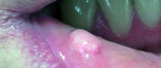
Manifestations of the disease depend on the nature of the tumor and the location. Typically, the affected area is either a local ulceration or an induration (bulge, nodule). The patient feels pain and discomfort in the corresponding area. Warning signs include the following:
- A long-term, non-healing mouth ulcer. The oral cavity is rich in blood vessels, the mucosal epithelium is well supplied with blood, and normally, in the event of injury, rapid healing occurs. Any damage to the oral mucosa heals faster than a similar injury to the skin of the arm or leg. Therefore, if there is an ulcer in the mouth and for unknown reasons it does not heal for a long time, you should seek medical help.
- Lumps, “balls” under the mucous membrane, regardless of the area of the mouth where they arose. A compaction under the mucosa can be a symptom of both a non-oncological pathology (for example, a cyst of one of the small glands) and a sign of cancer. Only a specialist can distinguish one from the other. The seal, which initially appeared as a cyst, may subsequently become malignant if left untreated.
- Impaired swallowing, chewing, discomfort at the root of the tongue - all this is a reason to suspect cancer in the corresponding area.
- Toothache and bleeding in a specific area of the gum may be signs of cancer. And, although similar symptoms are caused by much less formidable ailments, consultation with a dentist is necessary. Thus, any violation of the anatomical integrity or functionality of the organs of this zone carries a potential threat, and this is a reason to see a doctor.
Clinical manifestations
Symptoms of oral cancer may include:
- compaction and swelling of the affected area;
- long-term non-healing ulcer;
- white or red spots on the mucous membrane;
- weakening of tooth roots;
- bleeding gums;
- speech disorder;
- unexplained weight loss;
- soreness in the mouth;
- general malaise.
Most of these symptoms are nonspecific and are observed in chronic diseases of the dental system, and therefore are often ignored by the patient.
Stages of the disease
Stage I is characterized by the presence of a tumor up to 1-2 cm in diameter, not extending beyond the affected area (cheek, gums, palate, floor of the mouth), limited to the mucous membrane. Metastases are not detected in regional lymph nodes.
Stage II - a lesion of the same or larger diameter, which does not extend beyond any one part of the oral cavity, but extends into the submucosal layer. There are single metastases in regional lymph nodes.
Stage III - the tumor grows into the underlying tissues, but not deeper than the periosteum of the jaw, or has spread to adjacent parts of the oral cavity. In regional lymph nodes there are multiple metastases measuring up to 2 cm in diameter.
Stage IV - the lesion spreads to several parts of the oral cavity and deeply infiltrates the underlying tissues, in the regional lymph nodes there are immobile or disintegrating metastases, and the presence of distant metastases is also characteristic.
Classification by stages is periodically subject to revision; you can find a division of stages into subtypes - A and B. Currently, classification by stages is used less and less, the TNM classification is more relevant. Its principle is that this coding indicates the characteristics of the tumor itself, the condition of the closest (regional) lymph nodes, and the presence or absence of distant metastases.
In the diagnosis, the oncologist indicates the histological type of cancer, since different types of cells differ in growth rates, tendency to metastasize, and sensitivity to treatment. All types of classifications serve one purpose - to correctly assess the extent of the spread of the disease, the degree of damage and develop the right tactics of assistance.
Book a consultation 24 hours a day
+7+7+78
Diagnostics
Most often, the disease can be diagnosed only at the stage of metastasis due to the erased symptoms at the beginning of the disease.
X-ray diagnostics
Direct and lateral radiography of the jaw helps to detect a tumor in the bone. A neoplasm of odontogenic cellular material is also clearly visible on an x-ray. If there is a tumor, the image will show that the teeth are not in contact with the bone, and the alveolar edge does not have a clear enough contour.
General clinical examination
A patient with suspected jaw cancer must undergo a general blood test, urine test, and biochemical tests.
CT scan, biopsy, other diagnostic methods
To get an idea of the location and distribution of the tumor, a computed tomography scan of the jaws is done. A puncture biopsy of the submandibular lymph nodes and positron emission tomography are needed to detect metastases.
The most accurate diagnosis can be made by histological examination of the tumor material. Depending on the nature of the cancer cells, the analysis can be taken through the socket of a lost tooth or through trepanation of the jaw.
Additionally, you may need to consult an ophthalmologist, otolaryngologist, dentist, or surgeon.
Treatment
The following factors are important for optimal treatment:
- Timely appeal. The factor of advanced disease plays a huge role. Even now, when doctors have learned to successfully fight the disease even in later stages, the final prognosis in most cases is determined by how quickly the patient turned to an oncologist.
- High-quality diagnostics. There are many research methods that allow you to more accurately establish a diagnosis. In oncology, ultrasound, MRI, CT, PET-CT, as well as histological studies are widely used.
- Good contact between the patient and the treating doctor. The doctor’s ability to explain to the patient the essence of what is happening, support him and correctly organize joint work, as well as the patient’s ability to be disciplined, trust the specialist and not delay in carrying out the prescribed studies or treatment procedures.
For oral cancer, surgical treatment, chemotherapy, and radiation therapy (including brachytherapy) are used.
Surgery
The operation is a classic treatment tactic in oncology in general and in oral oncology as well. The difficulty of using oral surgery is that in some cases it is technically difficult to remove the tumor without damaging vital anatomical structures.
The doctor takes into account the following important points:
- The mouth area has a rich blood supply and is at risk of active bleeding.
- The organs and tissues of the mouth area are mobile, and the possibility of this displacement relative to each other determines such important functions as speech, swallowing, etc.
- The anatomical formations of this zone are small in size and closely adjacent to each other. In this area there are few ballast, functionally insignificant tissues; for every centimeter there are the most important vessels, nerves, and organs. This complicates the operation process. The lack of sufficient tissue makes it more difficult to close the defect created after tumor removal.
As in the case of radical surgical treatment of tumors in other areas, during operations on the oral cavity the following conditions are sought to be met:
- Removal of the tumor must occur within healthy tissues; the cut does not pass at the border of healthy and diseased tissue, but along healthy tissue, that is, the tumor is excised as if with a reserve. When the organs are close together, this poses a problem.
- When excision of a tumor, the importance of further restoration of the integrity of the integument is taken into account, as well as, if possible, preservation of function.
- When excising the tumor, in most cases, peripheral lymph nodes are also removed. This is because the lymph nodes closest to the tumor are a trap for metastases and can often contain tumor cells.
In classical oncology, surgical treatment was the method of choice; operations were used, as a rule, immediately after diagnosis. Currently, the approach has changed a little and has become more differentiated. Sometimes surgery is preceded by chemotherapy and/or radiation therapy.
Chemotherapy
A feature of tumor cells is rapid growth and division. This is what chemotherapy is based on. Special substances that are toxic to cells and inhibit their growth are introduced into the body. Chemotherapy affects the entire body, but most cells are insensitive to the substance, and the chemotherapy is harmful to tumor cells.
Chemotherapy is used as an addition to surgery, before or after it, as an independent method, and in combination with radiation therapy.
The appropriateness of chemotherapy is determined by the histological type of tumor. The type of drug is selected taking into account the sensitivity of a particular neoplasm to various types of relevant drugs.
The disadvantage of chemotherapy is its side effects. The body has a number of healthy cells that are frequently renewed and, accordingly, quickly divide. These are cells of the gastrointestinal mucosa, blood cells and others. Chemotherapy drugs affect them. The blood formula changes, the cells lining the stomach and intestines die. The result is nausea, stool upset, weakness, and decreased immunity. These phenomena are temporary and, as a rule, surmountable; however, the use of chemotherapy requires constant medical supervision.
Radiation therapy
The method is based on the damaging effect of X-ray radiation on cells, primarily on actively dividing cells, such as the cells of malignant tumors. Radiation therapy can be used as an independent method of treatment, or in combination with surgical methods and chemotherapy. Before the start of the course, markings are made to determine the direction of impact and ensure maximum impact on the tumor itself and minimal impact on surrounding tissues.
Brachytherapy is a subtype of modern radiation therapy, when a radiation source is introduced directly into the affected area. Brachytherapy allows you to treat the tumor with maximum doses of radiation and reduce the damaging effects on healthy tissue.
Prevention
Cancer can be prevented by following a number of recommendations from experts. First of all, it is important to properly care for your oral cavity and visit the dentist in a timely manner. The doctor keeps your teeth clean and monitors the condition of your gums and soft tissues twice a year.
Since HPV is a significant factor in the development of oropharyngeal cancer, it is important to reduce the risk of infection. To do this, it is necessary to exclude promiscuity and get a vaccine.
Experts recommend giving up bad habits and completely quit smoking. Immediately after eating, you should rinse your mouth and brush your teeth twice a day. A balanced diet is the key to health, so you should consume more vegetables and fruits every day, eat whole grain bread, and it is better to avoid processed meat, legumes and chicken.
Bibliography:
- American Society of Clinical Oncology. Oral and Oropharyngeal Cancer: Screening. 09/2017. Accessed at www.cancer.net/cancer-types/oral-and-oropharyngeal-cancer/screening on February 21, 2022.
- National Cancer Institute. Oral Cavity, Pharyngeal, and Laryngeal Cancer Screening (PDQ®)–Health Professional Version. February 9, 2022. Accessed at www.cancer.gov/types/head-and-neck/hp/oral-screening-pdq#section/_1 on February 21, 2022.
- International Union Against Cancer (UICC). Lip and Oral Cavity. In: UICC TNM Classification of Malignant Tumors, 7th Edition. 2009:328.
- National Comprehensive Cancer Network, Clinical Practice Guidelines in Oncology (NCCN Guidelines®), Head and Neck Cancers, Version I. 2022 - February 15, 2022. Accessed at www.nccn.org/professionals/physician_gls/pdf/head-and-neck .pdf on February 21, 2022.
- American Cancer Society. Facts & Figures 2022. American Cancer Society. Atlanta, Ga. 2022.
- American Cancer Society. Cancer Facts & Figures 2022. Atlanta, Ga: American Cancer Society; 2022.
- National Cancer Institute. Cancer Stat Facts: Oral Cavity and Pharynx Cancer. April 2022. Accessed at https://seer.cancer.gov/statfacts/html/oralcav.html on February 22, 2018.
- National Cancer Institute. Lip and Oral Cavity Cancer Treatment (Adult) (PDQ®)–Patient Version. January 29, 2022. Accessed at www.cancer.gov/types/head-and-neck/patient/adult/lip-mouth-treatment-pdq on February 22, 2022.
- Squamous cell carcinoma of the head and neck: new avenues of treatment? // Braunschweig1 , A. Lewandrowski1 , D. Smeets1 , MV Bolotin2
- Bolotina L.V., Vladimirova L.Yu., Dengina N.V., Novik A.V., Romanov I.S. Practical recommendations for the treatment of head and neck tumors. Malignant tumors. 2022.
Prognosis and survival
The prognosis of the disease depends on the stage, localization, and timely provision of assistance. In the early stages, with adequate treatment, the disease can almost always be overcome. When contacting a doctor was not timely, the prognosis is somewhat worse, and the percentage of cured patients is lower. In assessing the prognosis, the presence or absence of distant metastases is important. Despite enormous progress in oncology, distant metastases sharply reduce 5-year survival; this is a significant problem for a specialist. In addition, the prognosis depends on the histological type of the tumor, because The growth rate of different types of tumors can vary significantly. The age of the patient, the location of the tumor, the presence of concomitant diseases in the patient, the chosen treatment tactics - all these factors significantly influence the prognosis.
Chemotherapy and tooth extraction
Teeth that cannot be restored are subject to removal. Although patients receiving chemotherapy have a weakened immune system, the consequences of refusing to remove an inflammatory tooth are much more serious than the side effects of anticancer therapy. It is advisable to remove teeth before starting chemotherapy.
in dental treatment
is taken into account, the doctor takes into account the patient’s condition when choosing an intervention technique. Treatment by a dentist during chemotherapy is not advisable, but if there is an emergency and the patient is already taking chemotherapy, the intervention of a doctor will avoid big problems. Removal should be done as atraumatically as possible.
Dental problems and dental treatment for oncology
After chemotherapy, many are faced with the fact that they develop caries, gingivitis, stomatitis, periodontal disease, cysts begin to form, gums become more sensitive and bleed.
This condition creates additional difficulties for the dentist, but does not mean that you need to refuse dental treatment, cancer
not prevent. It is important to tell your dentist about your diagnosis and that you are currently undergoing treatment for cancer. This will allow him to choose the most gentle methods for solving dental problems.
You need to know that when treating teeth, oncology
does not progress and the disease itself is not a contraindication to dental intervention. Dental treatment will remove the source of infection, prevent its further spread, and defeat cancer.