20.07.2018
The author of the article is Dakhno L.A. Candidate of Medical Sciences, dental surgeon, radiologist.
Post-traumatic neuropathy of the trigeminal system is a sensory disturbance with or without neuropathic pain, often leading to functional and psychological consequences.
The trigeminal nerve is the largest sensory nerve in the body and is responsible for the orofacial region. Iatrogenic injuries of the trigeminal nerve (trigeminal nerve injuries -TNI) lead to pain in 70% of patients, which in turn leads to functional impairments in speech, eating, kissing, shaving, applying makeup, brushing teeth, etc., which means there is a negative impact on self-esteem, quality of life and patient psychology.
It must be understood that after injury to the trigeminal nerve, complete recovery is rare, except in cases of minor injury, so it is very important to maintain a trusting relationship between the dentist and the patient and not give false assurances of a complete recovery.
Nerve damage can occur during any dental procedure: injections of local anesthesia, wisdom tooth extraction, endodontic treatment, as well as at all stages of implantation - from the administration of anesthetic and preparation of the implant bed to implantation, bone augmentation and/or soft tissue swelling after surgery .
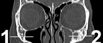
Rice. 1 Clinical case. CBCT computed tomography images show fragments of sealer that are directly adjacent to the lumen of the mandibular canal in the area of the mental foramen. The patient complained of paresthesia and pain from touch and wind in the area of the chin and lower lip on the left, which appeared after endodontic treatment of the 34th tooth. After 6 months - paresis (motor disorder) of the left half of the lower lip, which led to the inability to fully drink and eat (the patient holds the lip with her fingers while eating to prevent food and liquid from falling out of the mouth) and, as a result, a stress disorder accompanied by anxiety, fear and fits of anger.
Fig. 2. Removal of the sealer into the mandibular canal due to the absence of an apical stop
Regarding implantation, pain during bone preparation with a pilot drill can be an indicator of the proximity of the nerve and, if this is not addressed immediately (deciding to place a shorter implant), permanent nerve damage can occur.
Nerve injury during implantation is associated primarily with preoperative factors, including poor preoperative planning, which leads to inaccurate measurements and incorrect selection of implantation site and implant type (diameter and length).
Figure 3a. Planning error.
Figure 3b. Incorrect choice of implant length.
Figure 3-a, b. CBCT images from two clinical cases demonstrate implantation-related nerve damage. The implants are inserted directly into the canal of the inferior alveolar nerve, which is associated with errors in preoperative planning.
The habit of carefully planning implantation based on computed tomography data, performing implantation under infiltration anesthesia using surgical guides, performing intraoperative X-ray control and using drill stops (drill stops) can minimize possible nerve injury during implantation.
Any injury (penetration or compression) as well as hemorrhage into the mandibular canal results in acute and often severe intraoperative pain of the neuralgic type and it is imperative that the physician use an appropriate protocol of infiltration local anesthesia so that the patient can indicate the proximity of surgical instruments to the mandibular canal .
Since implantation is the surgery of choice, nerve injury, which leads to potentially irreversible consequences even after repeated surgery (implant removal), can always be avoided.
The physiological consequences of sensory nerve damage are immediate and often irreversible. The inferior alveolar nerve passes through the bony canal, which may be subject to compression and ischemic damage. Compression of peripheral sensory nerves for 6 hours can cause nerve fiber atrophy.
Ischemia itself, even without direct nerve damage, will cause sufficient inflammation and nerve damage that may result in permanent nerve damage.
Figure 4. Sagittal CBCT sections demonstrate an acceptable relationship between the implant and the lumen of the inferior alveolar nerve canal, but the clinical picture is consistent with ischemia, which caused pain and paresthesia. During the first 24 hours, a clinical decision was made to remove the implant and prescribe medication.
Three months after injury to the inferior alveolar nerve, permanent changes in the nervous system, both central and peripheral, will occur, which are unlikely to respond to surgical treatment or respond to drug treatment and peripheral interventions.
When nerve injury occurs, the clinician must be able to recognize the type and extent of injury, provide the most appropriate postoperative care, and be able to make recommendations.
Types of nerve damage:
- complete or partial resection of the nerve (cutting),
- compression, crushing, stretching, pinching, thermal and ischemic damage.
Total sensory deficits can range from minor sensory loss to persistent, severe, and debilitating pain dysfunction, but the most common combinations include anesthesia, paresthesia (painless altered sensation), dysesthesia (uncomfortable altered sensation), and neuropathic pain.
Currently, there is no standardized protocol for the dentist to diagnose and treat post-implant nerve injuries.
We will try to fill this gap.
What is mandibular nerve injury?
By this concept, dentists mean injury to one of the nerves:
- chin;
- lingual;
- alveolar.
Types of injuries include sprain, compression, crushing and rupture - partial or complete. The cause of the stretching is the long-term retraction of the mucoperiosteal flap, which is created by an implant of greater length than necessary. Crush injuries and compression are caused by needle injuries during the administration of anesthesia. Rupture occurs in two cases: when cutting the mucosa or during preparation of the hole for the implant.
Contraindications
The dentist will refuse to perform depulpation if:
- history of acute respiratory viral infection, sore throat, influenza;
- Stomatitis, acute leukemia, and hepatitis of infectious origin were diagnosed.
Contraindications to manipulation are heart disease in the acute stage and inflammatory pathologies of the oral cavity.
Remember: women in the 1st and 3rd trimester of pregnancy cannot have the nerve removed! This is due to possible psycho-emotional outbursts and (in the absence of pain relief) the painfulness of the procedure. Anesthesia during these periods is undesirable.
Causes and prevention of mandibular nerve injuries
The only cause of such damage is considered to be medical errors. Since in preparation for implantation, X-rays of the jaw are taken, which the doctor must carefully study so that when choosing an implant and a place for it, he does not injure the nerve, the injuries are caused by his unprofessionalism or negligence.
Damage to the mandibular nerve most often occurs when:
- improper administration of anesthesia - needle injury;
- choosing an implant that is too long;
- damage by an instrument - when preparing the site for the implant.
The only way to avoid such an injury is for a doctor to responsibly approach the stage of preparation for surgery, carefully studying the condition and structure of his patient’s jaw. The only way of prevention for the patient is to choose a trusted clinic and a highly qualified doctor. Specialists at the Implantmaster clinic have been able to reduce the number of injuries of this kind to 2%, since they carefully study three-dimensional photographs of a person’s jaw before implantation, and can correctly assess the condition of the bone tissue, the location of nerves and blood vessels, and select the optimal size of the implant.
Removal steps
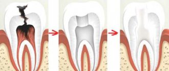
Depulpation is a dental operation that is carried out in stages:
- Examination and diagnosis. Radiography is often used. This research method helps determine the viability of nerve endings, the length and direction of canal branches.
- Anesthesia. Local anesthesia is preferably used, but sometimes general anesthesia is administered for medical reasons or patient preference.
- Isolation of the tooth using a rubber dam (special latex film). This will help prevent infection from entering the open cavity of the canal.
- Depulpation. After removing the destroyed layer of enamel and dentin, the pulp chamber is opened. The contents are removed using a pulp extractor.
- Filling root canals with gutta-percha.
- Installation of temporary or permanent filling.
- Repeated radiography to detect voids in the canal and double-check the accuracy of installation of the filling material.
This depulpation process is applied to premolars and molars, except for the number eight. Removing the nerve in a wisdom tooth is a more complex procedure. The location of the tooth does not always allow for high-quality filling of the canals.
Complications after surgery
The main causes of complications are fatigue, lack of professionalism of the dentist, and poor-quality equipment.
A complication from the aesthetic side is a change in the shade of the tooth crown. Its color depends on the material used to prepare the filling.
Care after removal
It is important to strictly follow the dentist’s recommendations and follow all his instructions:
- absence of stress for 2-3 hours after depulpation;
- It is forbidden to eat food and drink drinks for 4-5 hours;
- It is not recommended to chew on the side where the filled tooth is located for about a few days;
- Be sure to rinse and take medications prescribed by your doctor.
Our team of doctors
Maxillofacial surgeon, Implantologist
Bocharov Maxim Viktorovich
Experience: 11 years
Dental surgeon, Implantologist
Chernov Dmitry Anatolievich
Experience: 29 years
Orthopedist, Neuromuscular dentist
Stepanov Andrey Vasilievich
Experience: 22 years
Endodontist, Therapist
Skalet Yana Alexandrovna
Experience: 22 years
Orthopedic dentist
Tsoi Sergey Konstantinovich
Experience: 19 years
Dentist-orthodontist
Enikeeva Anna Stanislavovna
Experience: 3 years
Pharmacological therapy in the acute phase (first 30 hours) and intermediate phase (up to 4-8 weeks)
Pharmacological treatment of acute nerve fiber injuries includes the use of corticosteroids and non-steroidal anti-inflammatory drugs.
- Glucocorticosteroids – adrenocorticotropic hormone has been shown to inhibit central axon sprouting, reduce ectopic discharges in damaged sensory axons, and prevent neuroma formation. Seo K. et al.: Efficacy of steroid treatment for sensory impairment after orthognathic surgery. J Oral Maxillofac Surg 2004;62:1193-1201. Drug of choice - Dexamethasone - 8-12 mg/day for one week - Dexamethasone not only minimizes neuropathy after nerve injury when administered in high doses for one week after injury, but is especially recommended due to its significant anti-inflammatory effect compared to other corticosteroids. The recommendation is to prescribe a decreasing dose of dexamethasone (from high to low) for 5-7 days after a trigeminal nerve injury. Galloway EB, Jensen RL, Dailey AT, Thompson BG, Shelton C. Role of topical steroids in reducing dysfunction after nerve injury. The Laryngoscope 2000;110(10):1907-10. Kraut RA, Chanal O. Management of patients with trigeminal nerve injuries after mandibular implant placement. J Am Dent Assoc 2002;133:1351-1354.
- Nonsteroidal anti-inflammatory drugs (NSAIDs) are the best inhibitors of prostaglandin synthesis from damaged peripheral nerve endings. Prostaglandins released as a result of peripheral nerve injury sensitize peripheral nociceptor fibers and central neurons. Muller HW, Stoll G. Nerve injury and regeneration: basic insights and therapeutic interventions. Cun Opin Neurol 1998;11:557-559. Thus, maintaining therapeutic levels of NSAIDs in the blood as an adjunct to corticosteroids for 1-3 weeks after injury is very useful during the acute and intermediate phase of trigeminal nerve repair. Because any change in sensation may be caused by an inflammatory response, postoperative treatment with steroids followed by high-dose nonsteroidal anti-inflammatory drugs is given as soon as possible after any nerve injury. The drug of choice is Ibuprofen - 600 - 800 mg three times a day for three weeks (after finishing dexamethasone!). If necessary, two to three weeks after the injury, based on a repeat neurosensory examination, the doctor may prescribe an additional three weeks of taking NSAIDs if there are no signs of gastrointestinal disorders.
- Additional pharmacological agents - antidepressants, anticonvulsants, antisympathomimetic drugs, etc. Caution should be exercised with these types of pharmacological treatments, as they must be prescribed and monitored by a doctor who is familiar with the side effects of these drugs and has experience in treating nerve damage.
- Supportive pharmacological agents: - Neurorubin-Forte Lactab - one tablet twice a day for 4 weeks - contains high doses of vitamins B1, B6, B12, which play an important role in ensuring optimal metabolism in nerve cells. In high doses - has a weak analgesic effect; - Nucleo CMF Forte - one tablet twice a day for 20 days - Cytidine 5-monophosphate takes part in the synthesis of a complex of lipids that form the sphingomyelin membrane as the main component of the myelin sheath; - myogymnastics.
Symptoms and stages of damage
The symptoms by which this complication can be recognized are as follows:
- numbness of parts of the head - tongue, lips, chin, cheeks, etc.;
- biting lips and tongue;
- choking while eating or drinking;
- profuse salivation.
All this creates a number of inconveniences for the patient: it makes it difficult to eat and talk, disrupts facial expressions, and also prevents men from shaving and women from applying makeup. The severity of this injury is determined by its degree: a minor one goes away on its own or with the help of drug treatment, a severe one leads to irreversible processes of nerve degeneration and is not curable. Damage to the mandibular nerve, the symptoms of which the patient observes, requires immediate consultation with a doctor - only a specialist will be able to determine its extent and provide timely assistance.
Dentists distinguish the following stages of this implantation complication:
- minor - neuropraxia;
- more severe, but partial damage - axonotmesis;
- a serious injury that leads to complete loss of sensitivity - neurotmesis.
Radiation diagnostics
Of course, CBCT of the area of the impacted tooth provides the most complete three-dimensional picture of the position of the nerve and tooth. It is important to note here that any such examination must be supported by a written description from the radiologist. In this case, the surgeon has legal protection in case of a conflict situation and a lawsuit. The description must indicate the likelihood of such an injury: “the root of the tooth is adjacent to the wall of the mandibular canal,” “damage to the joint canal is possible when the tooth is removed,” “a narrowing of the canal at the level of the impacted tooth is detected.” OPTG is a more outdated research method; in the image it is not always possible to assess the location of the mandibular canal, especially in the buccolingual aspect. However, this classical diagnostic method is still widely used. A radiological sign of the contact location of the nerve and tooth is the following criteria: absence of the mandibular cortical plate at the level of the impacted tooth, narrowing of the lumen of the mandibular canal at the level of the tooth (Fig. 3,4). From a practical point of view, CBCT does not provide significant advantages and does not reduce the risk of developing paresthesia when removing an impacted tooth, since the extraction process is not computer-guided.
Prevention of complications. Before surgery to remove an impacted tooth, it is necessary to inform the patient about the possible risks of damage to the inferior alveolar nerve. It is important to convey to the patient that the timing of restoration of sensitivity of the lower lip depends on many factors and it is impossible to indicate the exact recovery time. The patient must be informed for what reasons the tooth is being removed; it is important to have a written description of the position of the tooth by the radiologist and a written opinion from the orthodontist. All this data must be attached to the patient’s medical record, and only after reviewing and signing the voluntary medical intervention can you begin to work with the patient.
Rice. 3. Narrowing of the lumen of the mandibular canal on OPTG is a sign of the anatomical proximity of the tooth root and the lower alveolar nerve.
Rice. 4. Darkening of the cortical pattern of the mandibular canal on OPTG is a sign of the anatomical proximity of the tooth root and the inferior alveolar nerve. After the tooth was removed, an open vascular nerve bundle was visible in the wound.
The technique of removing an impacted tooth requires sufficient lighting and good visualization of the surgical field. It is necessary to carry out a wide opening of the wound with skeletonized alveolar part at the level of the second molar and retromolar fossa and partially the branch. Small access significantly complicates the view of the wound. In most cases, tooth extraction begins with freeing the crown from the adjacent bone, then sawing the tooth along the dentino-enamel junction with a drill. After removing the crown and tooth, work is carried out on the roots. Based on the shape of the tooth cavity and the position of the orifices, the bifurcation of the roots is determined, and the roots are separated using a drill. When removing a single-rooted third molar, the socket is expanded on the side opposite to the position of the nerve (in most cases, on the buccal side). Root luxation with an elevator must be carried out without strong pressure, because a root fracture in the apical third with its subsequent removal increases the risk of nerve injury. In some cases, it is advisable to leave the tooth root in the wound as a “foreign body” in order to prevent nerve injury. The root fragment does not complicate wound healing, since the tooth does not have an apical infection. It is important to inform the patient that the tooth root “must be left” in order to preserve the integrity of the surrounding structures. Abroad, there is a generally accepted method for preventing nerve injury during tooth extraction - coronectomy, in which only the crown part of the tooth is removed. In fact, this removal can be considered incomplete. The root of an impacted tooth moves coronally over time, and bone tissue forms between the tooth and the mandibular canal. The second stage involves removing the roots of the tooth. This technique is not prescribed in domestic standards for the treatment of pericoronitis and is not described in the national guidelines, therefore it cannot be recommended for use in the Russian Federation.
Recovery and treatment
In the first case, self-recovery takes approximately 1 month; the help of doctors is not needed, since there is no anatomical damage. Symptoms of the second appear after a while - usually 6-8 weeks, so recovery can be painful and incomplete: it will take more than 2 months. In the third stage of damage to the mandibular nerve, treatment gives results only at the beginning and is performed surgically, since we are talking about degeneration with a violation of integrity. Loss of sensitivity, which is observed in a patient for more than 3 months, indicates a high probability of losing it forever. Damage to the mandibular nerve, the consequences of which is the lack of sensitivity of the nerve for a year, leads to irreversible changes. Only the professionalism and responsibility of the doctor, which is guaranteed by the specialists of our Implantmaster clinic, can protect the patient from such unpleasant injuries.
Author:
News
mandibular canola
Injury to the nerve of the mandibular canal
When implanting the lower jaw, the following complication may occur:
Perforation of the mandibular canal is in second place in terms of incidence after perforation of the bottom of the maxillary sinus when using dental implantation. According to the literature, among a number of factors of “failure” in implantology, that is, the occurrence of “complications,” trauma to the mandibular nerve ranges from 20 to 30%. Considering the fact that dentures on implants have become widespread, the patient hopes and demands high quality of this type of treatment. Understanding the mechanism of development of complications associated with mechanical compression of the inferior alveolar nerve allows us to conclude that early use of any treatment methods is necessary. Hence, the prevention of possible complications is of particular relevance and is increasingly becoming a matter of concern for implantologists. Therefore, the purpose of our study was to determine the doctor’s tactics after the occurrence of paresthesia during implantation of the lateral mandible. Materials and methods. Patients were selected - 16 people who, in the postoperative period, complained of impaired sensitivity in the skin of the chin area, lower lip and frontal group of teeth after surgery to install implants. The patients' age ranged from 26 to 69 years. Upon examination, an area of lack of tactile, pain and temperature sensitivity was revealed in all patients. Computed tomography revealed that the implants were adjacent to the mandibular canal or perforation of the upper wall of the mandibular canal (photos 1, 2).
| photo 1. Perforation of the canal wall. | photo 2. Nerve compression by implant. |
Results and its discussion. With atrophy of the alveolar height, the mandibular canal is located 5-7 mm from the crest of the alveolar process (Fig. 3). The operation of installing screw implants in such an area is fraught with perforation or compression of the mandibular nerve. An artery and a mandibular nerve pass through the canal. Perforation of the canal wall is accompanied by arterial bleeding, which can be stopped by tight tamponade or by introducing a hemostatic sponge into the bone wound. An implant installed in this location creates a precedent for nerve compression due to the formation of an intracanal hematoma (Fig. 4, 5). The complex conservative treatment carried out, regardless of drugs and duration, had no effect in any patient. Relief of the condition was noted after removal of the implant, and the sooner the implant was removed, the sooner the symptoms of discomfort disappeared. The wait-and-see tactic turned out to be wrong. When the apex of the implant was localized 1-2 mm from the upper wall of the canal, tactile sensitivity was maintained while pain and temperature were reduced. Restoration of sensitivity after removal of the implant occurred within a period of 5 to 8 weeks, if the implant was removed no later than 10 days after the onset of paresthesia. The mandibular nerve has a high regenerative potential, but if the rules of bone tissue preparation are violated during jaw atrophy, resection of the nerve is inevitable, which leads to persistent paresthesia. In cases where we adopted an erroneous wait-and-see approach, serious degenerative changes occurred in the nerve, which manifested itself in a persistent loss of sensitivity.
| photo 3. Localization of the canal with height atrophy. | photo 4. Damage to the canal with a cutting instrument leads to the formation of an intracanal hematoma, despite satisfactory installation of the implant. | photo 5. Intracanal hematoma. |
Conclusions.
Long-term unsuccessful conservative treatment of patients with severe dysfunction of the nerve allows us to conclude that it is impossible to eliminate paresthesia without removing the implant. In doubtful cases, it is necessary to clarify the location of the implant using computed tomography. If the localization of the implant is confirmed in the lumen of the canal, it is necessary to remove the implant as soon as possible.
www.alphadentural.ru link to the original source.