A convex formation on the eyelid in the form of a lump is not uncommon. The causes of this pathology may be different, but in any case, a lump on the eyelid should not be ignored. The formation can develop on both the lower and upper eyelids. It may not cause any inconvenience other than aesthetic discomfort, but it may hurt and fester. The bulge usually has a round or elongated shape. It may not change in size for a long time, and sometimes, on the contrary, it increases rapidly. Let's look at the reasons for the formation of lumps on the eyelid and how to treat them.
Chalazion
Cones of this type are quite common. They develop from a sebaceous gland whose duct is blocked. This formation is also called a “grading lump” or “cold barley.” The continued production of sebaceous gland secretion leads to the accumulation of a viscous mass in the capsule, which stretches and thickens, taking the form of a dense lump. On palpation, the contents under the skin feel like a moving ball.
Cold barley develops at a slow pace, so it does not cause pain. Only a formed hard capsule can cause pain when squeezed. If a chalazion is not treated, it can develop into a cyst. As the lump develops, the risk of complications increases: inflammation, formation of a purulent fistula, granulation.
Infections that cause bumps on the gums
Among the infectious diseases that contribute to the appearance of unusual gum growths:
- Periostitis. Inflammation of the periosteum of an acute or chronic nature. In advanced cases, it causes symptoms of general intoxication. The patient complains of a painful lump. It can either increase or decrease in size. Often there is pus inside it. Treatment consists of anti-inflammatory therapy, opening and draining the abscess. At the doctor's discretion, root canal treatment may be performed.
- Granulomatous periodontitis. Most often, this pathology is asymptomatic and is discovered accidentally during an x-ray for another disease. If the cyst is not localized in the area of the apex of the tooth, but is strongly displaced to the side (this is quite rare), a resemblance to a tumor can be observed.
- Radicular cyst. In advanced cases, its walls become inflamed. Then the infected contents of the tumor turn into pus. The necrotic masses are trying to find a way out. A lump appears, from which a foul-smelling liquid is released. During an in-person examination, hyperthermia and tenderness of the surrounding tissues are recorded. The situation may be complicated by symptoms of general intoxication: chills, fever, severe weakness.
- Gingivitis. Inflammatory gum disease. They bleed and become inflamed. Deformation of the gingival margin may occur. Sometimes the inflammation becomes so severe that small benign tumors appear.
- Periodontitis. A condition in which the tissues responsible for holding the tooth in its socket weaken. As a result, the unit becomes mobile. Deep “pockets” form at its base. If food residues accumulate in them, the situation is aggravated by the formation of small round seals.
Video from our specialist about the disease and its treatment
There are cases where chalazion spontaneously resolved without medical intervention. However, most often this formation does not develop back and requires prompt and conservative help. Treatment for such a lump on the eyelid is prescribed by an ophthalmologist. If the chalazion is small and not old, you can limit yourself to UHF therapy, ointments and eye drops. More severe cases are treated by injecting corticosteroids into the capsule cavity. Local drugs (ofloxacin, dexamethasone, sodium sulfacyl, hadrocortisone, levofloxacin, tetracycline ointment) can also be used as an addition to the injection.
If drug therapy is ineffective, the doctor decides on surgical treatment. The operation to remove a chalazion is performed under local anesthesia and lasts no longer than 15 minutes.
Barley
Styes are more common than chalazions. This type of lump on the lower or upper eyelid is caused by inflammation of the follicle (bulb) of the eyelash. This also clogs the sebaceous gland duct. Styes develop over several days or even hours and can occur in both adults and children. More often, the systematic appearance of barley is observed in people with weakened immune systems or who have changed their place of residence to an area with a more severe climate, as well as in people exposed to constant stress factors.
Based on their origin, there are two types of barley. Inflammation can be external (when the sebaceous gland suppurates) and internal (when the source of inflammation is located in the membolic gland).
The development of external styes is characterized by subjective sensations similar to a foreign body entering the eye. The initial stage may also be accompanied by stabbing pain. External stye visually manifests itself as redness and swelling of the eyelid. The internal one is usually not so noticeable, but it causes even more discomfort and pain.
Without treatment, barley develops within a few days into an abscess, which opens with the release of purulent contents. This brings relief, but an open wound is dangerous due to the possibility of re-infection.
It is better to start treating barley without waiting for the abscess to spontaneously break through. This allows you to get rid of the painful lump faster and with less risk of complications. If you still don’t have the courage or time to visit an ophthalmologist, you should remember that prolonged suppuration of the eyelid is very dangerous. If the stye does not open for more than two weeks, surgical treatment is necessary. An ophthalmic surgeon will remove the abscess under local anesthesia and give recommendations for further treatment of the eyelid. Most often, therapy for developing or already opened barley includes drops and ointments that contain antibiotics (albucid, gentamicin, erythromycin, tetracycline ointment).
When to see a doctor?
Many patients do not make an appointment with the dentist if the growth on the gum does not hurt. It is important to know that some diseases are asymptomatic in the initial stages. Unpleasant sensations appear when nerve endings become inflamed.
It is necessary to contact specialists in the following cases:
- a soft growth or hard compaction has appeared on the gum;
- I am worried about acute pain that is not dulled by painkillers;
- discomfort occurs when chewing or closing the jaws;
- a very swollen cheek or lip.
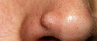
Millums (millet)
This type of bump occurs with equal frequency on the upper and lower eyelids. Millet grains can range in size from a poppy seed to a grain of rice and usually form in groups. Millums are the most harmless of all formations and cause only aesthetic discomfort. At its core, these are whiteheads localized in the eyelid area.
Millet removal should only be done by a cosmetologist. Since they do not carry the risk of complications, they are not considered an ophthalmological disease, but fall within the competence of a dermatologist-cosmetologist.
Prevention of millums includes caring for the eyelids, timely removal of dead epidermal cells, ensuring the cleanliness of the ducts of the sebaceous glands and pores of the skin around the eyes, as well as a balanced diet that excludes excessive consumption of fatty foods.
Xanthelasma
This type of eyelid bump looks more like a flat plaque. A connection between the development of such formations and chronic metabolic disorders has been revealed. Most often, xanthelasmas appear in women suffering from diabetes, hypercholesterolemia, and pathologies of the endocrine system.
These flat formations only partially rise above the skin and have a yellowish tint. They can be located not only on the eyelid, but also in the area around the eyes. In most cases, xanthelasmas appear on the skin in groups. They do not resolve on their own. Cosmetic removal is possible, but it should be understood that if the cause of their formation is not eliminated, it can lead to the appearance of new plaques. You should focus on treating the underlying disease, and only after that seek cosmetic help.
Content:
- Reasons for the formation of lumps on the gums
- Infections that cause bumps on the gums
- Bumps in the mouth of a non-infectious nature
- Lump at the base of the tooth after installing a permanent crown
- How do doctors treat a lump on the gum?
A change in the structure and shade of the oral mucosa is a reason to visit the dentist.
If a lump appears above the tooth - whether it hurts or not - it means there is some kind of problem. Such neoplasms do not just arise. This symptom may indicate various diseases. Most likely, the inflammatory process occurs in the gums, but without a preliminary diagnostic examination, the presence of systemic pathologies that manifest themselves in this way cannot be ruled out.
Papilloma
The causative agent of benign formations called “papillomas” is human papillomavirus. Most often, infection with this virus occurs during birth from mother to child, but the disease can also be acquired during life through contact with infected people. The virus can appear on almost any area of the skin and mucous membranes in the form of round growths. Papilloma is usually painless. However, it can hardly be called “aesthetically attractive”, so it causes significant discomfort.
You can distinguish papilloma from other types of formations by the following characteristics:
- the cone is more like a ball on a stalk or has a mushroom-like shape;
- the wart-like growth has a rough surface that resembles the surface of cauliflower to the touch.
It is worth noting that a growth with a smooth surface is not a papilloma, and it must be examined by an oncologist.
Treatment of papillomas should be comprehensive. Surgical removal is performed by a dermatologist. He also examines the type of virus and prescribes drug treatment. The fact is that the manifestation of a disease at one point does not mean that the entire body is not infected. The virus is suppressed by the immune system, but travels through the bloodstream. Some types of human papillomavirus are very dangerous with a high probability of degeneration of skin formations. Only an experienced dermatologist can prescribe adequate treatment based on diagnostic results. Measures to improve overall immune status are essential in the treatment of papillomas.
Reasons for the formation of lumps on the gums
When speaking about a lump under a tooth, dentists mean a dense, white or red growth. There may be purulent masses inside it. The disorder is usually caused by damage to periodontal tissues. What led to its occurrence remains to be determined by the dentist.
The described “growths” are of two types:
- infectious;
- non-infectious.
In the first case, they arise due to the abnormal activity of pathogenic microorganisms, in the second they are associated with mechanical damage to tissues, chemical burns and other unfavorable external factors.
More often than not, a lump on the gum forms precisely because of an advanced infectious process , but this does not mean that doctors can use standard schemes in their work. First, the doctor studies the features of clinical symptoms and draws up a detailed medical history, then carries out the necessary treatment measures and selects medications that will definitely help.