23.11.2019
The term “osteoblastoclastoma” was proposed by A.V. Rusakov in 1959. The International Histological Classification of Primary Tumors and Tumor-Like Bone Diseases uses the term “giant cell tumor.”
Until now, there are different names for giant cell tumors: giant cell tumor of the epulid type, brown tumor, gigantoma, giant cell sarcoma, osteoclastoma, local fibrous osteodystrophy.
In the etiology of this tumor, according to some authors, bone trauma plays a significant role. Giant cell tumors are classified as true tumors - blastomas. Currently, both surgeons and pathologists recognize these tumors as benign, although prone to recurrence. This led to the replacement of the terms “giant cell sarcoma” and “giant cell tumor” with the term “giantoma”. Giant cell tumors can have a sarcomatous structure.
Osteoblastoclastoma is a benign osteogenic tumor. Occurs in the jaw bones. The tumor develops mainly in young people, most often in women 11–30 years old. According to Yu. I. Bernadsky, osteoblastoclastomas account for 20.7% of the number of tumors of the facial bones.
Osteoblastoclastoma can be located along the periphery of the bone and in the thickness of the bone tissue. When localized on the alveolar process of the jaw, it is considered a giant cell epulid. In the lower jaw, the tumor is most often detected in the area of premolars and molars, in the upper jaw - in the area of premolars. Rare localizations include damage to the zygomatic bone.
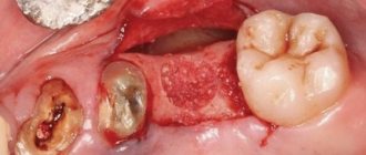
Osteoblastoclastoma—education clinic
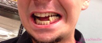
Unnoticed by the patient, a uniform thickening appears in one of the areas of the lower or upper jaw, often painless. Tumor growth continues for 3–10 years, but sometimes it is more intense.
When palpating the formation, its dense areas alternate with softened ones. Its shape is convex, dome-shaped, and sometimes thinning of the jaw walls occurs, which manifests itself as Dupuytren’s symptom (crepitus). The teeth in the formation zone are mobile, their electrical excitability is reduced, and resorption of tooth roots by 1/3 of the length is noted in the lesion. The mucous membrane above the tumor is either unchanged or slightly anemic; the venous network of the mucous membrane in the area of the tumor is expanded.
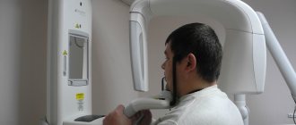
When an odontogenic inflammatory process occurs, signs of acute inflammation prevail. The diagnosis can be established through a comprehensive clinical and radiological examination.
Based on clinical and radiological data and the morphological picture, three main forms of osteoblastoclastoma are distinguished: cellular, cystic, lytic.
The cellular form is more often observed in adulthood and old age, and is characterized by very slow development. When examining the patient, a dense swelling with a lumpy surface is determined; it is not possible to clinically distinguish the tumor from healthy areas of the bone; the jaw is often spindle-shaped. Teeth located in the tumor area rarely change their position. The electrical excitability of the pulp of intact teeth is not impaired. The mucous membrane covering the tumor is somewhat anemic.
With the cystic form of the tumor, the first symptoms of the disease in most cases are complaints of toothache. During palpation examination, individual areas of the tumor are pliable when pressed (symptom of “parchment crunch”); the thinned bone above the tumor has a smooth-convex, dome-shaped shape.
The lytic form is rare, more often in childhood and adolescence, and accounts for 10% of all osteoblastoclastomas of the jaws. The development of this form of osteoblastoclastoma occurs quite quickly. In some cases, the first sign of a developing tumor is pain. When the cortical layer thins, along with independent pain at rest, pain appears on palpation. The venous network of vessels of the mucous membrane covering the tumor is dilated. Teeth often shift and become mobile, and the electrical excitability of the pulp decreases. Pathological fractures of the jaw may occur in the affected area; when localized on the upper jaw, it is possible to grow into the maxillary sinus, nasal cavity and other bones of the facial skeleton. When the formation is punctured, a brown or yellowish liquid is detected, which is associated with the breakdown of red blood cells and the formation of hemosiderin, sometimes with blood. The punctate does not include cholesterol crystals, which are usually found with cysts.
Etiology
The frequency of this tumor among all primary bone tumors is about 4–9.5%. The share of GCTs in the structure of all benign bone tumors is 18–23%. Most often, the disease is detected in the third decade of life; very rarely, the tumor is observed in children under 15 years of age and in patients with incomplete osteogenesis. Women are affected by GCT more often than men, in a ratio of 1.5:1. The most common location of the tumor is the metaepiphyseal sections of long tubular bones: distal femur - 23–30%, proximal tibia - 20–25%, distal radius - 10–12%, proximal humerus - 4–8% . Less commonly, GCT is observed in the sacrum - 4-9%, in the vertebral bodies - 3-6%, in the small bones of the hand - 1-5%, foot - 1-2%. In most cases, GCT is a benign tumor, but primary malignant forms occur in 1–1.5% and secondary malignant tumors are diagnosed in 10–25% of cases.
Osteoblastoclastoma - X-ray picture
Radiologically, with a cellular form of a tumor, a shadow from many small and larger cavities or cellular formations, separated from each other by bone partitions of varying thickness, is noted at the site of the lesion. No reaction from the periosteum is observed. The picture is in many ways similar to the X-ray picture of ameloblastoma.
The cystic form on the radiograph resembles an odontogenic jaw cyst and ameloblastoma. The difference between the cystic form of ameloblastoma is that its border with the bone often has fine scalloped outlines in the form of extremely small bays.
In the lytic form of osteoblastoclastoma, the tumor produces a structureless focus of clearing.
Causes of development of osteoblastoclastoma of the jaw
The basis of any tumor degeneration of cells is a change in the structure of cellular DNA. And today it is not completely clear what exactly causes such changes. A definitive answer has not been found to the question of why the immune system sometimes loses sight of tumor cells. At the same time, certain patterns of the occurrence of osteoblastoclastoma have been identified and it has been established what factors can provoke the development of this tumor. These include:
- Mechanical injuries - for example, osteoblastoclastoma can be caused by a bruise of the jaw bone or permanent injury to the mucous membrane of the oral cavity with a poorly processed dental filling or a poorly fitted denture.
- Infection in the socket of an extracted or fallen tooth.
- Chronic inflammatory processes in the periodontium, periosteum or jaw bone. Sinusitis or actinomycosis can also contribute to the development of a tumor.
- The presence in the tissues and cavities of the jaw of fragments of fillings, remnants of tooth roots and other foreign bodies.
Osteoblastoclastoma - microscopic picture
Microscopically, in osteoblastoclastoma, a large number of small, slightly elongated cells with a rounded nucleus (such as osteoblasts) are distinguished, among which massive accumulations of giant multinucleated cells (osteoclasts) are identified. In mononuclear cells, mitoses are observed; in multinucleated cells, they are absent. The formed elements of the tumor are also represented by fibroblasts and xanthoma cells. Areas of hemorrhage undergo a macrophage reaction with the corresponding phagocytic cells. In some areas of the tumor, islands of osteoid tissue are found. The tumor is saturated with decayed red blood cells and is imbibed by the blood pigment - hemosiderin, which gives it a brown color. The presence of the latter and multiple hemorrhages, often in the form of blood cysts, is due to the peculiarity of blood flow in the tumor.
Blood in osteoblastoclastoma circulates outside the vascular bed through intertissue gaps. The absence of endothelium allows it to penetrate the tumor tissue and accumulate there. In this case, the formed elements of the blood are destroyed, forming an accumulation of hemosiderin. Sometimes an abundance of fibrous tissue may be found in the tumor.
Osteoblastoclastoma - differential diagnosis
Osteoblastoclastoma (cystic and cellular forms) must be differentiated from ameloblastoma and radicular cyst, the lytic form - from osteogenic sarcoma (in contrast to osteolytic sarcoma, thinning and swelling of the cortical layer of the jaw are noted in osteoblastoclastoma) and fibrous osteodysplasia (characterized by slow growth and preservation of the continuity of the cortical layer on the radiograph). layer of the jaw), fibromyxoma, central fibromas of the jaws.
Clinical and radiological examination without histological comparison is sometimes insufficient to establish an accurate diagnosis of osteoblastoclastoma. The frequency of discrepancy between the clinical diagnosis and pathohistological diagnosis is 30%; the most common discrepancies arise when differentiating osteoblastoclastoma from ameloblastoma - 16%
Osteoblastoclastoma - treatment
Treatment for osteoblastoclast of the jaws is surgical. In cystic and cellular forms, careful curettage of the lesion is usually sufficient. If the tumor occupies large areas of the jawbone, it is possible to perform resection of the lower jaw, if necessary, with simultaneous bone grafting. In the lytic form, partial resection of the jaw without breaking the continuity of the mandibular bone (continuum resection) or resection of a fragment of the jaw with simultaneous bone grafting is more often used. Radiation therapy does not provide sufficient therapeutic effect. However, this method of treatment is prescribed to patients for whom any surgical intervention is contraindicated due to their general somatic status. Usually short-focus X-ray therapy, remote gamma therapy, bremsstrahlung and high-energy electron radiation are used. The average radiation dose ranges from 30–50 Gy/kg. The prognosis for treatment is favorable, but the possibility of malignancy and metastasis of the tumor due to the benign nature of the tumor cannot be excluded. Incorrect treatment tactics usually lead to relapses of the tumor process.
Published in Articles
Myxoma of the mandible
Myxomas are benign tumors that form on the maxillofacial bone. They make up no more than 1% of diseases of this type, which are localized in a specific area of the human body.
Causes
This disease is inherited, or is a continuation of the course of certain diseases of the oral cavity. With proper treatment, myxoma rarely becomes chronic. However, if you do not seek professional help from a specialist, it can reach impressive sizes.
The structure of the myxoma is formed from mucin and the blood vessels that penetrate it, which are, at the same time, nourishing. Due to the fact that the tumor has a fairly long period of time for its formation, which can be up to several years, it is most often possible to notice the tumor only during an external examination. Myxoma appears on the surface of the skin in the form of a smooth bulge, with a fairly dense base and has a cellular structure with several partitions.
The place of its localization is most often the lower jaw. The tumor leads to displacement or loosening of teeth with subsequent loss of teeth in the absence of necessary treatment. The lower lip may become numb or even lose mobility.
Diagnosis and subsequent treatment
To determine the presence of a tumor, the following methods are used:
- X-ray images. They indicate not only the presence of this formation, its size and structure, but also the presence of a structure of cells, which looks like a honeycomb in the image, instead of the normal bone tissue.
- Carrying out a puncture. A sample is taken from the patient from the affected tissues in the form of a light liquid. Although this method does not provide a complete picture, it does help in diagnosing myxoma. This type of research is carried out in cases where other methods have not brought satisfactory results.
- Histological examination. This type of diagnosis allows for the most complete study of the tissues of the emerging formation. This reveals the full clinical picture of the disease.
To treat myxoma at any of its stages, surgery is used. During the operation, the damaged area, along with healthy tissue, is completely removed under anesthesia. This helps prevent its further spread. This operation is simple and does not have any further consequences. However, this happens if the tumor is detected early. Therefore, if a person notices any tumor in the oral cavity, it is necessary to immediately seek diagnosis from a specialist. These steps will help prevent complications that can lead to the loss of a large number of teeth or even harm a person’s appearance.