13174
The expression “jaw asymmetry” is often used as a synonym for a broader concept – facial asymmetry. In fact, these are different, although closely related, pathologies.
An anomaly should be discussed when a violation of the proportions of the lower third of the face is caused by the dental factor. According to research, such cases account for 80% of the total number of imbalances in the space from the chin to the nose.
General information
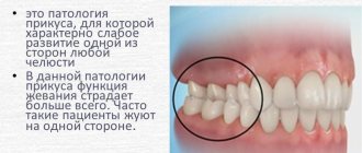
Jaw asymmetry is a pathology expressed in visually visible asymmetry of the lower third of the face. Displacement away from the midline of the entire upper or lower jaw, their segments or individual areas of the face - lips, chin, branches of the upper and lower jaw.
In addition to the above features, pathology can manifest itself as drooping corners of the lips, disruption of the oval of the face, widening or narrowing of the eyes, smoothed lip folds, and some specific features, for example, a pained expression.
To see whether there are deviations or not, you need to imaginarily draw the central line of the face - a vertical straight line passing strictly along the space between the central incisors of the upper and lower jaw. A wide smile makes asymmetry more pronounced.
Strictly speaking, there is no complete correspondence between the left and right parts of the face (ideal symmetry). A certain disproportion of them is present in all people.
The physiological norm is considered to be a position in which the asymmetry in the linear value does not exceed 2-3 mm, in the angular value - 3-5°. If these values are exceeded, they talk about pathology and look for the reasons for its occurrence.
To a greater extent, asymmetry is characteristic of the lower jaw. This is explained by the fact that it is located movably relative to the skull, and has more factors influencing its position than the upper jaw. This is the type of bite, the condition of the TMJ, habitual postures, manner of chewing food, etc.
Dental problems solved with mandibuloplasty and the results achieved.
Come here if you are wondering why your jaw clicks when chewing.
According to this address https://orto-info.ru/zubocheliustnye-anomalii/chelyustey/smeshheniya-sredney-linii.html we will discuss how to correct the displacement of the midline of the teeth.
Anomalies of dental development
The most common anomalies in the structure of hard tissues are systemic or local hypoplasia of permanent or temporary teeth. Systemic hypoplasia looks like a white or yellow spot with a smooth surface. It is impossible to determine it using x-rays. Pits or grooves appear in the image as areas of clearing.
Local hypoplasia develops most often due to injury to the follicle or against the background of chronic inflammation of the periodontium of a milk tooth - in a permanent tooth. Enamel hyperplasia is rare and appears on an x-ray as a more intense, dense shadow.
Reasons for the formation of anomalies in children
Often, an orthopedic doctor immediately after birth notes asymmetry of the skull in newborns. It is determined by the factors of the child’s development in the womb and the conditions of birth (the position of the fetus in the uterus, its passage through the birth canal).
Usually after 2-3 days the newborn’s skull takes the correct shape. If this does not happen, a thorough examination of the baby is necessary to identify the causes of the deformities.
In addition to the birth anomaly, most children under one year of age have facial disproportion due to infants being in one position for a long time.
Birth and infant asymmetry are natural types, and in most cases do not require medical intervention, except perhaps massage.
The pathological anomaly is more often observed in preschool children. It can be congenital or acquired, right-sided or left-sided.
Congenital asymmetry is a consequence of genetic factors and abnormalities in fetal development caused by the condition of the mother and embryo during pregnancy (intrauterine infections, abnormal position of the fetus, asphyxia, lack of nutrients, etc.).
These factors lead to disruption of the formation of soft tissues, cartilage and bones of the skull, uneven healing of sutures, etc.
The specific, most common congenital causes include the following:
- Hypertonicity of the masticatory muscles.
- Muscular dystonia (involuntary muscle contractions leading to impaired development of the TMJ and, often, crossbite).
- Crossbite (due to muscular dystonia or accelerated/slow development of one of the jaws).
- Cleft lip. An anomaly manifested by the presence of 1 or 2 clefts in the upper lip, leading to disruption of the shape of the nose and a depression in the middle area of the face.
- Stigmas of embryogenesis. Various etiological malformations of the fetus. They disrupt the symmetry of the dentofacial apparatus, nose, and skull.
- In addition to pathological factors associated with pathologies of the dentofacial apparatus, there are many other congenital diseases that lead to disruption of facial symmetry.
- These are torticollis, Sturge-Weber syndrome, craniofacial microsomia, etc. The dentist’s task is to differentiate them from pathologies of the dentofacial apparatus, and refer the patient to a doctor of appropriate specialization.
Acquired jaw asymmetry develops in the postnatal period under the influence of various external and internal factors:
- Malocclusion. The most common cause is crossbite, which develops due to bad habits and incorrect positioning of the child in the crib, stroller and at the table. A crossbite can result from slower or faster development of the upper or lower jaw.
- TMJ pathologies.
- Odontogenic and non-odontogenic tumors in the jaws (hard and soft odontoma, osteoma, salivary stones, etc.)
- Impacted teeth.
- Injuries to bone (fracture of jaw bones, skull and face) or muscle tissue.
- Birth injury.
- Periostitis.
- Periodontal inflammation.
Among the non-dental acquired disorders that can lead to an anomaly, the following should be mentioned: diseases of the ENT organs, eye pathologies, curvature of the spine, paresis of the facial nerve, stones in the salivary glands, tumors in the nasal cavity and paranasal sinuses.
Causes
Malocclusion can be caused by various reasons. This disease in most cases originates from childhood, and a common cause of the formation of a crooked jaw is a hereditary factor.
Children inherit the shape of their teeth from their parents, and pathologies can have very serious consequences.
There are several main reasons that result in the formation of malocclusion:
- Features of pregnancy. At a certain stage of intrauterine development, bone tissue is formed in the fetus - it is during this period that proper nutrition of the mother, enriched with calcium and other microelements, is important.
- Artificial feeding. The baby must develop the jaw from birth - this is why it is so important to prolong the baby’s natural nutrition as much as possible.
Sucking the breast is a necessary workout for muscles and joints. Without these exercises, jaw formation may go the wrong way. - Missing some teeth . For facial injuries associated with jaw injuries, you should consult a dentist as soon as possible, as teeth tend to take up as much free space as possible.
- “Bad” childhood habits. Even harmless thumb sucking or prolonged use of a pacifier can cause serious consequences (curvature of the jaw and teeth, as well as improper bite formation).
- Chronic runny nose. Mouth breathing has a negative effect on the muscles of the tongue when it stops putting pressure on the teeth from the inside and tends downward. As a result, improper formation of the upper jaw occurs.
- Mechanical injuries . Falls, blows and other actions sometimes lead to curvature of the lower jaw.
Based on all of the above, we can conclude that the sooner parents pay attention to the formation of the child’s jaw, the easier it will be to correct the defects.
For information about the causes of jaw displacement and methods for eliminating the problem, watch the video.
Provoking factors in adults
The cause of the anomaly in adults is acute and chronic diseases of the dentofacial apparatus and its injuries:
- Congenital pathologies of the dentofacial apparatus that were not corrected in childhood.
- Arthrosis of the TMJ (dystrophic destructive changes in the joints). They manifest themselves as a displacement of the LF towards the diseased joint, stiffness in the morning, and pain.
- TMJ ankylosis (joint immobility due to cartilage degeneration). The anomaly is noticeable at rest and increases when the mouth is opened, which in case of severe pathology does not exceed 1 cm.
The line drawn between the lower central incisors shifts towards the damaged joint. Movement of the jaw in the horizontal direction becomes impossible. - Contracture (restriction of movement) of the LF muscles. The midline is shifted towards the affected muscle.
- Odontogenic benign tumors (odontoma, ameloblastoma, steoma, osteoblastoclastoma). The jaw bones are destroyed by neoplasms, deformation occurs, which usually becomes the first manifestation of the disease.
- Malignant tumors of the upper and lower jaw (sarcomas, carcinomas). The resulting deformation is caused by tumor growth.
- Jaw defects (reduction in the volume of dental tissue) due to osteomyelitis, syphilis, tuberculosis, surgical interventions for oncological diseases. Bone deficiency causes receding of the cheeks and disruption of the oval in the lower jaw area. As the mouth opens, the asymmetry increases.
- Micrognathia – due to underdevelopment of the jaw or damage. Micrognathia HF manifests itself as a reverse overlap, LF - with a slanted chin.
- Diseases of the salivary glands.
- Periostitis. Asymmetry is caused by swelling and/or abscess under the periosteum.
- Periapical (at the root of the tooth) and periaxillary abscess. Facial deformation is caused by swelling that has spread to adjacent tissues. The pathology manifests itself as throbbing pain.
- Crossbite (transversal displacement of the jaws relative to each other). The chin appears to the side, the lip sinks on one side.
- Facial injuries, fracture of the lower or upper jaw. Temporary asymmetry occurs due to soft tissue swelling and/or displacement of the jaw bones.
In addition to the above pathologies, the anomaly can be caused by many other diseases that are not related to the dentofacial apparatus:
- Damage to the brain and meninges (meningitis, encephalitis, stroke).
- Nerve diseases (Bogorad syndrome, facial nerve damage).
- Pathologies of the salivary glands (mucocele, purulent parotitis, adenoma).
- ENT diseases leading to unilateral deformation (sinus cyst, atelectasis (collapse of the paranasal sinus)).
Facial asymmetry can occur due to hydrogen peroxide poisoning, botulism, actinomycosis (infection with actinomycetes fungi).
Treatment methods for small lower jaws in children and adults.
In this publication, read how to expand the dentition with braces on one side.
Here https://orto-info.ru/zubocheliustnye-anomalii/chelyustey/ukorochenie.html we will consider the reasons for shortening the dentition and methods for its correction.
Diagnostics
Only a qualified specialist can determine the degree of jaw curvature. This does not require high-tech equipment - the doctor just needs to look at the position of the dentition in several positions.
But, in order to decide which method to use to correct a particular defect, the orthodontic patient will have to undergo a series of examinations, including:
- clinical examination;
- X-ray examination;
- intraoral photographs and facial photographs from various angles;
- measuring the size of the jaws, dentition and teeth;
- assessment of the condition of the muscles of the maxillofacial apparatus;
- deviations in the position of the tongue during swallowing are examined.
In addition, a number of other examinations that are important for making a diagnosis are carried out, which make it possible to accurately and qualitatively determine the degree of neglect of the case.
In particularly difficult situations, radical measures may be required, such as removing several units and even surgery.
Diagnostic measures
The first doctor you should contact if an abnormality is noticed should be a dentist or neurologist. If you need to connect a doctor of another specialization to the diagnosis - an otolaryngologist, an oncologist, a neurosurgeon - the first receiving doctor will refer the patient to them.
Cosmetologists also claim to be the first to visit, claiming that in case of facial asymmetry caused by hypertonicity of the masticatory muscles, cosmetic procedures alone may be sufficient to solve the problem.
The list of dental diagnostic measures is as follows:
- Survey . The patient's complaints are listened to and the medical history is clarified.
- Physical examination . The degree of anomaly at rest and movement is assessed, probing, palpation and percussion are performed. The clinic of the dentofacial apparatus is assessed (presence of edema, tumors, suppuration, consequences of injuries, condition of the teeth).
- X-ray of the jaws . Photographs of individual teeth, dentition (orthopantomography and teleradiography) and nasal sinuses are taken. If necessary, radiographs of the skull and spine in the neck area are obtained.
- Ultrasound examination. Sonography of sinuses, tissues, salivary glands.
- CT or MRI. Used for detailed study and clarification of symptoms obtained by radiography.
- Orthodontic examination . Cephalography, determination of bite, occlusion, presence of supercontacts, analysis of diagnostic models in the articulator, etc.
When doctors of other specializations participate in the diagnosis, the following examinations may be carried out:
- From a neurosurgeon . A CT scan of the brain is done.
- See a neurologist . The innervation of the facial muscles is checked - by wrinkling the forehead, puffing out the cheeks, raising and lowering the eyebrows, and following the doctor’s movements with the eyes. The exit points of the nerves are palpated.
- See an otolaryngologist . Special examinations are carried out - diagnostic puncture, echosinusoscopy, tuning fork examination, audiometry.
If necessary, general laboratory tests are performed. The blood is examined for the presence of inflammation and bacterial infection. Cytological and histological analysis of punctates and smears is carried out.
Based on the diagnostic results, a conclusion is made about the causes of the anomaly, and a treatment plan is drawn up.
Anomalies in the number of teeth
The number of teeth may decrease due to the absence or death of rudiments, or increase. Primary adentia occurs mainly in the permanent dentition. With complete teeth, all teeth are missing; with partial teeth, the upper lateral incisors and lower second molars are missing. This happens with ectodermal dysplasia.
Supernumerary teeth are characterized by an irregularly shaped crown. They erupt for a long time and remain in the jaw. An x-ray - panoramic study - allows you to determine this or that anomaly.
Sometimes a tooth is missing from a row due to retention - retention of the tooth in the jaw bone. It is provoked by its incorrect initial position, lack of space or narrowing of the dentition. X-ray examination allows you to determine the position of the impacted tooth and its relationship with the surrounding anatomical formations. For this purpose, special techniques are used - tomography, oblique projections, etc.
Treatment methods
The variety of causes and nature of the disease determines a wide range of possible treatment methods - from cosmetic to surgical.
Depending on the root cause, cosmetic, conservative, orthodontic and surgical treatment can be used. As well as massage and therapeutic exercises.
Massage
Massage has multiple therapeutic effects:
- Relaxes and tones muscles, relieves hypertension.
- Activates blood and lymph circulation in the affected area, relieves swelling and inflammation.
- Stimulates nerve fibers.
- Reduces pain.
Manual therapy in children is more effective than in adults due to the plasticity of children's muscles. The earlier the anomaly is diagnosed, the more effective manual therapy is.
Cosmetology procedures
Cosmetic treatment consists of getting rid of hypertonicity of the masticatory muscles, which causes facial asymmetry. Technologically, the procedure involves injections of Botox into the masticatory muscles.
The drug relaxes overstrained muscle fibers, straightens the face, returns it to harmonious proportions, and gives a calm and confident expression.
Conservative therapy
Conservative therapy is determined by the nature of the pathology and provides for the treatment of pulpitis, caries, periodontitis, periodontitis and other diseases of the teeth and periodontium, which make it possible to do without surgical intervention.
Analgesics (for pain), antibiotics (for infections), antiseptics (for local inflammation and wounds on the mucous membranes), NSAIDs (for severe inflammation) may be prescribed.
Orthodontic correction
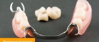
Orthodontic treatment is used for crossbites, narrowing of the jaws, and other dental anomalies. Treatment is carried out with the help of splints, mouthguards, extra- and intraoral devices, braces and other fixed and removable orthodontic appliances.
Surgical intervention
The type of surgical intervention is determined by the nature of the disease and is divided into:
- for dental (removal of tumors and cysts, removal of teeth, opening of abscesses);
- maxillofacial (lavage, arthroscopy, phlegmonectomy, TMJ endoprosthetics, rhinocheilognatoplasty);
- eliminating the consequences of injuries (splinting and regeneration of jaw bones, tying with a ligature);
- otolaryngological (ectomy of paranasal sinus cysts, etc.).
If asymmetry is caused by damage to the brain and meninges, according to indications, ectomy of tumors and abscesses, transcranial or endoscopic removal of hematomas, areas of the brain crushed by TBI, and other surgical protocols are performed.
How to fix?
Currently, there are a huge number of methods that allow you to straighten your jaw and get a beautiful smile. It all depends on the degree of curvature and the age of the patient.
The main problem of an older person is that the bone tissue has already been formed, which means that it takes more time to correct it.
In addition, “aged” patients are unlikely to be able to boast of absolutely healthy teeth - as a rule, many of them already have numerous fillings. This only means that the process of correcting defects will be significantly more difficult.
However, there are several ways to correct a crooked jaw that can help correct the situation.
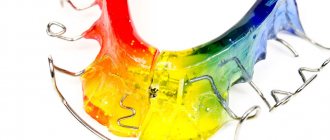
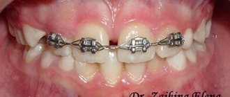
Correction with braces
One of the most common methods, the essence of which is to use special brackets or locks connected to each other by an arc. They are attached to the surface of the teeth using dental glue and are not removed until the end of the course of treatment.
Cases in which it is most rational to use braces:
- teeth straightening;
- the presence of wide gaps between adjacent units;
- pulling of teeth due to difficult eruption.
There are two types of braces: vestibular and lingual. The first type is more popular, as it allows the use of mechanisms made of different materials, various shapes and colors.
Lingual braces are attached to the lingual surface of the teeth, so they are less noticeable. With their help you can correct a deep bite.
The duration of wearing such systems depends on the initial position of the jaw, or more precisely, on the degree of its curvature. In some cases, the patient will have to wear braces for several years.
The cost of such treatment starts from 30 thousand rubles.
What is dental hypodontia and how to eliminate the problem.
In this publication we will talk about edentia.
Follow the link https://orto-info.ru/zubocheliustnye-anomalii/zubov/kolichestva/giperdontiya.html if you are interested in what the pathology called hyperdontia is.
Mouthguards
They are special plates made of silicone or polyurethane. First, the dentist makes plaster casts, on the basis of which a whole series of trays is made to be replaced during the treatment process.
The duration of the course varies depending on the degree of curvature. In adults, correction may take up to 2 years. This method has many advantages:
- Democratic price.
- Possibility of discreet wearing.
- The method is painless and lacks discomfort.
- Possibility of removing the aligners (before brushing your teeth, while eating, for important meetings, etc.).
- Safe for tooth surfaces.
- The mouth guards are easy to use.
For the entire course of treatment, the patient will have to pay about 160 thousand rubles.
Surgical intervention
Oral and maxillofacial surgery can also correct the shape of the face. Correction of the shape of the jaw and bite can only be indicated in the case of pronounced pathology of the dental system and deformation of the skull.
Oral and maxillofacial surgery can be used if it is impossible to correct the bite using the above methods.
Many patients use the possibilities of maxillofacial surgery when they want to correct their bite as soon as possible.
This operation can help in the following cases:
- A serious hereditary pathology that provokes improper formation of bone tissue.
- Asymmetry of shapes due to injury.
- Open bite type (lateral or frontal).
- Protruding chin.
Depending on the severity of the deformity and the reasons for its occurrence, jaw surgery can remove part of the bone or build it up. During such operations, part of the molars can be removed, which will create more space for the correct growth of the front units.
All maxillofacial surgeries are performed under general anesthesia. Any incisions and manipulations are made directly in the oral cavity, which means that there are no unaesthetic marks left on the patient’s face.
The cost of the operation can hardly be called affordable; it depends on many factors and can range from 8 to 50 thousand rubles. Do not forget that after the rehabilitation period, you may have to continue treatment with braces.
Possible complications
The consequences depend entirely on the underlying cause of the anomaly and the timeliness of contacting a doctor. In some cases, the problem can be eliminated without a trace with a quick cosmetic procedure or a light bite correction.
In others, if the problem is ignored for a long time, it can result in significant destruction of the temporomandibular joint, with no guaranteed hope for a complete restoration of its function.
Thirdly, create a serious threat to life itself (in case of malignant tumors).
Only a doctor can give an accurate answer to the question about the consequences of pathology after a thorough examination. In any case, the only correct action for the patient will be to consult a doctor as quickly as possible when a pathology is first detected.
Prevention and prognosis
Prevention of such a defect as crossbite should begin during a woman’s pregnancy. She should lead a healthy lifestyle, watch her diet, and exercise moderately. It is also important to avoid all kinds of infections and viruses. To do this, it is better to avoid visiting crowded places and communicating with carriers.
After the baby is born, he needs constant monitoring. At this stage, it is important to choose the right pacifier and pacifier, and, if possible, do without these devices. To do this, it is better to consult a pediatric dentist or pediatrician. It is also necessary to ensure that the child does not get carried away with thumb sucking or toys, and does not receive any injuries to the facial part of the head.
The prognosis of such a malocclusion depends on how well the treatment was carried out. Often the defect can be completely removed.
Important! Do not forget that at the initial stage only a specialist can identify dental deformation. Therefore, it is necessary to visit the dentist regularly from the moment the first baby teeth grow.
Preventive measures
Prevention of any dental diseases consists of following a number of rules that are relevant for almost every person. And although, unfortunately, they do not provide a complete guarantee of preventing dental diseases, they reduce the risk of their development. And if they have already appeared, they speed up and facilitate treatment.
This is the fight against bad habits of children, teaching them proper oral care. Quitting smoking and foods that are harmful to teeth. Using mouthguards when playing sports. And, perhaps most importantly, regular visits to the dentist for preventive purposes.
In the video, experts talk about the causes of asymmetry of the face and jaws.
Classification and stages of crossbite
Crossbite has several varieties:
- Lingual, in which the upper jaw expands and the lower jaw narrows. It can be observed on one side or both. It happens with or without displacement.
- Buccal – expansion of the lower jaw with a parallel narrowing of the upper. It may also have a displacement and be observed on one or both sides.
- Buccal-lingual is a combined type of bite that combines the characteristics of the previous two.
Also, the types of crossbites are divided based on the stage at which the pathology is at a particular moment. In this case, the following main stages are distinguished:
- milk stage, when malocclusion is observed in young children before the growth of permanent teeth begins;
- replaceable - occurs at the stage of replacement of the milk dentition with permanent ones;
- constant, which can be said to exist in the presence of a defect after the appearance of permanent teeth.
Depending on the type of defect and the stage of its development, the doctor can select the most effective treatment.
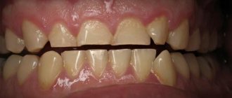
Mesial bite
Mesial occlusion or, as they also say, mesial occlusion is the second common malocclusion in the sagittal plane. And although mesial occlusion is less common than distal occlusion, there are no fewer questions and controversies surrounding its diagnosis and treatment.
Mesial occlusion is characterized by the fact that the closure of the dentition is visually observed in the so-called third class. When the anterior buccal cusp of the upper molar does not lie in the median fissure of the lower molar, but is located dorsally (posteriorly) from it. And the upper canine is positioned not between the lower canine and the lower first premolar, but also more dorsally. As a result, the normal relationships of the dentition are disrupted.
And we see that the lower row of teeth protrudes forward, more than expected, in relation to the upper teeth. But third grade closure is not a diagnosis. Just as the formulation itself is not a diagnosis - mesial occlusion or mesial occlusion. The closure of teeth, both in the third and, in principle, in any class, is just the closure of teeth. No more. Some external sign. It does not reflect the cause of any anomaly. Moreover, sometimes the closing of teeth even camouflages (hides, hides) the main, usually skeletal, problem. And therefore, just as in the case of treating distal occlusion, or any other problem, you should not make a diagnosis and build treatment tactics only by the type of closure of the teeth. This guideline is extremely unreliable and deceptive. Always and everywhere it is necessary to treat not the “third (or second) class”, but the very cause of the problem that led to this class of closure. Of course, this reason must first be determined. Well, more on that below.
For now, let’s look at the external signs that allow us to suspect mesial occlusion in a patient.
Mesial bite. Signs
At the level of the teeth, as already said, the lower teeth protrude forward in relation to the upper ones. The degree of their “protrusion” can vary from the closure of the upper and lower incisors “joint to joint” (the so-called straight bite)
or a slight “divergence” of the upper and lower incisors along the sagittal, or in other words, in the anteroposterior direction (reverse incisal overlap or reverse bite). Until there is a pronounced non-closure of the upper and lower incisors in the sagittal plane with the formation of a sagittal gap.
Only in this case, unlike distal occlusion, the “components” of the sagittal fissure (upper and lower teeth) have opposite (inverse) relationships. In the case of a pronounced sagittal gap and non-closure of the teeth, the diagnosis will sound like reverse incisal disocclusion (disocclusion - lack of closure). The vertical relationship of the teeth and jaws with mesial occlusion can be completely different: from a pronounced deep bite to, on the contrary, an open bite (vertical non-occlusion of the teeth).
On the face, as a rule, the lower third is enlarged due to the protruding lower jaw, and in some cases, the lower lip. And the upper lip, on the contrary, looks “sunken” (flat) because of this.
Also because of the protruding lower jaw, such a profile is called “energetic” or “masculine”. The most striking representatives of mesial occlusion with strongly pronounced facial signs of this anomaly are members of the Habsburg royal family.
Well, there are genetics, incest and other royal “pranks”... These issues will not be considered in this article... We are talking about other causes of mesial occlusion.
There is also a postural (at the level of posture) sign of mesial occlusion. This is the so-called posterior postural type of posture .
The body needs to somehow align itself in space and maintain balance when the lower jaw is forward. Read more about this in the article “Bite and Posture.”
But despite the general similarity of the external “pattern,” the causes of mesial occlusion can be completely different. Both by the root cause (which jaw, upper or lower, is to blame for the mesial bite), and by the level of occurrence (skeletal or dental).
Dental causes (dental-alveolar forms) of mesial occlusion
Let's start from smallest to largest. From the dental level. When can our lower teeth protrude in front of our upper teeth?
1. The lower dentition is elongated:
A) supernumerary (extra) tooth
B) lower macrodentia: the correlation between the sizes of the upper and lower teeth is disturbed - the lower teeth are larger in size than necessary for the given specific upper teeth
C) other reasons, for example, the tongue is located in the area of the lower incisors and brings them into protrusion (excessive forward tilt), lengthening the lower dentition
2. The upper dentition is shortened:
A) edentia (insufficient number) of upper teeth
Same patient, oral view:
B) upper microdentia: the situation is the opposite of that described in paragraph 1, subparagraph B, and the upper teeth are to blame - they are smaller than necessary for normal sagittal relationships with the lower ones
C) deformation of the upper dentition, its shortening, crowding of the upper teeth
These were “dental” reasons. But, as you know, teeth grow from the jaws. And therefore, the size of the jaws and their relationships, both among themselves and in relation to the skull, can also lead to various types of malocclusion. And mesial occlusion is no exception.
Skeletal forms (causes) of mesial occlusion
Let's consider the jaw (skeletal) causes of mesial occlusion. And I will deliberately start with the jaw... the upper one. Yes Yes. You heard right. In a huge number of cases of mesial occlusion, it is the upper jaw that is to blame. So. What could be the fault of the upper jaw with mesial occlusion:
1. Small, underdeveloped upper jaw. Usually the upper jaw is shortened in the anterior (premaxillary) region. This reason can always be suspected if there is crowding of the upper teeth. Or another indirect sign - the tongue is located behind the lower teeth or is laid between the dentition. Here even the appropriate diction may be “lisping.”
This is understandable: the tongue has no “native” place (on the palate), it finds its home wherever it happens to be.
2. The upper jaw is normal in size, but is in a posterior position. And this happens often. As always, the fault of the jaw lies either in size or position. Let us continue to list the jaw prerequisites for mesial occlusion. But now let’s list when the lower jaw is to blame. 3. Large lower jaw. Moreover, the increased size can be both in the chin and in the posterior segment (all this can be precisely determined by analyzing the lateral TRG according to Sassouni). But the branches of the lower jaw may also be enlarged...
4. The lower jaw is in an anterior position. As a rule, this position is forced. Due to the deformation of the dentition, the upper and lower teeth cannot normally close into a “lock” (it is simply inconvenient for them to close correctly). And the patient is forced to push the lower jaw forward. This very often happens in primary occlusion in children, when the primary fangs, with their sharp ends-tops, create such conditions for the forced position of the lower jaw.
Another reason for the anterior position of the lower jaw is rotation of the temporal bones of the skull. After all, as is known from anatomy, the head of the condyle of the lower jaw is “inserted” into the articular fossa of the temporal bone. And therefore, changes in the position of the temporal bone (articular fossa) affect, among other things, the position of the lower jaw.
And again, as we can see, the problem is either with the size (large) or with the position (front). Moreover, problems with the size and position of both jaws can be combined with each other in completely different variations. This creates, on the one hand, a great variety, and on the other, individuality of forms of mesial occlusion in each specific clinical case. And that is why there are no, and cannot be, universal (for all cases) methods of treating mesial occlusion. Just like any other malocclusion. Everything is decided individually...
By the way, when the jaws “diverge” along the sagittal, the sagittal gap described above does not always occur. Often it is simply absent due to protrusion (tilting forward) of the upper incisors. This is a kind of “dental” compensation for the mismatch of the jaws along the sagittal plane.
Diagnosis of mesial occlusion
The main causes of mesial occlusion were listed above. Both “dental” and skeletal (jaw). It remains only to add that these reasons can be combined with each other, well, in any variations. And these variations... well, count them yourself. Hence the moral: you always need to clearly know what is the cause of mesial occlusion in each specific case. Only then can we answer two original Russian questions: “Who is to blame?” and “What should I do?” Simply put, it immediately becomes clear what to treat and how to treat it. And the answer to this question can be given by analysis of TRG in the lateral projection. Because mesial occlusion is an anomaly in the sagittal plane. Well, analysis of the models, of course (the dental factor cannot be identified otherwise).
Mesial bite. Treatment.
Correction of mesial occlusion. This is where things get really messed up... And why? Because we don’t know how or don’t want to look for (identify) the reason for the teeth closing according to the third class (see above). And we strive to treat mesial occlusion without diagnostics, “by eye.” Relying only on its external manifestations. And that’s why we often “go in the wrong direction.” We are not treating what is needed. And not like that. For example, the upper jaw is short, and the lower teeth .
That's how it is in this case. The patient's mesial occlusion is caused by shortening of the upper jaw. And once, due to its shortening, the upper fangs, not fitting into the dental arch, “stuck out” from above. And the orthodontist removed the top 4 (which in itself is fraught with consequences, see here) to place the fangs. Thus, he “confirmed” and aggravated the shortening of the upper jaw by also shortening the upper dentition. The second orthodontist, when this patient came to him with a third class closure, instead of developing the upper jaw and upper dentition, also removed the lower 4ths. By adjusting the length (and number of teeth) to the defective length of the upper jaw. Thus, further aggravating the situation.
As a result, the teeth seem to be positioned correctly, first class.
But...Is this the result? And can this be called treatment ? Just take a look at the profile.
It is clearly flattened, which does not add aesthetics. The facial profile simply “screams” about the skeletal nature of the anomaly (you just need to be able to see it). So if you carry out a diagnosis, you will not be surprised that this patient needed to be treated not for a mesial bite, but for a distal bite camouflaged by the removal of the upper 4 teeth. This is how it happens...
Alas, this second would-be doctor was once me. Therefore, colleagues, do not step on the same rake, do not repeat the mistakes. Diagnose correctly. And do not treat according to the principle “what we see is what we eat,” basing your conclusions only on how (by what class) the teeth close. Everything is much more complicated and confusing. It’s a pity that you understand this only years later, having gained bitter experience...
How I identify and treat mesial occlusion (and other malocclusions) now is in the article “How this is done in our clinic.”
Or, again, the upper jaw (for example, it is in the posterior position ), and the “large” lower jaw . And she (the bottom one) has nothing to do with it at all.
But everything is simple in reality. Analysis of trg in a lateral projection (I really like the analysis according to Sassouni), competent interpretation of the data obtained - and it’s immediately clear what the reason is. This means that, in principle, there should be no difficulties with how to treat mesial occlusion.
Everything is logical. If the upper jaw is to blame (for example, it is short ), the mesial bite must be corrected by developing (increasing in length) the upper jaw. By the way, with development, the base of the upper jaw (read “living space” for teeth) will increase and their crowding and (or) protrusion will go away. Since both the first and second are options for placing teeth when there is a shortage of space on the jaw (this has been written about in detail here).
There are a great variety of devices for the development of the upper jaw. From a regular orthodontic plate.
Before fixed structures like this:
If you noticed, the patients in all the photos are adults. Not growing. Which, in general, did not affect the result in any way. Although it is clear that mesial occlusion is especially effectively treated in growing patients. In children and adolescents. So, moms and dads, if you notice a mesial bite in a child (the signs are listed above), you shouldn’t wait for the “sea weather” and that “it will go away on its own.” Don't hesitate to contact your orthodontist. After all, mesial occlusion in adults, however, is not treated as “fun” as in children.
If the upper dentition is short (for example, it is shortened due to edentia), then it can also be developed. And space for missing teeth can be obtained. As a result, the length of the upper dentition increases and this may be enough for the overlap of the front teeth to turn from reverse to normal.
If the reason for the shortening of the upper dentition is small teeth (smaller than necessary for a given patient), then the upper dentition should also be developed to the required length. And the resulting gaps can be closed by restoration or prosthetics. When the reason is precisely the violation of the size of the teeth , then the correction of the mesial bite should and is carried out due to the size (its increase) of the crowns of the teeth. Orthodontists should not be alarmed by this. It is better to approach the problem adequately than to try for years to close the gaps by “pulling” teeth with braces. If the problem (gaps between the teeth) lies “at the core” (the initial discrepancy between the sizes of the teeth and jaw, in favor of reducing the size of the teeth), then it is useless to “tighten” anything. All the same, the teeth will “scatter”.
If the upper jaw is normal in size, but is located posterior to the norm , and accordingly, this fact is the reason for the relationship of the jaws in the third class, then the position of the upper jaw must be changed. In other words, “pull it out” to the front. There are so-called face masks for this purpose. Dilyar's mask for example.
Well, a separate article has been written about face masks, their varieties and the nuances of their use (see here). I will only say that I personally use special masks to treat mesial bite. This is also written about in that article.
the lower dentition is to blame for third class occlusion , the logical conclusion suggests itself - to reduce its length. The length of the dentition can be reduced:
1. Distalization (movement back or dorsally) of the entire dentition. If there is somewhere to move, of course. For these purposes (so that there is somewhere to move) you can sacrifice your wisdom teeth. Separation of teeth (lower ones, of course, if we are talking about third class closure)
2. Removal of lower teeth , such as premolars. With further closing of the gaps. Unlike the removal of the upper premolars, the removal of the lower fours or fives does not have such dire consequences for the patient’s health. Provided that normal functional occlusion is formed at the end of orthodontic treatment, of course. And if such removal is justified.
If the lower jaw and its fault is in the anterior position . Here you need to work very carefully and thoughtfully. And not just “push” the lower jaw back, as in this video:
Such manipulations run the risk of driving the lower jaw (or rather, the temporomandibular joint through the lower jaw) into compression and causing TMJ dysfunction. With all the consequences...
In such cases (when there is an anterior position of the lower jaw), it is better to work together with a chiropractor. An osteopath, for example. So that it acts on the temporal bone (and we remember that it is the articular fossa of the temporal bone of the skull that is the natural “refuge” of the articular head of the lower jaw). And displacements (rotations) of the temporal bone (read articular fossa) can affect the position of the lower jaw. Or, if the reason for the forced position of the lower jaw is a block on the teeth (for example, on the primary fangs, as shown above), then this block must be eliminated and the lower jaw must be freed from the “trap” (for example, the same primary fangs must be polished)
Mesial bite. Operation
If the fault of the lower jaw is its large size (like the Habsburgs), then this is the very case when you can think about surgery. Think about it, colleagues. Just think for now. Instead of immediately sending the patient to a surgeon. Even if the “gap” in the jaws in the sagittal plane is very large and surgery is inevitable, the patient must be prepared for surgery for mesial occlusion. And preparation is the task of the orthodontist. Let's not forget about this.
Preparation for surgery for mesial occlusion should consist of a complete selection of all orthopedic (working with the jaws, their size and position) and “dental” capabilities (resources) of the patient. For example, along with an enlarged lower jaw, there may well be an underdeveloped or posteriorly positioned upper jaw. Before an operation to reduce the size of the lower jaw, it would not be a bad idea to develop or “pull” forward this “defective” upper jaw.
Firstly, for surgeons there is a completely different reference point (much more accurate).
Secondly, the patient is completely benefited: an underdeveloped upper jaw is a health problem in itself. Just as the upper jaw, “hammered” back (essentially into the base of the skull), is also a problem.
And thirdly, you have authority: they didn’t just discourage the patient from undergoing surgery, but did everything correctly.
And most importantly, very often in the process of such preparation it is possible to find compromise solutions when the patient, after looking at the result, changes his decision and refuses the operation. There are many such cases in my practice. And I consider this also a successful treatment. It is clear that we are not talking about a centimeter discrepancy between the jaws along the sagittal plane. But there were precedents. Not ideal, of course, in the end (because the “source” is far from ideal), but they refused the operation.
I repeat once again: practically the only indication for surgical treatment of mesial occlusion is the case when the closure in the third class (mesial occlusion) is caused by an increase in the size of the lower jaw. Moreover, the increase is significant, when no orthopedics (non-surgical work with the jaws) will help.
Mistakes in the treatment of mesial occlusion
Consider only the main errors and insinuations. Only those that are “widely distributed” among dentists and patients. Well, about the surgical option (and there are a lot of mistakes in treatment planning), they just talked about (when, in what cases is it appropriate). In the rest - no surgery. No problems with the position of the lower jaw. Neither “third class” problems caused by the upper jaw should be solved surgically. No matter how, all problems of closure in the third class should be solved with braces (or rather, only with braces).
Mesial bite. Treatment with braces
Photo of mesial bite correction with braces:
How many reasons are listed above that lead to teeth closing in the third class? A lot, right? But mesial bite with braces (or rather with the help of braces alone) can be corrected only when the teeth (dentition) are to blame. Or the top one is shortened. Or the lower one is elongated (macrodentia, supernumerary tooth). Those. in a very limited number of clinical cases. How many attempts do we see to cure mesial bite with braces alone? And can't be counted. Yes, take a look at any “bracket bearers” forum. There are dozens of them (if not more). And there are a lot of failures (now I hope it’s clear why). I can tell you from experience: there are an order of magnitude more skeletal causes of mesial malocclusion than “dental” ones. And the critical mass of skeletal causes is a “defective” (in size or position) upper jaw. By the way, have you noticed that in sagittal malocclusions (distal and mesial occlusions) the upper jaw plays a key “causal” role? And there is a reason for this - cranial disorders (but this is a topic for a separate article). But for the majority of us, mesial occlusion is still associated exclusively with a “large lower jaw.” With all the consequences... All errors are due to a lack of understanding of the reasons. And misunderstanding is due to poor diagnosis. Everything is simple, as we see.
Is it possible to restrain the growth of the lower jaw? Chin sling
The first common innuendo regarding the treatment of mesial occlusion in children is an attempt to restrain the growth of the lower jaw. All these chin slings, face masks and other things that are not very convenient for the patient...
Many people try and hope, but no one has succeeded so far. You can't go against nature. She (nature) will take her course anyway. The bones will grow as much as they are designed to grow. So “containing growth” is absolutely futile. If only because the patient obviously will not wear all these sling masks around the clock (unless, of course, he is also a patient in a psychiatric hospital). So there is no benefit from such actions. Just as there is not and cannot be strict control over the round-the-clock wearing of a face mask or sling. But the harm... The harm can be tangible and real. After all, what happens if we put an obstacle (literally lean on it) on the lower jaw? It, of course, will not stop growing (why suddenly?). But it will begin to rotate (rotate) around a new support point. And here you can easily get an “elongated” face with an open bite. And compression of the TMJ. And other bad things, such as changes in posture, for example (and not for the better, of course). And this is all in addition to the already existing mesial occlusion. In general, the sling clearly poses more problems than it is worth.
And most importantly, sometimes you look, there’s a chin sling. Something like “holds back”. You carry out diagnostics, and the lower jaw is absolutely normal. Both positionally and dimensionally. And the person (patient) suffers. And again, it all comes down to “UNDERDIAGNOSIS.”
Conclusions:
In class 3 closure, skeletal causes than dental ones
If this is so, then it is impossible to treat mesial occlusion in isolation from the “context” (general skeletal problems of the body). See “Bite and Posture” and “The Tale of How Dentists Correct Posture.” And also “How it’s done in our clinic.”
Of the skeletal causes, the upper jaw : either the size is reduced or the position is posterior. In the first case, the upper jaw develops , in the second, it extends forward (a combination is possible, since problems of position and size often coexist peacefully with each other)
Restraining the growth of the lower jaw is a futile task and of little use for facial aesthetics and health in general.
Since dental causes causing mesial occlusion are less common than skeletal ones , in the vast majority of cases mesial occlusion cannot be cured with braces alone . Therefore, correction of mesial occlusion with braces in adults ... is unlikely. And it has little prospects.
Removal of the lower premolars in the treatment of mesial occlusion does not entail such detrimental consequences as the removal of the upper premolars in the treatment of distal occlusion
Surgically, mesial malocclusion is treated only when the lower jaw . And her fault lies specifically in her size - she’s big . There are few Habsburgs among us, frankly speaking (I don’t know whether it’s fortunate or vice versa...)
And one more thing... The main moral. And the only correct logical message (aka conclusion): never neglect diagnostics. First you need to understand the cause of the anomaly. And only then make some efforts to treat (correct) any malocclusion. Only in this case the efforts will not be in vain. And only then can you hope for a positive result.
I hope this article will help my colleagues and their patients in the treatment of mesial malocclusion. I wish everyone good luck! Photos of examples of mesial bite correction (before and after results) can be seen here.
And here you can read an article about distal occlusion.
Why is correct diagnosis of malocclusion important?
- You will understand the essence of the problem with your bite and will know how to treat it - You will not waste extra time and nerves on incorrect treatment - You will not waste money on incorrect treatment - You will not be forced to use unnecessary services
The most accurate diagnosis of mesial occlusion in Moscow with Dr. Filatov
Sign up for diagnostics
Social Like
Comments for the site
Cackl e