What are the main causes of swelling with pain?
The main cause is inflammatory processes of periodontal tissues, which are caused by a number of pathological conditions.
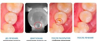
- Advanced caries, bad habits, ignoring hygiene rules or poor-quality root canal filling. In most cases, swelling of the cheek is preceded by pain localized on the side of the affected tooth. The source of inflammation is located in the root area and after a few days a flux can form, which without treatment transforms into an abscess.
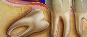
- Very often, swelling of the cheek occurs due to disruption of the eruption of wisdom teeth. In this case, purulent inflammation is caused by the formation of a pocket (hood) between the gum and the dental unit itself. In addition, due to lack of space in the jaw arch, molars can occupy an unphysiological position and injure the buccal mucosa.
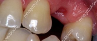
- Inflammation caused by infection of the socket after the removal of a molar very often leads not only to swelling of the cheek, but also to more serious consequences.
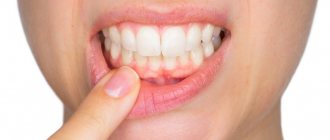
- A dental cyst is characterized by asymptomatic formation over a long period of time; when it increases to 5-7 mm, the neoplasm presses on the bone. The pathology is characterized by acute throbbing pain, accompanied by swelling and redness of the gums, as well as deterioration of the general condition.
The danger of inflammatory processes in the oral cavity lies in the close proximity of pathogenic swelling to the tissues of the face and brain. If left untreated, there is a high risk of phlegmon and abscess formation, which in extreme situations can lead to death.
Swelling in the cheek area, causes
The most common dental causes of cheek swelling are:
- advanced caries;
- complication of pulpitis;
- complication of gingivitis;
- periostitis, periodontitis and other diseases of teeth and gums.
If you suspect flux, call: 8 (495) 558-88-77
Flux is inflammation of tissues of various origins, for example, periostitis, an inflammatory process in the periodontal gap - periodontium. Such inflammation can occur in acute and chronic forms; the chronic form can periodically enter an exacerbation phase and then subside again.
By origin, periostitis is as follows:
- infectious;
- non-infectious;
- spicy;
- chronic;
The main role in the development of flux of infectious origin belongs to microorganisms, as well as their toxins. Microbes penetrate into the periodontium through the root canal, periodontal pocket, as well as through the blood or lymph flow.
Most often, flux is caused by an infection that enters through the root canal and is a consequence of acute diffuse and chronic gangrenous pulpitis, as well as pulp necrosis.
Microorganisms and their toxins, penetrating into periodontal tissue, cause dangerous acute inflammation
Non-infectious periostitis/periodontitis (fluxes) can develop as a result of trauma (blow, bruise, periodontal trauma after pulp extirpation, sharp, uncomfortable biting on a tooth; cracking nuts, gnawing bones) or chronic microtrauma (smoking pipe, brass band instruments, biting threads, pressing on the tooth with a pencil, pen, etc.); also as a result of the influence of medications - the ingress of potent substances into the periodontium during the expansion of root canals (trilon B, aqua regia), their sterilization (formalin, silver nitrate, etc.) and in the ingestion of arsenic paste.
The tooth does not hurt, but the cheek is swollen - the causes of the pathological condition
Very often, swelling is a consequence of an allergy to materials used for filling, prosthetics and treatment of dental units. In addition, the clinical situation may be associated with the body's reaction to food, pollen, dust or pet hair. In this case, the swelling does not have a clear localization and can spread to the area of the nose, lips and eyes. .
It should be understood that after the removal of a tooth, nerve or dissection of the gum, swelling and pain are considered normal and can persist for a week after the intervention. You should also exclude respiratory and viral pathologies accompanied by enlarged submandibular lymph nodes (mumps, pharyngitis), high body temperature, and chills.
Another reason is bruises and injuries to the face; in addition, the cheek may swell due to neurological disorders, for example, inflammation of the nerve. The problem is indicated by pain in the throat and parotid region, hearing impairment, facial expressions and diction.
The appointment includes consultation and drawing up a treatment plan with cost determination
sign up for a free consultation
Not ready for an in-person consultation with a doctor? Ask your question by phone
What to do for acute toothache
Its appearance signals the need to visit the dentist as soon as possible. The doctor will listen to complaints, examine the oral cavity, “knock” the teeth, and prescribe tests to evaluate the internal tissues: CT scan, radiography or ultrasound.
When a potential source of pain is found, it can be further examined using a radiovisiograph. A targeted image gives an idea of the degree of carious lesions, pathologies of bone tissue or the presence of neoplasms.
A more advanced diagnostic method is a 3D image. All pathologies are visible on the three-dimensional model, which allows you to make the correct diagnosis.
Important! If a trip to the dentist is postponed, under no circumstances apply heat to the sore spot. This can increase inflammation and lead to complications.
What are the main treatments for cheek swelling?
The ATLANTIS DENTAL medical center has created ideal conditions for the diagnosis and treatment of dental diseases of any complexity. If you find that your cheek is swollen, you should urgently visit a doctor. Experienced therapists and surgeons will quickly and painlessly eliminate the problem, which will avoid unwanted complications.
- During the consultation, the doctor collects anamnesis, performs a dental examination, and prescribes diagnostic measures (x-rays, CT scans, blood tests, etc.).
- If necessary, the patient is referred for consultation to specialized specialists: neurologist, otolaryngologist, endocrinologist, etc.
- Based on the results of the examination, a treatment regimen is determined.
- When cystic neoplasms, pulpitis and inflammation of the periosteum are detected, surgical intervention is necessary.
- It may also be necessary to remove the tooth or refill the canals.
Good results are shown by treatment with antibacterial drugs, rinsing the mouth with antiseptics and applying dental gels to the affected areas.
Why is acute toothache dangerous?
A person who develops dental diseases, suffers pain, or tries to cope with it by taking painkillers can not only lose teeth, but also have problems with the jaw bones.
Pain itself makes a person nervous, exhausted, and his reactions slow down, which often leads to domestic injuries. But much more dangerous is the effect on the body of serious inflammatory diseases in which it occurs (up to sepsis or meningitis).
How to relieve the condition at home?
To alleviate the condition before visiting a doctor, you can relieve pain and reduce swelling of the cheek with home remedies.
- A pronounced anti-inflammatory effect is exerted by rinsing infusions made from oak bark or calendula, chamomile, and sage inflorescences. To prepare the solution, two tablespoons of raw material are steamed with boiling water and infused for 15-30 minutes, after which rinsing is carried out three to four times a day.
- The soda-salt solution effectively relieves pain, reduces swelling and has a pronounced antiseptic effect. To do this, the components in the amount of 15 g (1 tsp) are stirred in a glass of warm water and used for their intended purpose.
- Dental ointments and gels, which are applied by application to the affected area 2-3 times a day, have proven themselves well.
- Pharmacy antiseptics are always effective, regardless of the causes of swelling of the cheek and gum tissue. Among the most popular: Chlorhexidine, Miramistin, Furacilin, hydrogen peroxide.
If your health worsens and you suspect an allergy, you need to take antihistamines, such as: Tavegil, Zodak, Suprastin, Kestin, etc. To relieve pain, take analgesics or NSAIDs - non-steroidal anti-inflammatory drugs.
Important! In case of severe swelling of the cheek, “tugging” pain, high body temperature and respiratory dysfunction, urgent hospitalization is necessary. Symptoms may indicate anaphylactic shock or sepsis.
The main symptoms of inflammation of the ternary nerve
What are the main symptoms of trigeminal nerve inflammation and tooth pain? The location of the pain and its nature will depend on which nerve endings are inflamed. If it is the lower trigeminal nerve, then discomfort will be felt in the lower jaw. With inflammation of the middle trigeminal nerve - in the upper jaw. Inflammation of the superior trigeminal nerve causes pain to spread to the upper part of the face (eyes, forehead, temples and even ears).
With neuralgia, teeth can hurt either constantly or periodically (paroxysmal pain). Unpleasant sensations can begin suddenly and end just as unexpectedly. Sometimes it can be very difficult to identify the exact location of pain.
How do teeth hurt with trigeminal neuralgia? Discomfort can manifest itself as ordinary toothache or migraine. But, characteristically, this pathology never causes edema.
How is trigeminal nerve inflammation diagnosed?
If you only feel aching pain in your teeth, then first you need to make an appointment at a dental clinic. Only a competent dentist can determine the nature of the pain, its cause and the exact source. Typically, an examination of the oral cavity is performed, followed by palpation and tapping of painful areas. To accurately verify the neuralgic nature of the disease, you will need to undergo a CT scan of the teeth (this will show the presence of inflammatory processes in the teeth and gums, and will also allow you to detect abscesses and other pathologies). You may need to consult an ENT specialist, since problems with the maxillary sinuses may also cause pain. Well, if you experience characteristic shooting pains, you should immediately contact a neurologist.
Once the presence of tertiary nerve inflammation is confirmed, the doctor decides how to treat the patient. You should not hope that the pain will go away on its own. These cases are extremely rare; moreover, it is important to determine the true cause of the discomfort and exclude other more dangerous and serious pathologies.
Shooting pain in face
Trigeminal neuralgia (facial Fothergilla pain, painful tic, neuralgic prosopalgia)
Among neurogenic pain syndromes caused by damage to the cranial nerves, the main place is occupied by trigeminal neuralgia (TN). NTN is one of the most common neurological diseases of the face, second in frequency only to facial neuropathy. The disease occurs in 3-7% of other types of headaches. Women (5:3) over 40 years of age are more often affected. It is characterized by a severe course and the absence of sufficiently effective treatment methods.
Pain with NTN is paroxysmal in nature, stereotypical, lasting from several seconds to 2 minutes, characterized by significant intensity, suddenness, reminiscent of an electric shock or lumbago. Pain can be provoked when eating, talking, as well as mechanical irritation when washing, brushing teeth, etc. Negative emotions can provoke a painful attack during an exacerbation. The highest frequency of exacerbations is observed in the autumn, less often in winter, periods of the year, which demonstrates the role of meteorological factors. Spontaneous pain may occur.
NTN is characterized by paroxysmal pain. Interictal pain of varying intensity of a dull or burning nature, paresthesia against the background of hypoesthesia, arising after injection-destructive treatment, are explained by associated neuropathy. Neuropathy of the branches of the trigeminal nerve can also be of odontogenic origin; it is a consequence of an inflammatory process or trauma during tooth extraction and is discussed in the section on symptomatic pain associated with dental damage.
The onset of NTN can occur in different ways: in some cases, typical paroxysms immediately appear, in others, local paroxysmal or constant pain first appears in the area of one or several teeth, which over the course of several months or years turns into typical neuralgia. The disease progresses with exacerbations and remissions. During the period of exacerbation of the disease, patients answer questions in monosyllables, barely opening their mouths, since the slightest movement of the facial muscles can cause a painful paroxysm. During an attack, patients freeze. Some try to squeeze the sore side with their hand or rub it roughly. During attacks, pain can be grouped in volleys; the interval between individual attacks is short, between volleys longer. Status neuralgicus is described when there are no gaps between individual volleys. As a rule, volleys can last for hours, and periods of attacks can last for days and weeks.
One of the reasons may be pathology of the dental system; Often the disease develops after tooth extraction. In most cases, the lower alveolar nerve is affected when the lower molar is removed, which is the terminal branch of the mental nerve. First, numbness and constant pain appear, which over time transform into typical neuralgic attacks. This form of neuralgia is called odontogenic.
In some cases, NTN may occur as a result of circulatory problems in the brain stem. In this case, the formation of the syndrome occurs at the level of the nucleus of the descending tract.
Postherpetic neuralgia is a complication of herpetic Gasserian ganglionitis and occurs in 10% of patients.
The most common form is the “idiopathic” form, the mechanisms of which have been studied quite well recently. The term “idiopathic” is used only conditionally in the 1988 international classification of headaches, which discusses the tunnel-compression mechanism of this most common form of neuralgia.
Treatment.
Prescribing analgesics for trigeminal neuralgia is practically ineffective. The fish drug is carbamazepine (finlepsin, tegretol) 600-800 mg per day. Other anti-epileptic drugs are used. With long-term use of antiepileptic drugs, their effectiveness is significantly reduced. Under these conditions, it is recommended to periodically change the drug or add gamma-aminobutyric acid derivatives to them - phenibut 0.25-0.65 g or pantogam 0.5-1 g 3 times a day.
Of no small importance is the treatment of patients with antidepressants, which soften the perception of pain, reduce fear of an attack, eliminate depression, and change the functional state of the brain.
In patients with vascular diseases of the brain (dyscirculatory encephalopathy), the treatment regimen includes vasoactive drugs: Trental, Cavinton, etc.
For the treatment of trigeminal neuralgia, glycine myeglinol (amino acid glycine) at a dose of 110 mg/kg body weight before meals has been proposed. The drug is dissolved in 50 ml of water. The course of treatment is 4-5 weeks. Myeglinol has a central effect on hyperexcitable neurons and a peripheral effect: it reduces the content of formaldehyde and thereby promotes remyelination.
In case of complete ineffectiveness of conservative therapy, there is a need to use surgical treatment methods: resection of the branches of the trigeminal nerve, decompression of neurovascular bundles, stereotactic operations to decompress the trigeminal nerve root. The most commonly used are thermoisotomy and microvascular decompression. The first is less traumatic, since it is performed without craniotomy, repeated operations are possible, has a minimum of complications and can be performed in older people. However, thermorizotomy produces more side effects: impaired chewing, painful dysesthesia in the facial area. Microvascular decompression consists of trephination of the posterior cranial fossa, revision of the relationships between the trigeminal nerve root, the superior anterior cerebellar artery, and, less commonly, the inferior anterior cerebellar artery and the superior petrosal vein. When the root is compressed by vessels, they are isolated, and a gasket is placed between the vessels and the root to prevent contact between them and the impact of the vessel on the root.
Despite the encouraging results of rhizotomy and microvascular decompression, especially in the early postoperative period (64 and 90%, respectively), the problem of pain recurrence remains.
In this regard, it is relevant to develop new methods of surgical treatment, the simultaneous use of surgical techniques at various levels of the trigeminal nerve system, depending on the predominance of distal or proximal tunnel-compression factors, as well as combination with pharmacological methods. To prescribe individual treatment and professional consultation, you can make an appointment with the specialists of the Pain Clinic. To do this, just call the phone number or fill out the feedback form, and you can also make an online appointment with doctors on our website. Health and prosperity to you and your loved ones!