Tooth root resorption is a physiological or pathological process that results in the loss of dentin and/or cementum [21].
Tooth resorption was first described by Joseph Fox in 1806 [26]. He associated the mechanism of the process with bone tumors.
There are physiological and pathological resorption of the tooth root.
Physiological resorption is characterized by the resorption of the roots of primary teeth during the period of occlusion change.
Pathological root resorption is the progressive loss of dentin and cementum through the continuous action of osteoclastic cells [57] and is further divided into external and internal root resorption.
The purpose of this review is to summarize the literature data regarding the etiology, pathogenesis and methods of treatment of pathological resorption of permanent teeth.
The analysis of literature sources was carried out using data from the central scientific medical library, the electronic medical library eLibrary and the database of medical publications PubMed and Scopus.
Etiology
Pathological root resorption can be initiated by many factors, acting separately or simultaneously.
Such factors may be chronic inflammatory diseases of the pulp, tooth trauma, chronic periodontal diseases or orthodontic treatment [27]. Resorption can also be caused by pressure from supernumerary teeth, surgical interventions near the root system, incorrect endodontic treatment and aggressive teeth whitening [5, 6, 19, 25, 27, 30, 43, 66].
Cases of resorption occurring against the background of somatic diseases such as scleroderma [23, 60], pathologies of the endocrine system [13, 42, 54] and viral diseases [7] have been described.
Cases of multiple idiopathic tooth root resorption have also been reported [10, 40, 41, 73].
Prevalence
Data on the prevalence of pathological resorption have significant discrepancies. Thus, E. Harris [31] in his study determined the frequency of apical root resorption in the permanent dentition in patients who were not treated orthodontically, and found that from 7 to 10% of 306 patients showed obvious apical resorption. F. Vier and J. Figueardo [68] showed that the prevalence of teeth with periapical lesions with resorption is more than 82%. A report by M. Haapasalo and U. Endal [29] estimated the prevalence of internal inflammatory root resorption to be between 0.1 and 1%, confirming that the estimate is quite rough and may be incorrect.
Differences in study methods, variability in pre- and post-treatment radiographic images, misdiagnosis, and undetected lesions are challenges in determining the overall prevalence of pathologic root resorption.
In some cases, it is difficult to determine what is causing the violation of the tooth structure: due to developmental disorders or resorption mechanisms. [52]
Symptoms of edentia
The main symptom of edentulism is the patient’s lack of teeth. The clinical picture of the disease can be supplemented by:
- pronounced reduction of both jaws;
- retraction of soft tissues in the oral area;
- the appearance of blunt exostoses or painful sharp bone protrusions on the jaws;
- formation of wrinkles in the perioral area;
- atrophy and laxity of the muscles of the lower lobe of the face;
- serious disorders of speech functions (the patient loses the ability to clearly and clearly pronounce dental-linguistic sounds - “ch”, “n”, “z”, “l”, “t”, “d”);
- lack of function to bite off and fully chew food;
- disturbances in the functioning of the TMJ (temporomandibular joint);
- disruptions in the gastrointestinal tract.
The development of adentia is one of the factors that significantly reduces the patient’s self-esteem, causing him to experience constant psychological and physical discomfort.
Diagnostics
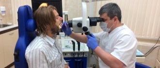
In most cases, pathological tooth root resorption is asymptomatic and is detected during routine radiographic examination. At the present stage, the number of detected resorptions has increased due to the introduction of cone beam computed tomography (CBCT).
In his study, K. Aziz [9] proved that a two-dimensional image often does not reflect the true nature or extent of the resorptive process.
K. Vasconcelos [67] in his report indicates that CBCT helps to determine the presence and topography of the resorptive area of the root, which determines treatment tactics.
Classification
Based on the analysis of the etiological factors of resorption, the authors proposed various classifications of this pathological process.
Thus, in particular, J. Andreasen [4] over the past 40 years has made a unique contribution to the understanding of tooth resorption after trauma. He proposed a classification that remains the most common to this day, and highlighted:
- internal resorption, which is divided into inflammatory and replacement;
- external resorption, which is divided into inflammatory and replacement.
However, this classification does not include other resorption processes that have been identified over the past two decades.
In 1999, M. Gunraj [28] proposed a more detailed classification:
Pathological tooth resorption:
1) External resorption:
a) external resorption caused by tooth trauma:
- superficial resorption;
- inflammatory resorption;
- replacement resorption;
b) external resorption caused by pulp necrosis and periapical pathology;
c) external resorption caused by pressure in the periodontal ligament;
2) internal resorption;
3) external invasive cervical resorption.
Today, hyperplastic processes leading to resorption of hard dental tissues are distinguished separately: invasive cervical resorption, invasive coronal resorption and internal replacement (invasive) resorption [32, 35].
The most recognized and convenient in clinical practice is the classification proposed by S. Lindskog [44] in 2006, based on the etiological factor causing pathological resorption of hard dental tissues. Three large groups were identified:
1) Resorption caused by trauma:
a) superficial resorption;
b) transient internal resorption;
c) resorption caused by pressure and orthodontic treatment;
d) replacement resorption.
2) Resorption caused by inflammation:
a) internal inflammatory (infectious) root resorption:
- apical;
— intrachannel;
b) external inflammatory resorption of roots;
c) combined internal and external inflammatory root resorption.
3) Hyperplastic invasive resorptions:
a) internal invasive (replacement) resorption;
b) invasive coronal resorption;
c) invasive cervical resorption.
Later, G. Heithersay [32] supplemented the classification of invasive cervical resorption, which includes 4 classes according to the degree of damage to hard tissues.
Class 1: small invasive lesion in the cervical area with slight penetration into dentin.
Class 2: Characterized by a well-defined invasive resorptive lesion that extends close to the coronal pulp chamber but has little or no involvement of the radicular dentin.
Grade 3: Defined by deeper invasion of dentin by tissue resorption, not only involving coronal dentin, but also extending to at least the coronal third of the root.
Grade 4: characterized by a large invasive resorptive process that extends beyond the coronal third of the root canal.
According to ICD-10, Pathological tooth resorption is designated by code K03.3 and is divided as follows:
- internal pulp granuloma;
— resorption of hard dental tissues (external).
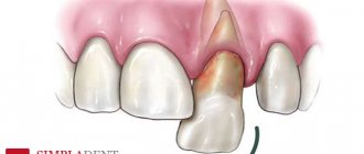
Primary and secondary edentulous teeth
Experts distinguish several types and subtypes of adentia. For greater convenience, below are tables showing the types of the disease, combined by certain characteristics.
Primary adentia
Caused by hereditary factors and diseases at the stage of fetal formation. With primary adentia, there is an absence of tooth germs.
Varies:
- partial primary adentia - the absence of one or more rudiments (for example, primary adentia of two incisors);
- complete primary adentia – absence of all tooth buds. It is extremely rare;
- true adentia – absence of tooth buds (without their destruction by diseases and infections);
- false edentia, when different teeth merge into one.
Secondary adentia
Secondary adentia occurs as a result of illness and injury during life.
Pathogenesis of resorption
The resorption process is associated with osteoclasts [14]. Osteoclasts are large multinucleated cells that are found on the surface of hard tissues in lacunae ( Howship 's lacunae
), or crypts.
They are very mobile and have visible pseudopodia. Osteoclasts differ from other multinucleated cells in that they are located on the surface of bone/dentin, in direct contact with the tissue. The osteoclast has a pronounced ruffle border
. This was first described in 1956 [61]. In the zona fimbriatum, intracellular vesicles fuse with the cell membrane and then release hydrogen ions and proteolytic enzymes into the resorptive space between the cell and the tissue surface [11]. This environment is very acidic, and as a result, calcium dissolves (leaches) from hard tissues. The zona fringe is tightly attached to the surface of the bone by integrin. Integrins are heterodimeric receptors involved in cellular processes such as migration, adhesion, proliferation, differentiation and cell survival [74]. Integrins in the zona fimbriae of osteoclasts interact with ligands to link the cells to the extracellular matrix of the root/bone, thereby isolating the resorptive region. Integrins also play an important role in transmitting information between cells [38]. The cytoplasm of the cell contains an array of organelles that are closely associated with active digestive roles, including the extensive endoplasmic reticulum, Golgi apparatus, ribosomes, and extensive collections of intracellular vesicles that migrate toward the zona fimbriae.
Osteoclasts are the main factor of resorption and can resorb bone, cartilage and, above all, in the context of this article, dentin. Odontoclasts and osteoclasts have similar modes of action but act in different locations. In this work, the term osteoclast will be used.
The exact origin and stimulation of osteoclasts are not fully understood. Several theories have been proposed.
It is known that the following signaling molecules take part in the formation and regulation of osteoclast activity:
M-CSF ( macrophage colony-stimulating factor
) - granulocyte-macrophage colony-stimulating factor. This cytokine is secreted by osteoblasts and affects hematopoietic stem cells, stimulating their differentiation into macrophages. M-CSF also has a paracrine effect on osteoclasts (binds to osteoclast receptors, inducing differentiation).
RANKL ( Receptor activator of nuclear factor kappa-B ligand)
- membrane protein, cytokine of the tumor necrosis factor family. Secreted by osteoblasts and stromal cells. Receptors for RANKL are found on the surface of monocytes. They are believed to be involved in stimulating macrophages and mononuclear cells to differentiate into osteoclasts. RANKL is important in the development and function of osteoclasts [49].
Osteoprotegrin or osteoclast inhibitory factor (OPG/OCIF) is a glycoprotein and also belongs to the tumor necrosis factor family. It is also secreted by osteoblasts and stromal cells, but inhibits RANKL [75, 76].
Thus, osteoblasts play a huge role in the activation of osteoclasts (secretion of RANKL and M-CSF activates osteoclasts and promotes resorption, and secretion of OPG prevents). Secretion of RANKL can be stimulated by parathyroid hormone, vitamin D3 and interleukin-1B [36, 37].
Interleukin-1 B is an integral part of inflammatory processes. It has been proven that interleukin-1B is closely related to the resorptive process of hard dental tissues associated with periapical and periodontal pathology [63, 64]. There is an assumption that patients with deviations in the interleukin-1 B allele have a genetic predisposition to resorptive processes [31].
Also, the resorptive process can be caused by the presence of bacteria. Complement proteins, bacterial toxins and antibodies from B lymphocytes attract white blood cells. Although not all chemical mediators of inflammation found in the pulp and periapical tissues are associated with osteoclastic activity, they are chemoattractants for leukocytes [39]. Leukocytes differentiate into osteoclasts in the presence of certain bacterial lipopolysaccharides. Treponema, Porphyromonas possess such surface lipopolysaccharide antigens
and
Prevotella
[16, 17, 55].
The destruction of the organic component (collagen matrix) of bone tissue and dentin occurs due to the activation of enzymes: collagenase, matrix metalloproteinase and cysteine proteinase. It is believed that the enzymes are initially incorporated by osteoblasts into the structure of dentin or bone tissue. However, their activation occurs only upon direct contact with osteoclasts, due to changes in the pH of surrounding tissues.
Osteoclasts are involved in maintaining normal homeostasis and tissue repair. For example, parathyroid hormone stimulates resorption to increase circulating calcium levels: a normal response to decreased blood calcium levels [22].
From a dental point of view, physiological resorption is an extremely important process in the replacement of temporary dentition. There is a constant balance between osteoclastic activators and inhibitors. When tissue damage occurs, cytokines are produced and the repair process involves osteoclast activity. The RANKL system is believed to be an integral part of restoration in dental hard tissues [45].
If extensive damage to tooth tissue has occurred, complete resorption may occur.
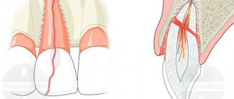
At the histological level, resorption of tooth root cement due to microdamage occurs constantly. However, when the provoking or causative factor disappears, it is restored. There are biological defense mechanisms to prevent normal tooth resorption (healthy tissues of the periodontal ligament, cementum, cementum and extracellular layer of predentin).
Cementum and periodontal ligament are barriers to external root resorption, and the presence of predentin is a barrier to internal resorption.
Damage to these barriers allows osteoclasts to attach to the root surface, and in the presence of inflammation, resorption of dental hard tissue occurs. Clinically significant damage can only occur with constant stimulation of osteoclastic activity.
Depending on the etiological factor, pathological tooth root resorption can manifest clinically in different forms and will require different treatment approaches.
Treatment of various forms of tooth root resorption
Tooth resorption caused by trauma
This group includes pathological resorptions of dental tissues resulting from acute or chronic trauma (high blood pressure). Damage to the cemento-periodontal complex occurs with activation of osteoclasts. This group of resorptions occurs without infection.
A. Surface resorption
Surface resorption occurs with non-acute tooth trauma.
It is almost impossible to detect x-ray.
From a clinical point of view, this option is the best. Resorption is limited by cementum. Over time, the resulting defect is restored.
Does not require treatment.
B. Transient apical internal resorption
Another form of traumatic, non-infected resorption was discovered by J. Andreasen [5] in 1986, which can occur with incomplete tooth luxation, lateral luxation, orthodontic treatment, and permanent occlusal trauma.
This process is associated with the phenomenon of transient apical breakdown. A transient apical breakthrough appears on a radiograph as a limited area of radiolucency in the apical region of the tooth root [18].
Visually, the tooth crown can be changed in color. The temperature test can be either positive or negative. Often the tooth pulp remains alive.
When the damaging factor is eliminated, the process will likely resolve itself within a few months. It is believed that a positive result is associated with good blood supply to the apical region and the reparative abilities of the pulp. Due to revascularization, self-liquidation of discoloration of the coronal part of the tooth is possible.
Treatment:
wait-and-see tactics. If the return to normal does not occur within six months or the clinic of periodontitis appears, then it is necessary to carry out endodontic treatment followed by obturation of the root canal.
B. Pressure-induced resorption and resorption during orthodontic treatment
An extensive group that includes resorptions caused by prolonged compression: unerupted teeth, neoplasms, incorrect orthodontic treatment.
It is characterized by a loss of hard tooth tissues from the side of the compressive object.
G. B. Ospanova [2], indicates that in adults, resorption of tooth roots during orthodontic treatment occurs more often than in adolescents; more often in women than in men. In patients who have previously undergone orthodontic treatment, resorption develops more slowly than in patients who are new to orthodontic treatment. The main cause of resorption is an overdose of force used to move teeth, the progression of which stops immediately after removal of the orthodontic appliance. With an intact periodontium, cement is formed again and the original shape of the periodontium is restored. However, the apical structure of the root is never restored.
Research by F. Weiland [72] shows that orthodontic forces up to 200 cN are probably not decisive for tooth root resorption.
Treatment
: When mechanical pressure is removed, resorption stops.
D. Replacement resorption (ankylosis)
The most serious form of traumatic, non-inflammatory resorption.
The phenomenon is caused by the death of periodontal cells and damage to the precement (trauma, improper treatment of the tooth during replantation).
There is a slow replacement of tooth tissue with alveolar bone according to the mechanism of typical bone tissue remodeling.
Clinically manifested by loss of physiological tooth mobility. A high percussion sound is possible. The X-ray shows the disappearance of the periodontal fissure. At other times the process is asymptomatic [24].
Treatment:
endodontic treatment does not stop this type of resorption. However, replacement resorption takes a long time. Complete replacement of tooth tissue with alveolar bone can take many years [65]. A wait-and-see approach is recommended, followed by decoronation (removal of the crown part) of the tooth, and after complete root resorption, implantation treatment.
In his works, M. Oliveira [56] coated the surface of the replanted tooth with Emdogain. The drug contains amelogenin and stimulates the restoration of periodontal tissue and non-cellular cement, which prevents the occurrence of replacement resorption.
Tooth resorption caused by inflammation
The reaction of the dentino-alveolar apparatus to infection, characterized by inflammation, can lead to resorption of hard dental tissues. The infection may be of endodontic origin or associated with resorption caused by trauma. Resorptions induced by infection (inflammatory resorptions) can occur inside the root system of the tooth, on the surface of the cementum of the tooth, and combined internal and external lesions are also possible. Resorptions caused by infection can vary widely in complexity but generally have a favorable prognosis if the infectious agent is removed.
A. Internal inflammatory resorption
Internal inflammatory resorption is a destructive process that occurs only within the root canal system and/or dental cavity, characterized by the loss of dentin as a result of the action of osteoclasts [1].
— Apical internal inflammatory resorption
A recent study by F. Vier and J. Figueiredo [69] showed that apical internal inflammatory resorption is much more common in teeth with various inflammatory periapical pathological processes than previously thought. The study showed that 74.7% of teeth with periapical lesions had varying degrees of apical internal resorption.
This study has important clinical implications for root canal preparation. There are two approaches to endodontic treatment with apical internal resorption. The first involves instrumental treatment of the canal to the level of resorption. It is assumed that when microorganisms are removed, the resorbed tissue will be restored.
The second approach is to enlarge and prepare the apical part of the root, including the resorbed area, and then fill the root to the root canal apex [15].
At the moment, there are no studies comparing the long-term results of the two approaches to treating this pathology.
— Intracanal inflammatory resorption
The histological picture of the development of internal resorption has two phases: The first is damage, the second phase is infectious stimulation or maintenance of the resorption process. The etiological factor leads to trauma to the tooth and initiates intrapulpal bleeding (tooth trauma, overheating of the pulp as a result of violation of the preparation rules). The hematoma is organized, that is, replaced by granulation tissue. Proliferating granulation tissue puts pressure on the dentin walls, damaging non-mineralized tissues covering the inner surface of the root canal, the odontoblast layer and predentin. Osteoclasts differentiate from connective tissue and resorption begins [1]. Maintaining this process is possible only if viable areas of the pulp are preserved inside the tooth cavity or if there are large-caliber lateral canals providing blood supply, as well as constant stimulation with an infectious agent [46].
There are studies explaining the transport of pulp resorbing cells from the viable apical part of the pulp [30]. Thus, for the progress of internal resorption, on the one hand, there is a need for a blood supply to viable pulp tissue, which ensures the transport of osteoclasts to the resorption zone, and, on the other hand, an infected part of the pulp, which provides stimulation or progression of resorption [50, 58].
Clinical picture: most often the process is painless; testing for tooth vitality can be positive or negative. Visually, you can see a pink spot within the crown of the tooth.
An x-ray reveals a vacuum in the area of the inner wall of dentin facing the tooth cavity. However, two-dimensional radiography does not cope well with the task of early diagnosis of internal resorption and its differential diagnosis. In cases where the doctor’s capabilities are limited only by this examination method, it is recommended to take a series of photographs of the area of interest in different projections. In the differential diagnosis of external root resorption, it is necessary to pay attention to the relationship between the shadow of the root canal and/or tooth cavity and the radiological focus of clearing, which represents the site of resorption. While maintaining the contours of the shadow of the root canal and/or tooth cavity, against the background of the shadow of the focus of enlightenment, resorption is external. In the case when the boundaries of the radiographic shadow of the root canal sharply disappear or are deformed against the background of a focus of radiological clearing, we are talking about internal resorption. Currently, it is preferable to use cone beam computed tomography (CBCT) to diagnose internal resorption [1, 9].
Treatment:
Features of the pathological process help to understand that it is possible to stop resorption only with the complete elimination of pulp tissue - both viable in the apical part of the tooth cavity and infected.
In the presence of internal resorption, we follow the standard protocol for endodontic treatment of the root canal with copious rinsing with sodium hypochlorite. The changed root system configuration must be taken into account. To influence untreated areas of the canal, temporary filling of the canal with liquid calcium hydroxide is proposed for a period of no more than 2 weeks [3, 46]. For obturation, a combined technique should be used: the apical part of the root below the resorption areas is filled using the lateral condensation method, and then using the hot gutta-percha method.
B. External inflammatory resorption
This type of resorption is extremely aggressive and usually occurs when infection superimposes trauma - after replantation or tooth luxation.
The presence of infection on the root surface causes a normal inflammatory response, including activation of clastic cells, leading to resorption. The process is asymptomatic.
Radiographically, external inflammatory root resorption appears as cup-shaped defects in the tooth root and adjacent bone [6].
The process can also be induced by endodontic pathology. This requires the presence of the following conditions: damage to predentin, cementum and cement with a violation of the sealing of the dentinal tubules, as well as complete necrosis of the pulp with infection of the root system of the tooth.
Bacterial toxins present from the dentinal tubules enter the periodontium [65]. A normal inflammatory response occurs, including activation of osteoclasts. The process is supported by a constant supply of microbial agents, which leads to resorption of both tooth and bone. This mechanism occurs more aggressively in young teeth due to the large diameter of the dentinal tubules [30].
Treatment
: The main effect should be focused on removing the infectious agent that supports osteoclast activity. Endodontic treatment is carried out using the method described above.
B. Combined internal and external inflammatory root resorption
Occurs when internal inflammatory resorption has spread to the outer surface of the root.
Radiographically, this pathology appears as a focus of radioluminescence in the internal structure of the tooth, extending to the outer surface of the root and surrounding bone.
Treatment:
On the first visit, we perform endodontic treatment of the root system with copious rinsing of sodium hypochlorite, close the perforation with MTA-based material and perform temporary filling of the canals with calcium hydroxide for 14 days.
On the second visit, provided that the MTA material has hardened and tightly covers the perforation area, we will obturate the root system using a combined filling technique.
Hyperplastic invasive tooth resorption
The third group of tooth resorptions is the most aggressive and difficult to treat. With this pathology, uncontrolled growth of resorptive tissue occurs, destroying the hard tissues of the tooth. The histological picture is similar to some fibro-osseous lesions, such as fibrous dysplasia [70].
A distinctive feature of hyperplastic resorptions is the fact that for treatment it is not enough to remove the causative factor. Only complete elimination of resorptive tissue helps stop the process. It is assumed that a high percentage of relapses is associated with invasive tissue growth—communications with the periodontium and the main source of resorptive tissue growth are formed in dentin [33].
Although these resorptions are not considered neoplastic processes, their aggressive characteristics are similar in some manifestations.
Hyperplastic invasive resorptions can be of pulpal (internal) or periodontal (external) origin.
Correct diagnosis of the type of resorption and the location of the resorption are critical to the choice of treatment to combat internal and external invasive tooth resorptions.
A. Internal invasive (replacement) tooth resorption
Internal replacement (invasive) tooth resorption is a relatively rare type of resorption [71], which clinically manifests itself as a pink spot on the crown of the affected tooth. The clinical manifestation is due to the fact that the resorptive tissue has already destroyed a significant amount of enamel and dentin, showing through the remaining hard tissues of the tooth.
The etiological factor of internal invasive resorption is trauma. The lesion can be isolated by the root system of the tooth, but in most cases there is communication with the periodontium.
On the radiograph it appears as a violation of the configuration of the root canal with uneven edges or its expansion in the form of an oval focus of radiolucency.
B. Invasive coronal tooth resorption
This rare condition usually develops during teething. The presence of a defect in the coronal enamel contributes to the invasion of hyperplastic resorptive tissue from the periodontium. The cause may also be trauma to temporary teeth with damage to the rudiments of permanent teeth.
Radiographically, it is defined as a defect of the coronal part in the form of an oval-shaped focus of radiolucency with uneven edges.
Treatment is aimed at complete removal of all resorptive tissue and restoration of the coronal defect. This can be achieved by preparing the defect with burs and hand instruments.
B. Invasive cervical tooth resorption
Invasive cervical resorption (ICR - invasive cervical resorption) is characterized by a cervical location. This resorptive process can occur in any tooth in the permanent dentition [34].
If left untreated, invasive resorption leads to destruction and significant loss of dental hard tissue.
Invasive cervical resorption, as a rule, is painless if there is no secondary infection.
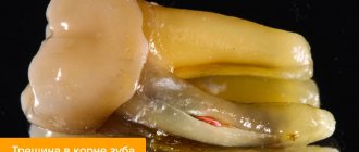
Resorption of coronal dentin and enamel often creates a clinically pinkish color in the coronal area. In the absence of external signs, diagnosing this pathology is difficult and most often occurs by chance during routine radiography.
When diagnosing this disease, a high percentage of errors remains. This is related to early descriptions by Gaskill in 1894 and Mummery [48] in 1920, which included teeth with “pink spots” in the group of internal resorptions.
The exact cause of invasive cervical resorption has not been identified, but the following predisposing factors have been identified: intracoronal bleaching, orthodontic treatment, trauma, periodontal treatment, defects in the development of hard dental tissues, bruxism.
G. Harrington and E. Natkin [30], in 1979, recorded intracoronal bleaching as the strongest risk factor, followed by other factors such as trauma, orthodontics, orthognathic surgeries, etc.
G. Heithersay, in his study of 222 patients with 257 teeth affected by ICR, analyzed the effectiveness of risk factors, causing them in isolation or in combination with each other. His analysis shows that orthodontics is the most common single factor causing ICR in 47 (21.2%) patients with 62 affected teeth, followed by intracoronal bleaching in 33 (14.9%) patients, trauma in 31 (14%) patients with 39 affected teeth and periodontal operations in 13 (5.9%) patients.
Other factors had very low frequencies. Bleaching combined with trauma was the cause in 2 (0.9%) patients, whereas bleaching with orthodontics was responsible in 4 (1.8%) patients [32].
Invasive cervical resorption expands first coronally and then apically, surrounding the root canal. The pulp usually remains imperforate and healthy due to the presence of a non-mineralized layer of predetiin [28, 34]. However, with long-term lesions, the canal can be perforated by advancing resorptive lesions [34].
Radiographic features of such lesions: a clear focus of radioluminescence, sometimes exceeding the contour of the pulp, while the contours of the canal remain clear [34].
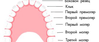
Classification of missing teeth according to the location of the disease
| By localization | Description |
| Missing upper teeth | The absence of teeth in the upper jaw can be either partial or complete, as well as single. |
| Missing lower teeth | A general designation for a disease indicating the absence of teeth in the lower jaw. |
| Lack of chewing teeth | May be referred to as missing lateral teeth. Lack or complete edentulism in the chewing region. In particular, we are talking about the absence of small molars (premolars) and molars. |
| Missing front teeth | Edentia of incisors or canines in the frontal zone. |
In addition to the classification described above, the type of teeth itself is also taken into account. Based on this, the absence of molars and edentulous primary teeth (not associated with the normal timing of their replacement) are distinguished.