Polyodontia (hyperodontia, hyperdentia) is the appearance of supernumerary teeth. Normally, an adult has 28-32 teeth (including wisdom teeth); with polyodontia, an indefinite number of extra teeth appear. About 2% of the world's population suffers from the pathology.
There are no methods to prevent the development of polyodontia, but it is possible to reduce the number of complications caused by the disease. Therefore, it is necessary to be attentive to your health and undergo a preventive examination with a dentist twice a year. If you suspect that an extra tooth has appeared in your mouth, you should immediately consult a doctor. Particular responsibility lies with parents, since children are not able to independently monitor the health of their teeth.
Polyodontia is an abnormal number of teeth.
In medicine, this disease is often called hyperdontia, and “extra” dental elements are called supernumerary teeth. Research is still being conducted into why this pathology occurs. Most scientists associate it with disturbances in the formation of tooth germs.
Nature provides that a person grows no more than 20 milk teeth and 32 permanent teeth in a lifetime, but exceptions occur, and in our time quite often. According to statistics, on average, dental anomalies occur in 2% of the world's population, most often in men.
In 2014 alone, two operations were performed, in one of which 80 teeth were removed, and in the other, a record 232 teeth. Until this time, the maximum figure was 37 teeth.
The most common hyperdontia (anomaly in the number of teeth) is an anomaly of the upper incisors. Supernumerary teeth are less common among the lower incisors and in other parts of the jaw. They can come in a wide variety of shapes and sizes. These are usually small, cone-shaped teeth.
Extra teeth lead to deformation of the dentition, so it is recommended to remove supernumerary elements. Another reason for removal is that most patients with this pathology have a lisp.
The formation of extra teeth is quite common today. According to statistics, 70% of patients have only one extra incisor, in 25% of cases – 2 supernumerary elements, and only 5% of all patients have 3 or more teeth during examination.
Supernumerary molar: review of a rare case
Supernumerary teeth or hyperdontia is a dental anomaly that is defined as the presence of a tooth or any dental tissue in excess of the set of 20 primary teeth and 32 permanent teeth. Supernumerary teeth can occur singly, in groups, unilaterally, bilaterally, they can erupt or be impacted on one or both jaws, both in the primary and permanent dentition. The frequency of occurrence in primary dentition varies from 0.1% to 3.8% and from 0.3% to 0.6%. In permanent dentition, the anomaly is more common in men than in women in a ratio of 2:1. However, this gender disproportion is not observed in the primary dentition. There is also evidence that the Asian population is more susceptible to the anomaly. Single supernumerary teeth occur in 76-86% of cases, double teeth in 12-23% and multiple teeth in less than 1%. Multiple hyperodontia rarely occurs in people without any other concomitant diseases and syndromes. Typically, this anomaly is part of systemic disorders such as cleft lip and palate, cleidocranial syndrome, Gardner syndrome, Fabry-Anderson syndrome, chondroectodermal dysplasia, Euler-Danlos syndrome, and tricho-rhinophalangeal syndrome.
Supernumerary teeth can be found in almost any area of the dental arch. Localization on the upper jaw is much more common than on the lower jaw, especially in the anterior region (80%). Somewhat less frequently, supernumerary teeth can be located in the distomolar zone, lower and upper premolars, in the area of the upper canines and lower incisors.
The crowns of abnormal teeth have a normal appearance or an atypical shape, and the roots are also fully or partially formed.
The position in the dental arch varies: mesiodens, paramolar, distomolar and parapremolar. Mesiodens is the most typical localization between the central incisors on the upper jaw, paramolar position is an additional molar, usually rudimentary, small in size and located on the buccal or palatal side in relation to one of the molars on the upper jaw. Most often found in the interdental space of the second and third molars on the buccal side; distomolar position is the fourth permanent molar; parapremolar localization is mainly found in the interdental space on the buccal side between the first and second premolars in the upper jaw. Variations in the morphological shape include different conical type, number of tubercles, and odontome. Supernumerary teeth may be small, conical with a normal root; teeth with multiple cusps are usually short, with a barrel-shaped crown and an invaginated rudimentary root. Another variant of a supernumerary tooth - an additional one - resembles one of the existing ones and is located behind it. Most supernumerary teeth in primary dentition are of the accessory type.
Odontomas are any tumors that develop from tooth tissue. Many authors are inclined to believe that odontomas are a hamartoma or malformation rather than a neoplasm. Compound and compound odontomas are two different types described. Complex odontomas are characterized by diffuse dentin tissue that is completely disorganized, while compound odontomas are malformations that have superficial anatomical similarities to a normal tooth.
According to their shape, supernumerary teeth are classified into additional (eumorphic) and vestigial (dysmorphic). If supernumerary teeth have normal morphology, they are classified as “additional”; if the morphology is abnormal, the teeth are classified as vestigial. The position of the supernumerary teeth can be between the central incisors, overlapping, and the orientation is described as vertical, inverted, or transversal.
This article presents a clinical case of the presence of an additional molar in a somatically healthy patient. A review of the literature regarding the incidence, classification, etiology, complications, diagnosis and treatment strategies of this pathology is also presented.
Description of a clinical case
A 22-year-old man came to the Department of Conservative Dentistry and Endodontics with complaints of pain in the posterior segment of the upper jaw on the left. Hereditary anamnesis and disease history are unremarkable; no signs of systemic diseases or syndromes have been identified.
Intraoral examination revealed Class I occlusion and no pathological tooth alignment. In addition to the full set of permanent teeth, one supernumerary tooth was found, located on the palatal side between the upper first and second molars on the left (Figure 1).
Figure 1: Intraoral photograph showing the paramolar position of the supernumerary tooth between the upper first and second molars on the left.
The supernumerary tooth is defined as a paramolar. The paramolar crown had two cusps and very much resembled the structure of a permanent premolar. The tooth is rotated axially, with the buccal surface distally and the mesial surface buccal. A carious lesion was found on the mesial side of the paramolar (Figure 2). Examination of the soft tissues revealed periodontal inflammation between the first and second molars and paramolars. X-rays were taken: panoramic, sighting and occlusal. Reading the panoramic image was difficult due to the palatal position of the tooth. On sighting and occlusal photographs, it was discovered that the supernumerary tooth was affected by caries and had one root (Photos 3 and 4).
Figure 3: Spot X-ray showing a paramolar with a fully formed tooth (indicated by arrow).
Figure 4: Occlusal radiograph of the maxilla showing the supernumerary tooth (arrow).
The patient was informed of the existing situation. It is recommended to remove the paramolar due to its inconvenient location for hygiene, possible food retention, recurrence of caries and damage to periodontal tissue. The patient was sent to the Department of Maxillofacial Surgery for paramolar removal.
The extracted tooth is cleaned, disinfected and analyzed. The morphology of the tooth is normal. The length of the root corresponds to the size of the crown. The root apex is fully developed. X-ray examination revealed type I canal configuration (Vertucci). Actual tooth dimensions: mesiodistal and bucco-palatal crown width 6 and 10 mm, respectively, crown length 6.5 mm, root length 12 mm. Morphometric measurements showed a high similarity of the supernumerary tooth with the premolar (Photo 2).
Photo 2: Photographs of the extracted tooth: (a) occlusal view, (b) mesial, (c) distal, (d) buccal, (e) palatal.
Discussion
The appearance of paramolars is a fairly rare occurrence. The etiology of this anomaly is not fully understood. Several theories have been proposed: phylogenetic, dichotomous, dental lamina hyperactivity theory, and a combination of genetic and environmental factors.
Phylogenetic theory refers to the process of atavism (evolutionary return). Atavism is a return to an earlier morphology or type. In past centuries, the third molar was almost always present in the permanent dentition; it was comparable in size to the second molar. Moreover, the fourth molar was also quite common. However, as a result of the evolution of phylogeny, the size of the dental arches gradually decreased, which led to a reduction in both the number and size of human teeth. This was one of the stages of preferential development of the cerebral skull over the facial skull. Thus, the appearance of additional paramolars can be considered an example of atavism, the genetic memory of the fourth molar in previous generations. It is worth saying that this theory was rejected by many authors.
The dichotomous theory explains the appearance of supernumerary teeth by splitting the tooth germ. The rudiment splits into two equal or unequal parts, from which morphologically normal independent teeth subsequently develop.
The lamina hyperactivity theory is the most accepted theory. She explains the appearance of paramolars as a result of local, independent, due to special stimulation, increased activity of the dental plate. According to the theory, lingual expansion of the accessory tooth bud leads to the development of a morphologically unchanged tooth, and the vestigial forms arise from the proliferation of epithelial lamina remnants, which is induced by the pressure of permanent teeth. Others are inclined to believe that hyperodontia is associated with multifactorial causes, which are still based on hyperactivity of the dental plate. Remnants of the dental lamina may remain in the jaws in the form of epithelial pearls or islands. When exposed to inducing factors, supernumerary teeth or odontomas can develop from additional rudiments. The best supported hypothesis is that the development of supernumerary teeth is associated with a complex of genetic and environmental factors. This is confirmed by the presence of similar anomalies in close relatives. However, despite the literature data, similar pathology was not found in the relatives of the described patient.
A careful analysis of the literature revealed very little information about the appearance of paramolars. Paramolars are somewhat less common in the upper jaw, very rarely bilaterally and almost never in the primary dentition. They are usually vestigial and located buccally between the second and third molars, although in some cases they can be located between the first and second molars. Fusion of paramolars with normal teeth is also incredibly rare. The literature describes the only case of endodontic treatment of a fused second left molar in the lower jaw and a paramolar with a split crown.
Diagnosis also requires differentiation of other structures that may appear in the molar area, such as an additional cusp or a fused supernumerary tooth. Bolk in 1916 first described an additional cusp on the buccal surface of the upper and lower permanent molars, which he called the paramolar cusp. Dahlberg in 1945 proposed the term paramolar cusp to refer to any abnormal cusp, supernumerary inclusion, or elevation on the buccal surface of both maxillary and mandibular premolars and molars. He presented a paleontological nomenclature in which he classified these structures as “protostylid” if they are on the lower jaw and “parastylid” if on the upper jaw. It is widely accepted today that such formations originate from the cervical region of the tooth and are variable in appearance. Often these structures appear on the buccal surface of the mesiobuccal tubercle and quite rarely on the distobuccal tubercle. It is believed that the paramolar tubercles may originate from the remains of their own epithelium or be a genetic remnant from mammals and lower primates.
Supernumerary teeth may erupt normally, remain impacted, or appear axially rotated or with other abnormalities. Supernumerary teeth with a normal position in the bone usually erupt. However, only 13-34% of supernumerary teeth in the permanent dentition erupt normally, compared to 73% in the primary dentition. The rest may remain impacted and cause complications.
The development of complications can cause a delay in the eruption of associated permanent teeth, retention, ectopic eruption, disposition, rotation of adjacent teeth, crowding due to insufficient space for eruption, malocclusion due to a decrease in space in the dental arch during the eruption of paramolars, tremors in the molar area, traumatic bite and ulceration of the buccal mucosa with buccal placement of paramolars, difficulties in orthodontic treatment, pathological development of the root of associated permanent teeth, formation of follicular cysts from the follicular sac of a supernumerary tooth, trigeminal neuralgia due to compression, pulp necrosis and root resorption due to excessive pressure of the paramolar , caries due to plaque accumulation, gum inflammation and localized periodontitis. As can be seen from the described case, due to plaque retention, carious lesions of the paramolar and inflammation of the surrounding periodontium occurred.
Most supernumerary teeth are impacted and are usually discovered incidentally during radiographic examination. However, if a patient presents with complications that are often associated with the presence of a supernumerary tooth, the dentist should consider this anomaly in the differential diagnosis and insist on appropriate x-ray examination.
The most valuable radiographic examination is the OPG with additional targeted photographs and photographs of the upper and lower jaw in the occlusal plane. To clearly localize an unerupted tooth, use the vertical or horizontal parallax technique. Parallax is a change in the view of an object against a specific background based on the movement of the viewer. This technique can be carried out by taking images of the same area, but from different angles, with two different devices. When using this method, as a rule, the reference point is the root of the adjacent tooth. In addition, cone beam CT may be used. This technology provides a three-dimensional image of the structures of the specified zone and is incredibly informative for the described anomaly.
Clinical management of patients with paramolars depends on the position of the tooth and its effect on surrounding tissues and important anatomical structures. Treatment offers two options: removal or observation. Observation does not include any manipulations other than clinical and radiological monitoring of the patient. This method is preferable if the presence of a paramolar is asymptomatic and does not cause any inconvenience. If any complications arise, tooth extraction is advisable. In the described case, we resorted to tooth extraction in order to maintain the proper level of hygiene, prevent the carious process and preserve the surrounding periodontium.
Conclusion
The dentist needs to know about the different types of supernumerary teeth for proper diagnosis and timely detection of this anomaly. Each such case requires careful diagnosis and subsequent appropriate treatment that causes minimal complications.
Authors: Gurudutt Nayak, Shashit Shetty, Inderpreet Singh, Deepti Pitalia Department of Conservative Dentistry and Endodontics, Kanti Devi Dental College and Hospital, Mathura, Uttar Pradesh, India
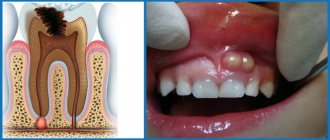
Causes of polyodontia
Medicine has not yet found an exact answer to the question of what are the causes of supernumerary teeth. Scientists put forward several hypotheses:
- Atavism. Supernumerary teeth are explained by the fact that the dental system strives to return to the original number of elements laid down by nature. There is evidence that our ancestors had 6 incisors on both the lower and upper jaws. As a result, many doctors consider atavism to be the cause of the development of polyodontia in humans.
- Splitting of the tooth germ. Even in the embryonic period, the activity of the dental plate is disrupted in the child, as a result of which hyperdontia is formed. Violations can be caused by viruses, poor ecology, drugs, medications prohibited during pregnancy, alcohol and other factors. This hypothesis is increasingly supported today, because recently the disease has been rapidly progressing due to bad habits and poor ecology.
The causes of hyperdontia continue to be researched. Scientists cannot give an exact explanation for this anomaly, but most of them are inclined to the second hypothesis - the splitting of the tooth germ at the embryonic stage.
Spreading
Normally, a person should have 20 baby teeth in childhood, which are replaced by 32 teeth, starting in adolescence. But for the reasons stated above, hypersets with two, four or more redundant teeth can be formed. 2% of people susceptible to this pathology is the total number. But among them it is possible to identify groups in which supernumerary teeth erupt in numbers of no more than two.
The location of such extra teeth (mainly incisors) is the upper jaw, with a shift of the supernumerary units towards the palate. Such people, of the total number of people susceptible to hyperdontia, are about 48%.
The second group is characterized by the growth of canines or premolars, also on the upper jaw. Incomplete teeth appear in them in the amount of one or two.
More than four, six, eight, etc. extra teeth grow only in a rare minority, with hypersets located in the lingual part of the oral cavity.
The remaining cases occur due to the unsystematic appearance of teeth in the lower jaw, on the side of the tongue, lips and cheek.
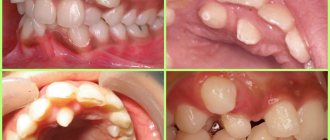
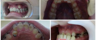
What does polyodontia look like?
Quite often, extra teeth are almost indistinguishable from normal ones. It is not uncommon for them to grow in the form of a drop or a thorn. These dental elements can appear either individually or fused with permanent ones. They can form tooth-like formations and entire arrays of teeth.
Also in medical practice, there are cases where polyodontia was hidden and was detected only by radiography. There are many different cases of abnormal development of the number of teeth, and if you notice symptoms, you should definitely contact the dentist.
Types of polyodontia disease
Polyodontia in the oral cavity manifests itself in different ways. By studying the statistics, signs and symptoms of the disease, dentists were able to classify the types of this anomaly.
Depending on the origin, the disease is divided into two types:
- False polyodontia. Provides for a baby tooth that does not fall out, regardless of the person’s age. At the same time, it fulfills its functions, does not create discomfort to the bite, and is firmly fixed in the patient’s jaw. In addition, teeth fused together and other anomalies are classified as a false type of disease.
- True polyodontia. It can be caused by genetic predisposition, as well as terogenic factors. At the same time, extra molars begin to form in the human jaw.
Diagnostic methods
Diagnosis of polyodontia is carried out exclusively by a specialized doctor - an orthodontist, who conducts a preliminary examination of the oral cavity and dentition, refers the patient to x-rays (orthopantomography) and analysis of anthropometric casts of teeth made of plaster. Such models are made for a more detailed study of a specific anomalous phenomenon, on the basis of which a diagnosis is then made and treatment is prescribed.
Table of normal human teeth. Deviation from the norm - polyodontia
When examining plaster models made on the basis of dental casts, the doctor measures the size of the teeth, and then analyzes the data obtained based on a special table by Wetzel and Ustimenko. The length of each row of teeth must be measured. This indicator may fluctuate depending on such characteristic individual characteristics of a person as crowding of teeth or the presence of large gaps between them. In the first case, the length will be less than normal, and in the second, a little longer. To diagnose asymmetry, the distance from the midline of the palate (or hyoid frenulum) to the far point of the dentition in one direction or the other is determined.
As for the placement of extra teeth, dentists distinguish the following types of disease:
- Typical hyperdontia. Applies to those patients in whom extra teeth appear only in the dentition and do not extend beyond it. Many scientists are confident that this is simply heredity, because our ancestors had a more developed dental system than modern people.
- Atypical hyperdontia. It occurs much less frequently and is characterized by the appearance of teeth outside the dentition.
In case of anomalies with baby teeth, the latter pose almost no threat. On the contrary, such a tooth can last a lifetime. But the permanent molars, over which the supernumeraries grow, should be removed, if only because it is not aesthetically pleasing.
Often, the patient grows extra fangs or incisors, or even several front teeth at once. In addition to a ruined smile, the disease can cause serious complications if the necessary measures are not taken in time.
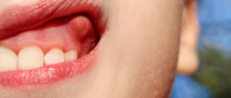
Symptoms of the disease in children
The first supernumerary teeth in children appear before birth or in the first six months of life. The main inconvenience they cause is difficulty in feeding.
Polyodontia of primary teeth in older children occurs with symptoms similar to the eruption of regular teeth. In this case it is observed:
- temperature increase;
- swelling of the gums in the place where the tooth should erupt;
- pain;
- excessive salivation;
- swelling of the nasal mucosa;
- loose stool.
Symptoms are especially severe when extra teeth appear in the upper palate.
If hyperdontia makes itself felt in a two-year-old child, this can interfere with the formation of normal speech. In turn, due to injury to the tongue and mucous membranes, some kind of inflammation constantly appears in the oral cavity.
When supernumerary teeth appear in very noticeable places in school-age children, ridicule towards the patient may occur, which is fraught with the development of psychological problems and complexes in the future.
Complications caused by polyodontia
Supernumerary teeth appear more often on the upper jaw in the permanent dentition, and are less common on the lower jaw and in the primary dentition. Their appearance threatens with the following complications:
- Development of anomalies of the dental system: change in the position of complete teeth: their displacement to the sides, forward or backward, rotation around an axis;
- bite deformation, as a result – disruption of chewing and speech functions;
- disruption of the eruption of permanent teeth, up to the loss of their ability to erupt;
- the appearance of a diastema - a pathological distance between the central incisors;
- the formation of three spaces between the remaining teeth.
- chronic gingivitis;
The eruption of supernumerary teeth is often accompanied by the following symptoms:
- increased body temperature;
- pain and swelling in the eruption area;
- increased salivation;
- swelling of the nasal mucosa.
Supernumerary teeth affect not only the visual component of the smile, but also cause systemic disorders that lead to deterioration in chewing food and disruption of the stability of the main teeth. All this also affects a person’s psychological comfort: he becomes withdrawn, does not smile, so as not to demonstrate smile defects.
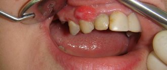
Symptoms of hyperdontia in adults
Polyodontia affects permanent teeth more often than baby teeth. An adult usually develops dystopic and impacted supernumerary teeth.
Dystopic teeth are those that appear outside the dental arch. Most often they erupt on the lingual surface of the gums and in the palate. With this form of the disease, the patient typically:
- poor pronunciation of sounds;
- noticeable malocclusion;
- change in the usual arrangement of teeth: curvature of the angle at which they grow, as well as their rotation
- around its axis;
- frequent injury to the oral mucosa and, as a result, its inflammation;
- disruption of chewing processes, resulting in digestive problems.
Among other things, dystopic teeth often cause psychological problems. Due to a non-aesthetic, and sometimes completely unattractive smile, the patient becomes withdrawn and uncommunicative. Psychological problems, in turn, cause chronic diseases of the endocrine, digestive and nervous systems.
Impacted supernumerary teeth are teeth that do not erupt, but continue to remain in the bone tissue of the human jaw. Often they hardly make themselves felt until complications begin. Dentists diagnose this anomaly during a routine examination of the patient.
This abnormality in the number of teeth is accompanied by the following symptoms:
- normal teeth begin to loosen (the condition is considered pathological);
- the bone begins to protrude (if the impacted tooth is too close to the edge of the jaw);
- Aching pains appear periodically.
One of the most difficult situations is when extra teeth grow in place of impacted third molars. Wisdom teeth cannot grow and begin to negatively affect the roots of other teeth, which in turn can lead to serious complications.
Consequences of polyodontia disease
Polyodontia in humans can often be the cause of retention. This is a phenomenon in which normal teeth are unable to erupt due to the interference of supernumerary teeth. The former may remain in the jaw or take an abnormal position.
In addition, even if the complete incisor grows before the supernumerary one, the latter will be able to displace it. This will lead to the person being unable to chew food normally. And if several extra incisors grow at once, they can cause the loss of permanent teeth.
Diagnostics
Examining supernumerary teeth during an x-ray is not as easy as it seems. They can be superimposed along the contour onto the permanent ones and remain invisible. In such cases, patients are recommended to undergo a computed tomography scan, which shows a more accurate picture of the disease.
If the extra dental elements have already erupted, the dentist can easily detect them. In practice, the patients themselves find the erupted supernumerary teeth and already at the initial appointment with the dentist they complain about the pathology.
Mouthguard as a preventive measure for the second dentition
In dental practice, mouth guards are worn by children over four years of age. At this age, the child is already consciously approaching wearing a trainer and can wear it for a long time. The device is made according to individual casts, taking into account the characteristics of jaw development. The material from which the mouthguard is made is soft and does not injure the mucous membrane.
From the age of two, children can wear removable plates to correct bad habits. If a child often sucks a pacifier or fingers, his bite does not develop properly. These children are more likely to have teeth growing in two rows. A soft plate will help avoid this.
In our clinic, parents trust doctors with their most valuable asset – their children. You can rely on our specialists in any situation. To confirm these words, we publish reviews from our grateful clients.
Natadent specialists know how to win over the youngest patients. Most guys leave the dentist's office without the same fears. True professionals in their field not only master their tools flawlessly, but also make every trip to them as comfortable as possible.
Symptom relief
Most often, in adults, extra teeth erupt without any symptoms, but for children this can become a problem that needs to be addressed.
Supernumerary teeth erupt with the same symptoms as regular teeth, so the treatment for them is the same.
- To lower the temperature, it is recommended to give your baby Paracetamol or Ibuprofen. If the child is very small, these drugs can be used in the form of suspensions or rectal suppositories. In addition to lowering the temperature, these medications do an excellent job of treating pain and inflammation.
- To relieve gum pain, local anesthetics are used - ointments and gels (for example, Kalgel, Dentinox, Solokoseryl). These remedies cope well with painful sensations and slightly relieve inflammation.
- Adults and children over 2 years of age can be treated with folk remedies: propolis, honey, decoctions of calendula, chamomile and lemon balm. Some decoctions help reduce pain and relieve inflammation. Traditional methods of treatment should be used only after consultation with your doctor.
- If primary supernumerary teeth have partially erupted, stimulation of eruption is prescribed. For this purpose, vibration and electrical stimulation, as well as special massage, are used.
Normal deletion
If the dentist decides that in a particular case, polyodontia can only be treated by removing an extra tooth, the patient should count on the following procedures:
- First of all, the patient should be sent for radiography. This is necessary in order to determine the size and number of roots, as well as the ratio of supernumerary and normal teeth.
- After collecting research, the doctor gives the patient anesthesia and removes excess teeth.
- In some cases, soft tissue sutures may be necessary after surgery.
Removal of impacted teeth
In order for the operation to be successful and polyodontia to be cured without any complications, the doctor must fully examine the patient and plan his further actions.
- To begin with, X-rays and/or computed tomography are performed to determine the exact topography of the anomaly.
- Removal is performed under local anesthesia, but there are cases when general anesthesia can be used on the patient.
- First, the mucous membrane is peeled off, then the bone tissue is opened and the root and crown parts of the tooth are removed.
- If necessary, bone defects are covered with osteoplastic material, and the mucous membrane is sutured.
After tooth extraction, the patient continues treatment at home: takes antibiotics (if prescribed by the attending physician), rinses the oral cavity with antiseptic solutions.
Until the wound heals after surgery, it is not recommended to eat too hot, hard or spicy food. You should also brush your teeth carefully, especially on the operated side.
Treatment
Relief of teething symptoms
Teething may be accompanied by the usual symptoms for this process: pain, itching, swelling of soft tissues, and sometimes fever; Children are especially sensitive to the teething process. To relieve symptoms use:
- Nonsteroidal anti-inflammatory drugs: paracetamol, ibuprofen. They are taken orally to reduce fever and reduce inflammation.
- Products for topical use: gels Dentol, Dentinox-N, Kamistad. The products are applied to the teething area and have analgesic and anti-inflammatory effects.
- From the age of three, rinsing with propolis tincture is allowed: 15 ml of tincture is diluted in 100 ml of warm boiled water, rinse the mouth up to five times a day.
- It is advisable to consult a doctor.
Tooth extraction
As a rule, the treatment of polyodontia consists of the removal of supernumerary teeth, which is carried out under local or general anesthesia. Extraction of an erupted, evenly positioned supernumerary tooth is usually not difficult; removal of impacted teeth is difficult. In this case, the surgeon makes an incision in the soft tissue to access the tooth, then saws it out of the bone tissue, completely removing it. After this, biomaterial is placed into the hole to restore bone tissue, and the wound is sutured.
Removal of a supernumerary tooth can be complicated by the proximity of its roots to the roots of permanent teeth, the large depth of the tooth and the irregular shape of the tooth being removed. Data from studies conducted using computed tomography provide great assistance in choosing a removal technique and making a treatment prognosis.
To avoid serious disorders of the dental system, it is advisable to visit an orthodontist. And children are recommended to have a consultation for prevention from the age of 3.
Sometimes, in order to prevent injury to the tooth germs of adjacent teeth, it is necessary to delay the removal of supernumerary teeth until they have fully or partially erupted. A delay is indicated if the extra teeth, when erupting, do not cause changes or deformations in the jaw bone.
It happens that supernumerary teeth have a normal shape and a well-developed healthy root, while the permanent tooth is damaged, incorrectly positioned or susceptible to retention (delayed eruption). If an X-ray examination confirms the futility of a complete tooth, it is removed, leaving the supernumerary one.
Orthodontic treatment
This type of treatment is usually required for children and adults with existing pathology to restore normal jaw development and correct the bite. Also, the help of an orthodontist will be required to place a supernumerary tooth into the dentition if a decision was made to replace a complete tooth that was removed for some reason.
To correct any abnormalities, braces are used, which the doctor selects individually for each patient. The braces system consists of locks that are attached to the teeth and connected to each other by a metal arch that maintains a given shape. While wearing an orthodontic device, under the influence of the arc force, the teeth take the correct position, the smile becomes even and beautiful. Dental plates are made for children at a young age. The sooner the problem is identified and measures taken, the faster the solution will be.