Most wounds heal due to the formation of granulation tissue. This is a kind of primary defense against bacteria that penetrate from the outside, provoke inflammation and interfere with healing. Patients do not identify this type of tissue, but doctors clearly distinguish it from pathological deposits so as not to remove the thinnest film during dressings.
Stages of the wound process
A wound is considered a violation of the anatomical integrity of the skin and deeper tissues. Even a paper cut or a needle prick is also a wound in which three sequential processes occur.
Inflammation
The inflammatory reaction is caused by the penetration of bacteria from the outside into the formed wound. Inflammation always develops, but sometimes it is not noticeable due to the shallow depth of the wound or the small area of the affected area. The inflammatory process allows you to destroy bacteria, and also leads to the melting of dead particles and cleansing of the wound.
Granulation
The formation of granulation tissue begins during the course of the inflammatory process. In areas of the wound where there is no longer dead tissue, small granules form (translated from the Latin granulum
- “grain”), which fit tightly to each other, forming a temporary protective film. In the future, this special type of connective tissue will turn into a denser permanent covering - skin or scar.
By the way! The granular structure of granulation tissue allows purulent exudate to come out of the wound. The pus just comes out into the spaces between the granules.
Scarring
New skin at the site of the wound surface does not grow on its own, but is created from granulation tissue. Depending on the nature of the wound, it will be either a scar or ordinary smooth skin.
The wound healing process proceeds without problems in most cases. He goes on his own, and the doctors only control him. If the wound is very severe and deep, and the patient’s condition and his life depend on it, then the treatment is approached more responsibly. Doctors can even measure the size of the wound and examine it under a microscope to determine the presence and amount of granulation tissue. If it does not form, they speak of a pathological process, the cause of which must be sought in concomitant diseases (blood, immunity).
Wound healing
Granulations always form at the boundaries between living and dead tissue. They form faster when there is good blood circulation in the damaged tissue. There are cases when granulations form at different times and develop unevenly. This depends on the amount of dead cells in the tissue and the timing of their rejection. The faster granulation occurs, the faster the wounds heal. After cleansing the wound of dead tissue and inflammatory exudate, the granulation layer becomes clearly visible. Sometimes in medical practice it is necessary to remove granulation tissue; most often this is used in dentistry during gingivotomy (gum incision).
If there are no reasons preventing healing, the entire wound cavity is filled with granulation tissue. When granulations reach the skin level, they begin to decrease in volume, become a little paler, then become covered with skin epithelium, which grows from the periphery to the center of the damage.
Structure of granulation tissue
It is based on fibroblasts - cells of the future connective tissue. Fibroblasts form type 3 collagen, which is found in the soft tissues of the body. It is eventually replaced by the stronger type 1 collagen, which is also found in bones, tendons and organs.
Granulation tissue also contains leukocytes, which are necessary to destroy destroyed cells and protect the body from pathogenic microflora. Another essential component is microscopic blood vessels that provide the wound with oxygen and nutrients, promoting regeneration.
There are 6 layers of granulation tissue in total:
- superficial leukocyte-necrotic (deepest);
- layer of vascular loops;
- layer of vertical vessels;
- ripening;
- layer of horizontal fibroblasts
- fibrous (external).
Tissue granulation function
Granulation tissue forming on a wound means the body is healthy and working to form a new layer of skin over the part that was torn during the injury. It gets its red color from new blood vessels that form to deliver nutrients to the tissues. It also contains many cells that help create new structure, form new extracellular matrix, destroy damaged cells, protect against infection, and provide nutrients through blood vessels.
Clinicians measure how much of the wound has undergone granulation to determine where the wound is in the healing process. They can measure the length, width and depth of a wound and classify the tissue that forms in and around the wound.
What does it look like
Granulation tissue has a predominantly reddish color, which is given to it by blood vessels. Sometimes it may be covered with yellowish fibrin, which should not be confused with pus. The presence of fibrin indicates the wound healing process, because these are future fibroblasts that are necessary for the formation of connective tissue.
If you look closely, you can see a grainy structure. The granules are quite dense and sometimes even extend beyond the boundaries of the wound. When there is a lot of granulation tissue, it is called “proud flesh” (from the English proud
flesh
). Such a growth often bothers the patient, and he sometimes picks it off on purpose or accidentally. In this case, the wound process begins from the beginning: inflammation, granulation, scarring.
By the way! After the scab breaks off the sore, you can often notice slight bleeding. The blood comes from the small vessels of the granulation tissue.
If granulation tissue grows too quickly and intensely, it may turn out to be a pathology. Cells can be infected, so they need to be dealt with. But you cannot remove them mechanically (cut them off, pick them off). To get rid of overgrown granulation tissue, bandages with special solutions, for example, silver nitrate, are used.
Healing by primary and secondary intention
Wound healing can occur by primary or secondary intention, depending on its nature.
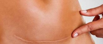
Primary intention is characterized by a reduction in the edges of the wound due to the connective tissue organization of granulation. It firmly connects the edges of the wound. After initial tension, the scar remains almost invisible and smooth. Such tension can tighten the edges of the wound slightly if the opposite sides are at a distance of no more than one centimeter.
Secondary intention is characteristic of the healing of large wounds, where there is a lot of non-viable tissue. Significant defects or all purulent wounds undergo healing by secondary intention. Differing from the primary type, secondary intention has a cavity, which is filled by granulation tissue. The scar after secondary intention has a pale red color and protrudes slightly beyond the surface of the skin. As the vessels in it gradually thicken, fibrous and scar tissue develops, keratinization of the skin epithelium occurs, the scar begins to turn pale, becomes denser and narrower. Sometimes scar hypertrophy develops - this is when an excess amount of scar tissue forms.
On trophic ulcers
Granulation tissue can be seen especially clearly on extensive wounds such as trophic ulcers. They clearly show a coating with a bubbly structure, a pinkish color and a yellowish coating (fibrin). But since a trophic ulcer involves very deep tissue damage (sometimes it reaches the bones), the granulation stage is very long.
The healing of a trophic ulcer is associated not only with careful care of the wound, but also with its blood supply. And the reason for the formation of such a sore is precisely the insufficient trophism (nutrition) of the tissues. And the granulation tissue that appears on ulcers may not have blood vessels. Therefore, treatment necessarily involves wearing compression garments to improve blood supply to the wound.
Functions
In addition to protecting the wound surface from external bacteria and contaminants, the granulation stage is vital for several other reasons. Thus, granulation tissue performs the following functions:
- fills the defect cavity for a normal aesthetic appearance of the damaged area;
- rejects necrotic tissue;
- inhibits the vital activity of microbes that have managed to penetrate the wound;
- destroys toxins;
- is a framework for the formation of connective tissue.
The faster granulation begins, the sooner the wound will heal.
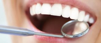
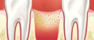
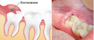
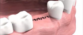
In dentistry
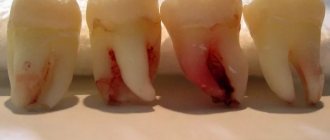
Dental manipulations also often lead to damage to the gums. This is common practice after tooth extraction. During treatment, the gums may be damaged by the partition that the dentist inserts between the teeth so as not to fill the natural interdental gap. Dentistry also involves excision of gums in order to accelerate the eruption of wisdom teeth or in the treatment of gumboil, for example.
After inflammation, the granulation process is preceded by another process - the formation of a blood clot. This mandatory stage is due to the fact that a large number of blood vessels approach the gums, and to stop bleeding, the blood begins to thicken at the site of the wound. Approximately 4-5 hours after this, granulation tissue begins to form, which replaces the clot.
If the damage is shallow and the blood does not ooze too intensely, then there may not be a clot. And the granulation stage will pass almost unnoticed: the patient will not see the granular structure and fibrin due to the humidity and high temperature in the oral cavity, as well as due to the inaccessibility of the affected area (especially if a wisdom tooth located at the end of the dentition was removed).
Pathologies of granulation
If the wound process is disrupted, pathological granulations may form. Insufficient or excessive growth of granulation tissue, disintegration of granulations, and premature sclerosis are possible. In all these cases, and also if the granulation tissue bleeds, special treatment will be required.
The development of granulations and epithelization processes fade away if there are such unfavorable factors as deterioration of blood supply, decompensation of any systems and organs, oxygenation, or repeated purulent process. In these cases, granulation pathologies develop.
The clinical picture is as follows: there is no contraction of the wound, the appearance of the granulation tissue changes. The wound looks pale, dull, loses turgor, becomes cyanotic, and becomes covered with a coating of pus and fibrin.
Tuberous granulations are also considered pathological when they protrude beyond the edges of the wound - hypergranulations (hypertrophic). Hanging over the edges of the wound, they interfere with the process of epithelization. In these cases, they are cauterized with concentrated solutions of potassium permanganate or silver nitrate. The wound continues to be treated by stimulating epithelization.
In gynecology
Nature arranges it in such a way that the inner walls of the female vagina, uterus and other gynecological cavities heal at an accelerated rate. And granulation in such areas occurs almost simultaneously with the process of inflammation.
Sometimes the growth is so intense that a polyp is formed from the granulation tissue, which can subsequently degenerate into a malignant tumor. Therefore, after gynecological injuries or operations, a woman is recommended to regularly visit a doctor to constantly monitor the growth of the tumor.
How to promote healing
It is to facilitate, and not to accelerate, because healing takes a certain time. Immediately after injury, the most important point is disinfection. This makes it possible to destroy bacteria that have entered the surface of the wound from the surface of an object on which a person was injured (knife blade, needle point) or from the environment (for example, from a burn). Wound healing directly depends on the quality of granulation tissue. To improve it, you should do regular (every 24 hours or more often) dressings. After removing the next bandage from the surface of the wound, it is necessary to remove the pus, but preserve the granules, so hydrogen peroxide or a weak solution of potassium permanganate is usually used for disinfection.
The use of auxiliary agents in the form of ointments or gels does not improve the growth of granulation tissue. External preparations are used to destroy bacteria (antibiotics), eliminate prolonged inflammation (anti-inflammatory) and relieve pain (painkillers). When there are no bacteria on the wound, and the inflammatory process is already coming to an end, nothing prevents granulation tissue from forming. And that means it’s not far from healing.
Healing under the scab
The third type of wound healing is the simplest - the wound heals under a scab. This is typical for minor wounds and damage to the skin (abrasions, scratches, abrasions, 1st and 2nd degree burns). A scab (crust) on the surface of the wound is formed from blood that has coagulated there and lymph. The role of the scab is a protective barrier that protects the wound from the penetration of infections; skin regeneration occurs under this shield. If the process goes well, no infection has occurred, and after healing the crust comes off without a trace. There are no signs left on the skin that there was once a wound there.