For professional dental care, various methods and tools are used, among which one of the main places is grinding the enamel with subsequent polishing. The technique is used independently or simultaneously with other manipulations, for example, after removing large stone deposits. Often the method is used during preparation for other processes, for example, prosthetics or restoration with veneers.
Grinding is performed for primary processing, removal of irregularities and roughness, microcracks. At this stage, stone and soft plaque are eliminated, and caries is prevented. Polishing is carried out after grinding. They completely smooth out enamel unevenness and eliminate minor imperfections. Additionally, this step has a mild whitening effect by removing deposits. But there is a slight increase in color, as the natural shade is restored. If the purpose of the procedure is serious whitening, then the doctor uses other methods and drugs.
Tools and procedure features
There are four types of procedures, including grinding and polishing of enamel:
- traditional mechanical;
- ultrasonic;
- air;
- laser (one of the safest).
The traditional method is used when there is a large volume of deposits. To do this, the dentist uses different types of handpieces and a drill. Under the influence of an abrasive coating, deposits are removed, exposing the enamel and restoring the color of the teeth. The cost of the procedure is affordable, but the patient may feel discomfort during cleaning. Therefore, such manipulations are not recommended for people whose tissue sensitivity is increased. If other methods fail to remove stones, local anesthesia may be used.
The ultrasound method is painless and effective. The procedure is performed using a special apparatus and replaceable tips. Ultrasonic exposure allows you to work with hard-to-reach areas and remove plaque from the subgingival area. The patient does not feel discomfort or pain, the procedure is carried out quickly.
The air method is one of the most common. The surface is treated with a strong air flow under pressure using an abrasive mixture. Little time is spent on work, the process does not cause pain or discomfort. A slight lightening effect is achieved.
Laser enamel resurfacing is an expensive procedure. To perform the manipulations, special equipment is used, with which you can remove complex plaque from all areas of the row. The enamel whitens and deposits accumulate much more slowly.
When performing work, a dentist uses special tools. With laser and ultrasound methods, additional funds are not needed; the equipment used, already equipped with everything necessary, including attachments, is sufficient.
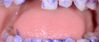
The air method uses abrasive powders with grains of different fractions. To remove stone and large volumes of plaque, compositions with large particles are selected. For final polishing - fine powders. There are also tips and processing pastes. On flat areas, rubber cup-shaped nozzles are used, for hillocks - with a cone-shaped nozzle. Hard-to-reach places are cleaned using pins and strips. When choosing pastes, you need to pay attention to the color, which indicates the graininess. Yellow pastes are intended for soft finishing polishing, blue pastes are rough, used for initial removal of deposits. If necessary, intermediate compositions of green and red colors are used.
Stages of tooth formation in children
Few expectant parents know that the foundation for their child’s baby teeth begins to form as early as the 7th week of pregnancy, and from the 5th month the formation of permanent teeth begins. The future dental health of the baby depends entirely on the habits, lifestyle and nutrition of the mother.
At the age of 6-8 months, babies' first teeth erupt - the two lower incisors. Then, at the age of 8-9 months, the top two appear. The timing of teething is quite individual and depends, among other things, on genetic factors. The normal range is considered to be the eruption of the first teeth at 5-9 months. The third, fourth and fifth teeth gradually erupt. The formation of a temporary bite is completed by the age of 2.5-3 years, when the child has 20 milk teeth - 5 teeth on each side on the upper and lower jaws.
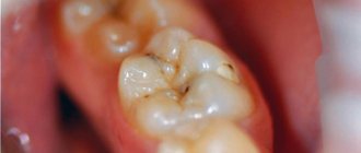
In this case, the process of maturation of the enamel of primary teeth occurs for another 2 years after the tooth erupts in the oral cavity, i.e. The enamel on the central incisors finally matures by about 3 years, and on the lateral teeth, accordingly, even later. In its anatomical structure, a child’s baby tooth differs from a permanent tooth: its walls are much thinner. For example, on the lateral surface the thickness of the hard tissues of a baby tooth (enamel, dentin) is only 1 mm. Such a tooth, compared to a permanent one, is much more vulnerable to various types of problems - first of all, the development of caries and its complications. And if the tooth is still forming, the enamel is in the maturing stage, then it can be destroyed very quickly, especially against the background of a sharp decrease in the child’s immunity: during and after a viral infection, after an illness with antibiotic treatment.
At the age of 6-7 years, the physiological change of temporary teeth to permanent teeth begins. The order of their appearance is the same as during the eruption of primary teeth. The first to appear are the lower and upper central incisors, and at the same time the so-called sixth teeth appear: first the lower, then the upper. The sixth teeth do not change, but grow. The pattern of appearance of all teeth is similar: first, the tubercles become noticeable above the gum, then the chewing surface. At the moment when a tooth erupts, its root is approximately half formed, it is short and has a wide canal opening. During its formation, the root grows in length and its walls thicken.
The last milk tooth changes by the age of 11-12 years. At the same time, for two years after eruption, the enamel matures, and the formation of the root of a permanent tooth lasts even longer: from 2 to 4 years. Thus, only in adolescence can we say that the situation has stabilized.
It is important to know!
From the moment the first teeth erupt at the age of about 6 months and up to 3 years, the child develops a temporary bite. The anatomical structure of a baby tooth differs from a permanent tooth; its walls are much thinner, which means the risk of caries is higher. Therefore, we recommend having preventive examinations with a doctor once every 3-4 months. It is not easy for parents to spot the beginning of caries in a hard-to-reach place, and the process that has begun can develop too quickly and lead to undesirable consequences. At the same age, we recommend visiting an orthodontist for the first time in order to, if necessary, correct the formation of the bite in a timely manner. At the age of 6-7 years, the process of physiological replacement of temporary teeth with permanent teeth begins, which is completed by 11-12 years. At the same time, the maturation of enamel and the formation of the root of permanent teeth continues for another 2-4 years after the appearance of the tooth. Thus, teeth are finally formed and take their place in the oral cavity only by the age of 16. Until this point, it is important to pay increased attention to hygiene and prevention.
Indications and contraindications
The procedure for grinding and subsequent polishing of the enamel surface is indicated in the following cases:
- presence of hard and soft deposits;
- condition after filling;
- as a preventive measure when wearing braces;
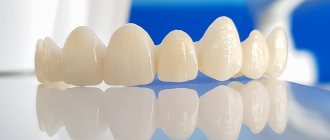
- before prosthetics to exclude complications, the development of caries, soft tissue diseases;
- for regular care, restoration of the natural shade of enamel, improving the aesthetics of the range.
Contraindications to the procedure include:
- state of deep caries damage, multiple defects requiring preliminary treatment;
- inflammation of the gums, bleeding, other soft tissue diseases;
- increased enamel sensitivity;
- inflammatory processes in the acute stage;
- The Patient has an individual intolerance to the abrasive pastes used.
Most contraindications are relative. They must be eliminated, after which you can polish, restoring the natural color. But only a doctor can make a decision about the possibility of such manipulations after a visual examination and some diagnostic measures, if the need arises.
The structure of tooth enamel -
The main structural unit of enamel is the so-called “enamel prisms”, between which there is an interprismatic substance that glues the prisms together. In this section we will analyze their structure, and also talk about the structure of apatite crystals, the structure of the interprismatic substance, as well as such formations as enamel plates and bundles, enamel spindles.
1) Enamel prisms and their structure -
Enamel prisms are formed from apatite crystals that are adsorbed onto an organic matrix. The latter has a fibrillar structure - in the form of a thin protein mesh, which evenly penetrates all the prisms and the interprismatic substance. The prisms themselves have the shape of thin elongated formations that pass through the entire thickness of the enamel - from the enamel-dentin border to the tooth surface (Fig. 4-5). The enamel of one tooth consists of a total of several million enamel prisms.
Longitudinal section of enamel prisms –
The thickness of the prisms ranges from 3 to 6 microns, and as they approach the enamel-dentin border to the surface of the tooth, their diameter increases by approximately 1.5-2 times. This is due to the fact that the area of the enamel-dentin junction (where the prisms begin) is significantly less than the surface area of the tooth enamel. The prisms have a radial direction and lie in relation to the enamel-dentin border - almost at a right angle. But, as for the enamel surface, in the area of occlusal surfaces they will lie parallel to the long axis of the tooth, and on the lateral surfaces of the crown - perpendicular to the tooth axis.
As for the length of the enamel prisms, it will depend on the thickness of the enamel layer on different surfaces of the tooth crown, and the length of each prism will in any case be greater than the thickness of the enamel layer. The latter becomes possible due to the fact that the bundles assembled from enamel prisms have wavy bends along their course (in the shape of the letter S). The appearance of such a radial structure with pronounced S-shaped bends in the enamel is associated with a functional adaptation that prevents the appearance of radial cracks under the influence of occlusal load (Fig. 6).
Cross section of enamel prisms –
On transverse sections of teeth, prisms can have oval, hexagonal, polygonal, but most often - the shape of arcades that resemble fish scales or a keyhole (Fig. 7). According to R. Frank, this form of prisms occurs due to the uneven mineralization of enamel prisms that occurs during their development. Thus, one side of the prisms mineralizes and becomes hard earlier than the other, which causes compression of the softer part of the prism. According to research by J. Saot and N. Symons, only 2% of prisms had a regular hexagonal shape, 57% had an arcade shape, another 31% of prisms were polygonal or oval, and another 10% had an irregular shape.
It is worth noting that the surface of each enamel prism is surrounded by a shell, which is called the “prism cortex”. However, such a shell is not considered as an independent formation, and is distinguished by the fact that it is less mineralized and contains significantly more enamel proteins than the rest of the prism. As a result, the shell is more resistant to acids (compared to the prism core). Below you can see an electron microscopy image showing enamel subjected to acid demineralization for 5 days (Fig. 8). As we see, only the outer shell of the prisms and the interprismatic substance have been preserved.
Enamel prisms after demineralization –
Prismatic enamel –
However, the innermost layer of enamel, adjacent to the enamel-dentin junction, does not contain enamel prisms. This layer is often called the term “initial enamel” (the thickness of this layer is only 5-10 microns). This layer of enamel consists exclusively of small crystals of hydroxyapatite, only 3-5 nm thick. The formation of prismless enamel is due to the fact that in the initial period of its formation, enameloblasts still lack Toms fibers (Toms processes). Only later do enameloblasts begin to form short protoplasmic processes, which give rise to enamel prisms.
The outer layer of tooth enamel is formed in a similar way (at the final stages of its development). During this period, Toms' processes in enameloblasts already disappear, and therefore the most superficial layer of enamel (terminal enamel) will also be devoid of enamel prisms. The layer of the so-called “final enamel” is more pronounced in permanent dentition teeth, but in primary teeth, electron microscopy shows a predominantly prismatic structure of the surface layer of tooth enamel.
2) Features of apatite crystals –
We have already said above that of the different types of crystalline apatites, enamel contains the most hydroxyapatite [Ca10(PO4)6(OH2)], the share of which is 75%. Hydroxyapatite crystals are covered with a hydration shell 1 nm thick. The microspaces between apatite crystals are filled with water, which is called enamel fluid. The water content in enamel is about 2-3%, and its function is the transfer of ions, which ensures the processes of mineralization/demineralization.
In turn, the hydroxyapatite crystals themselves have the form of hexagonal plates - with an average length of about 200 nm (but crystals with a size of 500-600 nm, and even up to 1000 nm can be found), as well as a width of 40-90 nm and a thickness of 25-40 nm . The direction of the crystal axis in relation to the long axis of the prism differs in its different sections. In the central part, the crystals will lie parallel to the long axis of the prism, and on the periphery, they move away from this axis, forming an increasingly larger angle with it. For example, with an arcade-shaped enamel prism, this angle will be about 40–65°.
3) Interprismatic substance –
We have already said above that enamel prisms are, as it were, cemented in a thin layer of interprismatic substance, the thickness of which is less than 1 micron (Fig. 10). By the way, it is worth noting that with the arcade configuration of enamel prisms, the latter are in such close contact with each other that the interprismatic substance between them is almost completely absent. The interprismatic substance also consists of apatite crystals, which are located at an angle to the enamel prisms (often even at an angle of 90°).
The interprismatic substance is less mineralized than the enamel prisms themselves, and therefore, in comparison with them, it has less strength. As a result, cracks that occur in the enamel usually pass through the interprismatic substance, without affecting the prisms themselves. Below you can see longitudinal and transverse sections of the enamel, on which crystals of the interprismatic substance are located between the enamel prisms (at an angle to the enamel prisms).
Tooth enamel (electron microscopy image) –
4) Enamel plates and enamel bundles –
Such formations are characteristic of mature enamel (Fig. 12-13). Both plates and bundles are low-mineralized areas of enamel prisms and interprismatic substance, differing from each other only in position and shape. Enamel plates are very thin “sheet-like” structures that pass through the entire thickness of the enamel (most of them are in the neck of the tooth). They contain enamel proteins as well as organic matter from the oral cavity.
On enamel sections, they look exactly the same as enamel cracks, but are only filled with organic matter. It should be noted that they can serve as entry points for the development of caries. In turn, enamel tufts are small cone-shaped formations (resembling the shape of “spearing tufts of grass”) that extend from the enamel-dentin border. The distance between the beams is approximately 30 to 100 μm.
5) Enamel spindles –
Enamel spindles are flask-shaped structures that extend from the enamel-dentin border at a right angle (Fig. 14). Their formation is due to the fact that during tooth development, some of the processes of odontoblasts penetrate the enamel-dentin junction, which is apparently necessary for communication between odontoblasts and secretory anameloblasts. Thus, enamel spindles are structurally dentinal tubules.
In addition to odontoblast processes, enamel spindles also contain tissue fluid and other organic components. According to most authors, enamel spindles play an important role in the mineralization of deep layers of enamel from the dental pulp side. Below you can see exactly what enamel spindles look like:
6) What are Gunter-Schräger stripes –
We have already said above that enamel prisms have a wave-like curvature along their course (in the shape of the letters S). This leads to the fact that on a longitudinal section of a tooth it is impossible to cut each enamel prism strictly longitudinally along its long axis and along its entire length. Therefore, it turns out that some sections of the prisms will in any case be ground in the longitudinal direction, and their extensions – in the transverse or oblique directions. The areas of the prisms that will be dissected longitudinally look light (parazones). Sections of prisms cut transversely will appear dark (diazons).
As a result, a correct alternation of transverse and longitudinal sections of bundles of enamel prisms appears on the tooth section. When studied in reflected light, they appear in the form of dark and light stripes, crossing the entire thickness of the enamel in an arc in the radial direction. They start from the enamel-dentin junction and end in the superficial layer of enamel. Such stripes are called Gunter-Schräger stripes, and they can be clearly distinguished even with low magnification (Fig. 15-16).
Doctor's recommendations
After completing the procedure, you must follow the dentist's advice:
- correction of the diet for the first time, refusal of coffee or tea, coloring foods or fruits that contain active acids;
- inclusion of medium-temperature dishes in the menu, which will protect against soreness and discomfort in the first couple of days;
- Limit alcohol intake and smoking.
You should also constantly pay attention to personal hygiene. This is cleaning using special brushes and pastes, using irrigators and rinses. Preventive appointments at least once every six months will help monitor the condition of the oral cavity, identify problems in a timely manner and begin treatment if necessary.