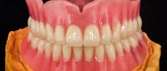
In order to give a person back a beautiful smile, modern dentistry uses artistic dental restoration. This term refers to a whole range of manipulations aimed not so much at restoring the functionality of teeth, but at improving their appearance. This can be done using filling composite materials or veneers.
Bringing the dentition to ideal aesthetic results is a rather complex and labor-intensive process that requires the use of expensive materials and modern technologies. That is why prices for artistic dental restoration are quite high. However, the result obtained, as a rule, fully justifies the money spent.
Anatomical modeling according to Schultz
The basic principle of the technology is the division of each tooth into segments.
Anatomical modeling involves creating wax models of teeth that respect natural morphology. The main attention is paid to those elements that are involved in closing the jaws (occlusion). The basic principle of this technique is to divide each tooth into segments and accurately reproduce them on a wax model. Work on creating models of anterior teeth is carried out according to the following algorithm :
- Applying a separation agent to the plaster stump, then placing a polymer cap on it.
- Marking the edge of the cap with wax in a contrasting color.
- Modeling of cones on a plaster stump, the dimensions of which are based on the parameters of adjacent teeth.
- Applying wax along the incisal edge and checking for correct function in the articulator.
- Creation of models of convex medial and distal ridges.
- Checking the inclination of the teeth relative to the distal edge.
- Removing excess wax along the edges.
Using a similar algorithm, models of teeth of other groups are recreated, taking into account their natural features and functions.
Lomiashvili teeth modeling
The author of the technique is Larisa Lomiashvili, who developed the technology of modular modeling using the direct method, that is, directly in the oral cavity. The main principle is the most accurate reproduction of natural forms, especially for the teeth of the upper jaw, which are the most difficult for the dentist due to difficult access.
Lomiashvili's textbook gives a detailed description
This technique makes it possible to model a tooth directly in the oral cavity.
features of the anatomical structure of all teeth, with tubercles, depressions, ridges, grooves. The restoration of this building is carried out using modular technology. A composite material of the same shape, but in a larger volume, is applied to the central module. Thus, filling the free space of the tooth is carried out according to the “matryoshka” principle, when larger forms absorb smaller ones.
In this case, it is not necessary to use one material. Various composites can be used, differing, among other things, in color, to achieve maximum identity with a natural tooth. When using this technology, it is always possible to correct the shortcomings of the previous layer with the subsequent one.
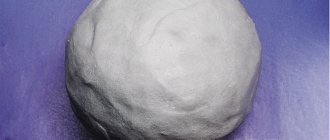
Modeling teeth from sculptural clay
Sculpture clay has long been used in art to recreate shapes. This inexpensive material is ideal for sculpting teeth. It’s pleasant to work with clay in your own way; it’s soft, doesn’t stick to your hands, and hardens gradually. Sculpting clay requires longer preparation for work than plasticine, which is most often used for modeling teeth. If the clay is dry (photo 11), then first you need to mix it with water to the consistency of sour cream (photo 12), leave for some time until it dries and forms a plastic mass (photo 13). After this, the clay becomes hard only after a few hours, this time is quite enough to successfully complete the work. The smaller the volume of available material, the faster it hardens. When the desired consistency is obtained, the clay is shaped into a ball (photo 14).
Let's consider modeling the first right molar of the upper jaw (tooth 16). The overall outlines of the model of tooth 16 are set (photo 15, 16), the location of the main surfaces is outlined: medial contact (M), distal contact (D), vestibular (V) and palatal (P). The tops of the main hillocks are determined. Markings are applied on the chewing surface (photo 17), corresponding to the first-order fissure of the H-shape. Completion of the formation of the external contours of the model and the equator (photo 18) is done by hand. Until this stage, all actions were performed by hand (photo 19).
To model the chewing surface of the tooth, it is better to use tools. The first-order fissure is deepened with a spatula (photo 20, 21). When modeling, you do not need to draw fissures, but rather divide the main tubercles so that an H-shaped fissure appears between them (photo 22). Tools for work are chosen that are more convenient to work with, this could be a spatula, ironing ka. A fissure shaping tool, such as a probe, is required. Two or three tools are enough.
After completing the work, the model can always be corrected by cutting off excess with a scalpel or spatula. The resulting tooth model can be stored for a long time, reminding of the results of the work.
Vetchinkin method
The author of another technique aimed at solving issues of functional occlusion coupled with the aesthetics of the dentition is Anton Vetchinkin. He created a technology called “Aesthetic foundations for the formation of teeth,” which is based on three principles of bionics (the science of connections and patterns in living nature):
- The law of the cone, which implies that the stability of objects is provided by two multidirectional forces - the force of growth and the force of gravity, which balance each other.
- Law of the logarithmic spiral. The strength of natural objects is ensured by the fact that their structure experiences twisting (shells are an example).
- The law of differentiation of form, according to which individual objects can achieve greater efficiency in their development by combining into complex systems.
Expert opinion. Dentist Verbitsky A.L.: “ The author of the technique proposes to use these laws of bionics when modeling artificial teeth. Thus, the listed principles make it possible to determine control points and lines of the future model and thereby recreate the shape of the tooth as accurately as possible. Thus, auxiliary landmarks are added to the points that are already used in the practice of dental technicians (medial and distal edges and others).
Method for restoring the crown part of teeth
The invention relates to medicine, namely to therapeutic and orthopedic dentistry, and can be used in dental restoration. The technical result is the improvement of the aesthetic and functional qualities of the tooth being restored through the development of an algorithm for the clinical restoration of dental crowns. The method is carried out by modeling the anatomical features of the tooth surface using a mesh. The number of mesh cells depends on whether the modeled tooth belongs to a functionally oriented group of teeth. A tubercle is formed in each cell, oriented towards the first-order furrow. The construction of the tubercle is carried out on the basis of the canine-odontometer, which is the basic unit (module) of any dental modeling. 1 ill.
The invention relates to medicine, namely to therapeutic and orthopedic dentistry, and can be used in dental restoration.
There are known methods for restoring teeth (S.V. Dmitrienko, A.I. Krayushkin, M.R. Sapin. Anatomy of human teeth. M.: “Medical Book”, 2000, pp. 166-180; V.N. Radlinskaya, S. O.V. Radlinsky “Modern technologies for dental restoration.” Poltava, 2002, 57 pp., A.V. Vetchinkin “Aesthetic principles of tooth shaping.” Journal “Maestro of Dentistry” No. 3/12, 2003, p.54-56), including modeling of missing coronal tissues by using various techniques for applying filling material.
However, the known methods do not allow restoring the natural surface of the tooth, because do not take into account the individual characteristics of the human dentofacial apparatus.
There is a known method for restoring tooth crowns (Enrico Steger. Anatomical shape of the chewing surface of the tooth. Quintessence Publishing House, 1996, 93 pp.), including modeling the stereotypes of the crown parts of teeth developed by the author.
However, the known method of modeling the coronal part of a tooth does not take into account the individual microrelief of the restored surface, which affects the aesthetic and functional results. In addition, the implementation of restoration techniques by constant rotation of the manufactured model is carried out only in a dental prosthetic laboratory. It is impossible to perform the corresponding manipulations in the patient’s oral cavity.
The objective of the invention is to develop an algorithm for clinical restoration of dental crowns.
The result of solving the problem is to improve the aesthetic and functional qualities of the tooth being restored.
The problem is solved due to the fact that when restoring the coronal part of the teeth by modeling the anatomical features of the tooth surface, a modular reconstruction of the tooth monolith is carried out, while the modules are formed in the form of fangs - odontometers, each of which is built within the boundaries of a cell of a conditional grid simulating a first-order fissure , which is previously applied to the chewing surface of the simulated tooth.
Analysis of the distinctive features showed that the proposed method differs significantly from the known one in that an algorithm for the clinical reconstruction of any tooth crown is proposed: modules (odontomer canines) are used as the basic unit, the number of which for each specific tooth directly depends on the shape of the first-order fissure, and accordingly, on the number of cells formed by it. All together determines the shape of the tooth surface depending on its belonging to a functionally oriented group of teeth (molars, premolars, etc.).
The essence of the invention is illustrated by the drawing.
The proposed method is carried out as follows.
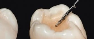
The hard tissues of the coronal part of the tooth are subjected to preparation in order to remove demineralized dentin. The carious cavity is treated with medication, dried, and adhesive technology is applied. After that, a conditional mesh 1 is applied to the chewing surface of the tooth, simulating the first-order fissure of the given tooth, thanks to which the nth number of cells 2 are identified. Before modeling the crown part of the tooth, the overall outline of the tooth, the relationship between the height, width and length of the tooth crown are taken into account. Then, in each of the cells, one tubercle 3 (odontomeric canines) is laid out, fitting into the overall outlines of the crown and orienting them towards the fissure of the first order.
Layer-by-layer modeling of the missing tissues with filling material is carried out, with the restoration of the expected modules, forming a microrelief of the chewing surface of the tooth. Grinding and polishing of the restored tooth crown.
EXAMPLE. Patient L. came to the dental office with complaints of a damaged crown of the lower right first molar (46 tooth). According to the patient, 5 years ago she sought help from a dentist, where she was treated for complicated caries. As a result of the treatment, the canals were sealed with endomethasone paste, an insulating lining made of phosphate cement, and a filling made of Evicrol were placed. 1 month ago, the patient noticed a filling falling out and food retention in the tooth.
An objective examination reveals 2/3 destruction of the tooth crown; probing and percussion of hard tissues are painless.
Diagnosis: 46 Chronic periodontitis, filling defect.
Treatment was carried out according to the proposed method:
The hard tissues of the coronal part of the 46th tooth were subjected to preparation in order to remove demineralized dentin. Then the carious cavity is treated with medication, dried, and adhesive technology is applied. Before modeling the crown part of the tooth, the overall outline of the 46th tooth and the relationship between the height, width and length of the tooth crown are taken into account.
A conditional mesh is applied to the chewing surface, simulating a 1st order fissure, in this case F-shaped. Within the boundaries of the formed cells, the main tubercles (canine odontomeres) are laid out, fitting into the overall outline of the crown of the area of the contact marginal ridges. A light-curing composite, macrofil of the Filtek family, is used as a filling material. Layer-by-layer modeling of the missing tissues with filling material is carried out with the restoration of the expected modules, forming a microrelief of the chewing surface of the 46th tooth. Grinding and polishing of the restored crown of the 46th tooth.
The condition of the restored crown of tooth 46 and hard tooth tissues and enamel was monitored after 1, 3, 6 and 12 months using aniline dyes. The results of control examinations in the immediate and long-term periods of completion of treatment of the hard tissues of the 16th tooth confirmed the integrity of the restored tooth crown.
Thus, the proposed method unifies the modeling process, providing improvement in the aesthetic and functional qualities of the restored teeth.
A method for restoring the coronal part of multi-rooted teeth by modeling the anatomical features of the surface, characterized in that a modular reconstruction of a tooth monolith is carried out, while the modules are formed in the form of fang-odontomeres, each of which is built within the boundaries of a cell of a conditional grid simulating a fissure of the first order, which is previously applied on the chewing surface of the simulated tooth.
3D modeling
3D modeling technology allows you to create a three-dimensional model of the jaws.
Modern technologies have firmly established themselves in dentistry. One of them is 3D modeling, with which you can create three-dimensional models of jaws. For this, a so-called three-dimensional tomograph is used, consisting of a computer itself and a scanner.
The scanner continuously takes pictures over a certain period of time and transfers them to the computer. Thanks to them, the doctor can determine not only the structural features of the jaws, but also the location of blood vessels, nerves, and maxillary sinuses. In this case, there is no need to make casts and impressions: based on the information received, the doctor can create a three-dimensional model of the future structure on the computer screen in a few minutes.
The information obtained is transmitted to a special milling machine, which automatically grinds the prosthesis. The technology is used for the restoration of both anterior and chewing teeth: thanks to the high precision of the method, it is possible to achieve ideal functional occlusion. Using 3D modeling, prosthetics, ceramic crowns, and veneers can be made.
Despite the introduction of high technologies into dentistry, the work of a dental technician continues to remain not only a craft with precision calculations and honed skills, but also an art. It is not without reason that it is believed that technicians are a kind of sculptors, artists who create real works of art in the form of beautiful smiles of patients.
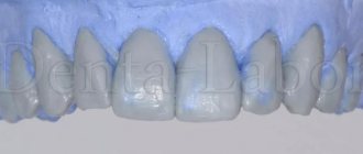
Restoring the structure using the indirect method
In this case, veneers are manufactured in a specialized laboratory. They are special overlays for teeth, which are made in the form of plates made of composite materials. The use of veneers allows you to provide the proper level of protection for the outer layer of the tooth. Note that restoration of the tooth surface with veneers involves covering the front part of the tooth with a plate.
Regardless of whether the restoration of the lateral group of teeth or the restoration of permanent teeth is carried out using the indirect method, the procedure in any case will consist of several stages. First of all, we are talking about performing preparatory work when a person’s teeth are treated and filled, as well as creating an impression based on the photograph taken. The final stage of the work is installing the plates on the teeth.
Dental restoration using this method is relevant in cases where the teeth are not very white or straight. Choosing veneers allows tooth restoration to be done very quickly and painlessly. The result of the procedure will be a perfect smile. At the same time, dental restoration doctors most often do not undertake the installation of plates if a person has an incorrect bite. In addition, there are also cases when a person has a crown installed on some teeth, and veneer plates on others. In this case, it is extremely important that the material of the crown and veneer is the same. Then the person will not have any problems with the oral cavity.
Artistic restoration and modeling of teeth
Artistic modeling and restoration of teeth make it possible to restore damaged dental tissues using artificial materials. For this purpose, composite filling compounds or special overlays – veneers – can be used. The procedure allows you to fully restore the correct shape, aesthetic appearance and chewing functions of the tooth. Thanks to the use of modern technologies, discomfort for the patient is minimal. Artistic dental restoration in Moscow is performed at a first-class professional level by experienced dentists working in clinics of the Medline-Service network.
Prices
| Aesthetic restoration | 6800 rub. |
| Aesthetic restoration with nanohybrid composites | 12000 rub. |
Modeling teeth using various techniques using modern composite materials
Other articles
Professor L.M. Lomiashvili, Doctor of Medical Sciences, Head of Department Associate Professor L.Yu. Zolotova, Candidate of Medical Sciences Assistant S.G. Mikhailovsky Assistant D.V. Pogadaev Student E.G. Tkach Student A.G. MOOC Resident Doctor V.V. Logunov Resident Doctor K.K. Gritsenko
Department of Therapeutic Dentistry of the State Medical University (Omsk) of the Ministry of Health of the Russian Federation
Summary. Achieving a high-quality result when restoring lost hard dental tissues is possible only if a harmonious combination of shape, color and functional characteristics of the restorations is ensured. Changes in color parameters were studied using various combinations of dentin and enamel shades of the Estelux NK nanohybrid material (StomaDent, Russia). A comparison of the resulting restorations with the VITA 3D-Master color chart showed that by sequentially adding certain shades of a given thickness, it is possible to create an anatomical shape of the tooth crown with varying degrees of brightness, saturation, and tone. Perceiving and analyzing color is a skill that can be learned and then improved with practice. The cooperation of manufacturers, teachers, and clinicians contributes to solving current issues, including the reproduction of the natural harmonious beauty of teeth using artificial materials.
Key words: restoration technique; modeling; color options; transparency; nanocomposite
A high-quality result when restoring lost hard dental tissues is possible only if a harmonious combination of shape, color and functional characteristics of restorations is ensured [2, 4, 7, 8]. Translated from Greek, “harmony” means integrity, unity, “the interaction of all parts and elements of the form, that is, it is considered as an aesthetic category. Animated harmony, filled with human meaning and feeling, is called beauty. A piece of sculptural clay begins to come to life as it forms the delicate silhouette of a child or the courageous profile of a warrior. It is the harmony of forms that occupies a leading place in the concept of beauty. The harmony of color seems to fade into the background. It would seem, what is special about a piece of clay that has a uniform structure and a single color? How can he attract attention? At what point does clay come to life in our hands?
Of course, the overall outline of the object is initially set, then priorities are set in relation to the whole to the particular, and then the microrelief of the surfaces is drawn. The more the surface is differentiated, the more we receive light rays reflected from it. The clay comes to life when the process of surface differentiation is completed. The reflection of light from many intertwined faces makes it possible to see the beauty of the newly created object. Famous Spanish architect of the 19th century. Antonio Gaudi said: “Light is the main architect.”
All these intricacies of craftsmanship are taught to students of art and graphic faculties, but, unfortunately, the process of restoring harmony remains a secret for dentists. But it is in the 21st century, having mastered modern technologies, that members of the professional dental community can afford to create harmony in the dental system and get closer to aesthetic dentistry. The right to see, realize and reproduce the harmony of teeth and dentition currently belongs to both dentists and their colleagues, dental technicians [4]. For this purpose, dental restoration materials are constantly being improved, and students and doctors are continuously trained to work in compliance with the rules of art.
It has been noted that the size, shape of teeth and their proportional relationship in the dentition are the main aesthetic parameters in dentistry [2, 4, 7]. According to many authors, when creating aesthetic restorations, it is necessary to take into account (in descending order): shape, transparency, saturation, surface texture, brightness of the enamel layer, individual characteristics, shade [6]. Recreating each parameter requires doctors to know the anatomy of teeth, the physical and mechanical characteristics of the material used, the optical properties of hard dental tissues, and the peculiarities of color perception by a visual analyzer [1, 2, 8].
Accurate visual reproduction of the color of a restoration is not an easy task. To facilitate color determination, many ready-made colors are offered to help the practitioner. The selection of shade depends on the dentist’s ability to sense the differences in tooth color and shade patterns. The main criteria for color are brightness, saturation, hue.
Tone (according to Munsel) is the quality with which we can distinguish one color from another. For example, red from yellow. Brightness is the lightness of the object being studied: the darker the tooth, the less brightness it is. Saturation is the intensity, concentration of a certain tone. When choosing the color of teeth in practice, the difference in brightness (lightness) is the most important, and the difference in tone is the least important. A small hue mismatch is not as noticeable as even a minimal luminance mismatch [1].
When choosing a color, a number of important conditions must be observed [6, 8]. Color perception and analysis is a skill that can be learned and then improved with practice [1, 3, 5].
Goal of the work
Improving the process of teaching teeth modeling by developing the skill of analyzing the color of the resulting restorations from different combinations of dentin and enamel shades of the nanohybrid material “Estelux NK” (StomaDent, Russia).
Materials and methods
Determination and analysis of the color of the resulting restorations on phantom teeth (incisor, canine) was carried out using the VITA System 3D-Master scale.
The technique consisted of several stages.
1. Determination of brightness (lightness). In the scale, color standards are divided into five brightness groups - from 1 to 5. The higher the number, the darker the color.
2. Determination of color intensity (saturation). Each group is divided into subgroups: M - medium shade, L - more yellow, R - more red. Saturation was determined by the average hue M in the selected group. All standards on the M bar are of the same color tone and lightness, but differ in intensity and are coded with a numerical value.
3. Determination of tone. In the same brightness subgroup, the hue is adjusted if necessary, that is, code L or R is selected (Fig. 1).
Results and its discussion
| Rice. 1 VITA System 3D-Master scale | Rice. 2 Canine: stages of complete restoration of the crown with composite DA3 (MONO) |
| Rice. 3 Stages of creating the palatal wall with enamel shade EA3 "Estelux NK" | Rice. 4 Adaptation to enamel shade EA3 dentin shade DA3.5 |
| Rice. 5 Incisor: adaptation to dentin shade DA3.5 layers of composite shade DA3 | Rice. 6 Final view of the restoration made using shades EA3, DA3.5, DA3, EA3 |
Initially, reconstruction of the canine of the phantom tooth was performed with one dentin shade DA3 of the Estelux NK nanohybrid composite (Fig. 2). The color of the resulting restoration was closest to the color code 3M2. The shape of the crown part of the tooth really matches the canine, but the color is perceived as lifeless and does not cause aesthetic satisfaction. Shade DA3 is intended to restore the “dentin mass” in the cervical region of the crown of the tooth and in the future it is expected to cover it with “enamel mass”. However, there are patients with a fairly rich yellow color of the coronal part, the absence of zones of transparency, and a filling material with a shade of DA3 may be the only one (the first and the last), if it suits the patient.
Depending on the wishes and capabilities of the patient and the qualifications of the doctor, the material can be used both in the MONO method and in multi-layer restoration.
To study the transparency, possible changes in brightness, and saturation of the Estelux NK composite material, a complete restoration of the crown part of the phantom tooth was carried out using a “silicone key” and layer-by-layer introduction of a combination of different shades of enamel and dentin of the same thickness in all samples.
Stages of working with Estelux NK when restoring the coronal part of a tooth using layer-by-layer application of material
1. Restoration of the palatal wall using a “silicone key”, layer EA3 (Fig. 3). The thickness in the area of the cutting edge was 0.5 mm, in the area of the neck - 1.2 mm.
| Rice. 7 Final view of the restoration made using shades EA3, DA3, DA2, EA3 | Rice. 8 : a) formation of the palatal wall with shade EA2; b) adding dentin shade DA2; c) application of enamel layer EA2 |
| Rice. 9 Restoration of the palatal wall with enamel shade EA2 “Estelux NK”: a), b) view from the contact surface; c) view from the palatal surface | Rice. 10 Layer-by-layer restoration of a composite tooth: replacement of the bulk of dentin with shades DA3 and DA2 “Estelux NK” |
| Rice. 11 The final result of layer-by-layer restoration of a composite tooth with shades EA2, DA3, DA2, EA2 “Estelux NK” | Rice. 12 : a) depth of preparation for veneer of phantom teeth with a marking bur; b) prepared surface of the phantom tooth for veneer |
2. Application of opaque saturated dentin shade DA3.5 in the form of mamelons with a thickness of 2.2 mm in the area of the tooth neck and 1.5 mm in the body area. This layer is absent in the upper third of the crown (Fig. 4). If the thickness of this layer increases, the restoration may appear grey.
3. Next, the dentin shade DA3 was applied, repeating the relief of the underlying layer. The thickness was 0.4 mm in the area of the tooth neck, 1.8 mm in the body area, gradually thinning towards the cutting edge (Fig. 5). The inner layer, which replenishes dentin, determines the saturation, dullness, fluorescence and tone (color) of the future restoration, and also occupies the bulk of the introduced material.
4. The last layer is enamel shade EA3 (Fig. 6).
The restoration made with shades EA3, DA3.5, DA3, EA3, according to the VITA System 3D-Master scale, corresponded to the 3R 2.5 coding (3 - brightness: the darkest of all shade combination options, R - in tone closer to a reddish tint, cm Fig. 6).
| Rice. 13 a) dentin shade DA3 is adapted to the surface of the phantom tooth; b) layer DA2 is applied, repeating the relief created by layer DA3 |
| Rice. 14 A layer of composite enamel EA2 has been applied: view from the vestibular (a) and contact surfaces of the incisor (b), canine (c) |
The restoration made with shades EA3, DA3, DA2, EA3 was assigned code 2M 3 (Fig. 7), that is, it was brighter than the restoration made with shades EA3, DA3.5, DA3, EA3.
When restoring the coronal part of the canine with Estelux NK material using only two lighter shades (DUO) - enamel EA2 and dentin DA2 - the resulting brightness result corresponded to group 2, medium shade with the letter M, degree of saturation 2 (2M2) on the VITA scale 3D-Master (Fig. 8).
The combination of three shades EA2, DA3, DA2, EA2 with layer-by-layer application of Estelux NK made it possible to obtain a restoration that was also close to 2M2 according to the VITA 3D-Master scale (Fig. 9-11).
To study the effect of the thickness of the applied filling material on the brightness and saturation of phantom teeth, direct veneers were made from the Estelux NK nanocomposite material by layer-by-layer application of the material in various combinations of enamel and dentin shades:
1) DA3 + DA2 + EA2
2) MONO DA3
3) DUO DA3 + EA3
4) DA3.5 + DA3 + EA3.
The thickness of the introduced layers was also controlled using a micrometer. Measurements were taken in the cervical area, in the middle part and in the area of the cutting edge of the tooth. The color was determined in the middle part of the tooth, where a slight thickness of the enamel layer was noted, using the VITA 3D-Master shade scale.
Stages of working with Estelux NK material in the manufacture of veneers
1. Phantom teeth (incisor and canine) were prepared for veneer using a marking bur to an equivalent depth (Fig. 12).
2. After adhesive preparation, the first layer of composite is added - dentin shade DA3 with the simultaneous formation of mamelons, then shade DA2 (Fig. 13).
3. An enamel shade has been added (see Fig. 3).
Dentin layer DA3 of Estelux nanocomposite
NK", introduced into the cervical area, and the DA2 layer, replenishing dentin to 1/2 of the volume, created the basis of the restoration. A layer of EA2 enamel with a minimum thickness in the area of the tooth neck and the greatest thickness in the area of the cutting edge modified the final color, imparted brightness and transparency. This layer formed the relief of the vestibular surface of the tooth (Fig. 14).
Analysis of veneers made by the direct method from the Estelux NK nanocomposite material using various combinations of enamel and dentin shades showed that the veneer looked brighter (lighter) when using shades DA3 + DA2 + EA2. It corresponded to the sample with code 1M2. The veneer made with a combination of layers DA3.5, DA3, EA3, code 3R (Table 1) was more saturated (the color is more intense).
The veneer made using the MONO technique (DA3) turned out to be less attractive. In this case, there was no transparency of the restoration. On the contrary, in samples where enamel shades were used, transparency was well visualized. An excellent shine of the filling material after polishing was noted.
The nanohybrid structure of the filler ensures the strength characteristics of the material. The specific gravity of the fill indicator with inorganic filler is 82%. The advantage of such a high degree of filling should be a clinically significant reduction in polymerization shrinkage of the composite and an increase in the hardness of its surface. The introduction of an antimicrobial additive into the Estelux NK composite makes it possible to prevent the development of secondary caries.
Taking into account these characteristics, crowns were made using the indirect method for teeth 26 and 27 from the Estelux NK composite with the crowns reinforced with Glasspan fiberglass tape. This method was chosen as an alternative restoration option - a temporary restoration for a long time.
Clinical case
Patient K., 51 years old, came to the clinic.
Complaints: destruction of the crown part of tooth 26.
From the anamnesis: tooth 26 was previously covered with a restorative crown.
Objectively: the coronal part of tooth 26 is made of GIC, after removing the old filling, probing at the mouths of the root canals is painless, percussion is painless, the tooth is motionless. On the intraoral radiograph of tooth 26, foci of bone tissue destruction are visualized in the area of the apexes of the buccal roots, the lumen of the root canals is narrowed, intermittent, the radiological shadow corresponding to the filling material is not detected in the lumen of the root canals.
In tooth 27, no changes in the periapical tissues are detected; the periodontal gap can be traced throughout. EOM 14 µA (Table 2).
When working with this material, its good handling properties, ability to retain its shape, and good adaptation to hard dental tissues were noted.
Table 1. Various options for color combinations of veneers made with Estelux NK shades using the layer-by-layer method
| Rice. 1 Cutter, shades DA3, DA2, EA2 | Rice. 2 Cutter, shades DA3, DA2, EA2 | Rice. 3 Fang, shades DA3, DA2, EA2 | Rice. 4 Fang, shades DA3, DA2, EA2 |
| Rice. 5 MONO cutter, shade DA3 | Rice. 6 MONO fang, shade DA3 | Rice. 7 MONO fang, shade DA3 | |
| Rice. 8 DUO cutter, shades DA3 + EA3 | Rice. 9 DUO cutter, shades DA3 + EA3 | Rice. 10 DUO fang, shades DA3 + EA3 | Rice. 11 DUO fang, shades DA3 + EA3 |
| Rice. 12 Cutter, shades DA3.5, DA3, EA3 | Rice. 13 Cutter, shades DA3.5, DA3, EA3 | Rice. 14 Cutter, shades DA3.5, DA3, EA3 | Rice. 15 Cutter, shades DA3.5, DA3, EA3 |
conclusions
Thus, knowing the algorithm of actions, the sequence of introducing certain shades, a given thickness, it is possible to create the anatomical shape of a tooth crown with varying degrees of brightness, saturation, and tone from the Estelux NK nanohybrid material.
Few people master the art of choosing colors, but this can be learned and then improved in further work. The first steps, the basis of knowledge about choosing colors, are laid in practical classes. According to the Federal State Educational Standard, after graduating from a university, a graduate has the right to work independently, without an additional postgraduate educational certificate. In this regard, it is very important to improve the learning process, to set the goal of intellectual, manual and aesthetic education of the student.
Table 2. Stages of preparing teeth 26 and 27 for indirect restoration of the coronal part with the Estelux NK material
| Rice. 1 Tooth 26: coronal part restored by GIC | Rice. 2 Intraoral radiograph of tooth 26 | Rice. 3 Filling i from the GIC was removed, hard tissues of the tooth were prepared, unsatisfactory crown I on tooth 27 | Rice. 4 Access to the orifices of four root canals is provided. Formation of root canals with rotating Ni-Ti instruments (FKG Race system), medicinal treatment according to the recommended protocol |
| Rice. 5 Intraoral radiograph, filling control | Rice. 6 The tooth stump was restored using FlowCor Duo material (StomaDent) with a light-conducting pin, and a ledge was created for a composite crown | Rice. 7 Fixed indirect restoration from the Estelux NK composite on tooth 26 | Rice. 8 Temporary-permanent crowns made of the Estelux NK nanohybrid composite were fixed on teeth 26 and 27 |
The joint work of manufacturers, teachers, and clinicians can lead to solving pressing issues in aesthetic dentistry, including the reproduction of the natural harmonious beauty of teeth and dentition using modern artificial materials.
Coordinates for contacting the authors: +7, [email protected] - Lomiashvili Larisa Mikhailovna, Zolotova Lyudmila Yurievna, Mikhailovsky Sergey Gennadievich, Pogadaev Dmitry Vladimirovich, Tkach Ekaterina Gennadievna, Mook Anastasia Gennadievna, Logunov Vitaly Vladimirovich, Gritsenko Ksenia Konstantinovna
BIBLIOGRAPHY
1. Goldstein R. Aesthetic dentistry. — M.: STBOOK, 2003, 492 p.
2. Lomiashvili L.M., Ayupova L.G., Mikhailovsky S.G. et al. The art of modeling and restoration of teeth. — Omsk: Polygraph, 2014, 436 p.
3. Lomiashvili L.M., Zolotova L.Yu., Zolotov A.N. Experience of using interactive teaching methods at the Department of Therapeutic Dentistry of Omsk State Medical Academy as part of the implementation of the third generation Federal State Educational Standard.//Professional competence of a medical university teacher as a condition for improving the quality of education.//Sb. materials of the All-Russian scientific and practical. conf. with international participation. — Omsk: OGMU, 2014, p. 168-172.
4. Lomiashvili L.M., Sedelnikov V.V., Postolaki A.I. From unity to diversity of forms in nature. — DentArt, 2015, No. 178, p. 58-66.
5. Lopanova E.V., Lomiashvili L.M. Medical teacher training university to the formation of communicative competence of a dentist. — Dentistry, 2015, No. 5, p. 76-79.
6. Manauta J., Salad A. Layers. Atlas of layer-by-layer composite restorations. — M.: Azbuka, 2014, 444 p.
7. Mikhailovsky S.G., Lomiashvili L.M., Pogadaev D.V. Methodological approaches to the process of developing aesthetic skills among dentists. modeling in dentistry. - Cathedra - Department. Dental Education, 2015, No. 52, p. 38-42.
8. Ryakhovsky A.N. Shape and color in aesthetic dentistry. - M.: Avantis, 2008, 208 p.
Direct full restoration
The advantage of direct artistic restoration of a tooth is the ability to restore its functions and shape in one visit to the dentist. The procedure is also minimally invasive. Modeling and restoration of destroyed dental tissues is performed directly in the patient’s oral cavity. Indications for direct restoration are:
- the need to change the shape of fangs and incisors; the presence of chips and cracks in the enamel; the need to restore the position of the crowns by extension; change in tooth shade.
Materials
Composite filling materials are used to model and restore dental tissues. They are selected taking into account the shade of the patient's enamel in order to provide the necessary aesthetic effect. Composite materials have such advantages as affordability, speed of production, and ease of adjusting the shape of teeth restored with their help. Pins can also be used during restoration. They are made from various metals (titanium, silver, etc.).
Stages
- Preparation. Before starting the restoration, the doctor removes plaque and stone; if there is caries, the affected tissue is drilled out. If necessary, anesthesia is performed. The tooth to be restored is isolated from saliva and wet breath. Selection of material . For this purpose, a special scale is used to determine the color of the patient’s enamel. In accordance with it, the doctor selects a composite material of the appropriate shade for extensions. Restoration of dental tissues . If a tooth is destroyed by 50% or more, a pin is installed in the root canal before restoration. Then layer-by-layer application of the composite material is performed. Final simulation. The tooth is ground to the desired shape, then its surface is ground and polished.
The correctness of the newly created forms is the key to unraveling harmony! It is interesting to note that at the turn of the 19th and 20th centuries, societies were created in Western Europe whose members paid great attention to the harmony of forms. These were people belonging to various professions: architects, sculptors, artists, builders, engineers. It is known that, translated from Greek, the term “harmony” is considered as an aesthetic category denoting integrity, unity, and interaction of all parts and elements of form. Animated harmony, filled with human meaning and feeling, is called beauty. A piece of sculptural clay begins to come to life when the delicate silhouette of a child or the courageous profile of a warrior is formed from an unshaped object. It is the harmony of forms that occupies a leading place in the concept of beauty.
The harmony of color seems to fade into the background. It would seem that a piece of clay that has a completely definite homogeneous structure, a single color - what is special about it, how can it attract the eye of the average person? How does the amazing miracle of sculpting happen, at what point in our activity does the object we are working with become animated?
Of course, the overall outline of the object is initially set, then priorities are set in relation to the whole to the particular, and then the microrelief of the surfaces is drawn. The more the surface is differentiated, the more we receive reflected rays of light from it. A piece of clay comes to life in the hands of the sculptor when the process of surface differentiation is completed. Obviously, the sanding of surfaces, the process of reflecting visible light from many different intertwined facets, allows us to see the beauty of the newly created object. These intricacies of mastery are taught to students in higher educational institutions, in art and graphic faculties, but, unfortunately, the process of restoring harmony remains a secret for dentists. But it is in the 21st century, having mastered modern technologies, that members of the professional dental community can afford to create harmony in the forms of the dentofacial apparatus and get closer to aesthetic dentistry. The right to “see, realize and reproduce the harmony of the shapes of teeth and dentition” currently belongs to both dentists and their colleagues, dental technicians.
Nature is amazing and unique! Initially, the tooth as an organ was created in such a way that in an area of insignificant size there is a huge number of grooves, depressions, hollows, cavities, crevices and many other morphological formations. There is a harmony of forms created by nature. One can imagine how highly the surface of the teeth is differentiated. The greater the degree of differentiation of the surface, the more voluminous the working surface of the tooth, the better the chewing efficiency of the dentofacial apparatus, the faster the process of formation of the food bolus. Children instantly chew and grind hard, coarse-fiber food without making any effort; subsequently, all systems of the human body function in a decent manner, the gastrointestinal tract accepts a well-mechanically processed food bolus. Over the years, the surfaces of the teeth wear out, the process of food processing becomes difficult, the food bolus is swallowed mechanically insufficiently processed, after which a series of problems arise at various levels, the functioning of many organs and systems is disrupted.
Unfortunately, many reproaches are expressed against dentists, which are not always unfounded, and the restoration of lost tissue does not stand up to criticism. A clinical case is demonstrated where the previously restored coronal part of tooth 36 does not match either the shape or color of the lower jaw molar.
A patient came to the therapeutic dentistry clinic with complaints of food retention in the interdental space in the area of teeth 35, 36, as well as an aesthetic defect in the crown of tooth 36 (Fig. 1, 2).
Figure 1. Tooth 36, initial clinical situation.
Figure 2. The crown of tooth 36 does not correspond to the main aesthetic parameters: shape and color.
From the medical history it was found that in 2011, the crown part of tooth 36 was restored using the direct method using composite material. An objective examination reveals a discrepancy between the shape and color of the first molar of the mandible and its group affiliation.
Diagnosis: tooth 36 - restoration defect, chronic periodontitis, tooth 35 - dentin caries.
In Fig. 3 and 4
Figure 3. Tooth 36, stage of preparation of the old restoration.
Figure 4. Tooth 35, carious cavity on the distal contact surface. The stages of preparation of hard tooth tissues are depicted; attention is drawn to the contact point between teeth 35, 36 and, as a consequence, caries of the dentin of tooth 35.
Next, the composite material in the area of tooth 36 is completely removed, the distal canal is prepared for the LuxaPost (“DMG”) light-fiber pin, which is subsequently fixed to the double-curing material Luxa Core Z (“DMG”), and a tooth stump is formed (Fig. 5, 6) .
Figure 5. A LuxaPost (“DMG”) light guide post is installed in the distal canal of tooth 36.
Figure 6. The coronal part of tooth 36 was restored with Luxa CoreZ (“DMG”) material.
The tooth stump is covered with a temporary crown Protemp Crown (“3M ESPE”) (Fig. 7, 8).
Figure 7. Temporary crown Protemp Crown (“3M ESPE”).
Figure 8. The Protemp Crown (3M ESPE) composite crown is fixed in the oral cavity.
After receiving the collapsible plaster model, we begin to work with the plaster stamp of tooth 36 (Fig. 9).
Figure 9. Plaster die of tooth 36.
Filtek Flow material (“3M ESPE”) is applied along the perimeter of the ledge to ensure close contact of the future restoration with the tooth tissue (Fig. 10).
Figure 10. Filtek Ultimate Flow (“3M ESPE”) composite was applied in the ledge area.
Next, a thin layer of composite material is applied to the opaque plaster stamp (Fig. 11).
Figure 11. Lining a gypsum die with the composite material Filtek Z550 (“3M ESPE”).
To strengthen the future structure, GlasSpan tape (“GlasSpan Inc.”) is used, in which the resulting composite cap is wrapped (Fig. 12-14).
Figure 12. Preparing the required length of GlasSpanan tape (GlasSpan Inc.).
Figure 13. Reinforcement of a composite coping with GlasSpanan tape (GlasSpan Inc.).
Figure 14. Composite material cap produced.
A filling material is applied to the resulting composite cap, the overall outlines of the future restoration are set, the equator and the tops of the main tubercles are formed (Fig. 15, 16).
Figure 15. Modeling of the lingual cusps of tooth 36.
Figure 16. The overall outlines of the future restoration have been specified and the tops of the five main tubercles are outlined.
The chewing surface is modeled (Fig. 17, 18).
Figure 17. Modeling the chewing surface of tooth 36.
Figure 18. A color corrector was used to draw fissures.
Modeling of the coronal part of tooth 36 is carried out using modular technologies for dental restoration (L.M. Lomiashvili, 2004) (Fig. 19, 20).
Figure 19. Appearance of the composite restoration before the polishing stage.
Figure 20. The coronal part of tooth 36 was restored using the author’s method “Modular technologies for dental restoration” (L.M. Lomiashvili, 2004).
To make the restoration look natural, a color corrector is used (see Fig. 18).
The perimeter of the resulting restoration is polished using the Sof-Lex system (“3M ESPE”)
(Fig. 21).
Figure 21. Polishing using the Sof-Lex system (“3M ESPE”).
Microcontouring and polishing of the chewing surface for finishing are carried out with burs (SS White) of various configurations (Fig. 22).
Figure 22. Finishing and microcontouring of the occlusal surface of the restoration.
Before the final fixation of the restoration of tooth 36, treatment of tooth 35 is carried out. To control the quality of the preparation of the hard tissues of tooth 35, the Life-DT, “SoproLife” (Acteon) technique was used (Fig. 23-28).
Figure 23. Distal contact surface of tooth 35 in normal mode. Application of the Life-DT technique using the SoproLife lamp (“Acteon”).
Figure 24. Distal contact surface of tooth 35 in diagnostic mode. Application of the Life-DT technique using the SoproLife lamp (“Acteon”).
Figure 25. Quality control of the preparation of hard dental tissues 35. Life-DT, SoproLife (“Acteon”) technique.
Figure 26. Quality control of the preparation of hard tissues of tooth 35 in diagnostic mode. Methodology Life-DT, SoproLife (“Acteon”). Further preparation is required.
Figure 27. Dynamic observation of the condition of the hard tissues of tooth 35 during the preparation of hard tissues of tooth 35. Methodology Life-DT, SoproLife (“Acteon”).
Figure 28. Quality control of the preparation of hard tissues of the 35th tooth in diagnostic mode. Methodology Life-DT, SoproLife (“Acteon”). No further preparation of the hard tissues of tooth 35 is required.
Rice. 29, 30
Figure 29. Tooth 36, restoration completed.
Figure 30. Appearance of tooth 36 from the lingual side. demonstrate the final result of dental treatment 35, 36 using modern technologies.
Thus, dental reconstruction is a fairly important link in the chain of restoration of the functional and physiological interaction of organs and tissues of the oral cavity. Possessing modern technologies, as well as knowledge and skills in the field of reconstructive therapy, dentists must restore lost tooth tissue in a harmonious manner, restoring the individuality of the forms intended by nature.
Indirect full restoration
When performing artistic restoration of teeth using the indirect method, pre-fabricated individual microprostheses are used - veneers, inlays, crowns. Their modeling is carried out in a dental laboratory. Due to the multi-stage nature of the restoration, you will need to visit the dentist at least twice. Indications for tooth restoration are:
- significant destruction of the coronal part; large chips of the cutting edge; unaesthetic appearance after caries treatment.
Indirect artistic restoration of anterior teeth is often prescribed to restore/correct the shape of a group of incisors. The procedure allows you to eliminate various functional and aesthetic defects: chipped cutting edges, enlarged interdental spaces, discoloration of enamel.
Materials
To restore dental tissues, microprostheses made of metal, ceramics, and metal-ceramics are used. These materials are superior in strength and abrasion resistance to composite compounds used to restore dental tissue using the direct method. After removing a large amount of hard tissue during caries treatment, the dentist may recommend a restoration using ceramic inlays as an alternative to prosthetics.
Stages
- Preparation . At your first visit to the dentist, the doctor will determine the type of structure suitable for restoring the shape of the tooth and, if necessary, draw up a treatment plan. Then the specialist will make an impression from which a microprosthesis will be made. Making veneers or inlays . Modeling of the structure is carried out taking into account the shape and shade of the dental tissues in contact with it. The production of microprostheses is carried out by a dental technician. Installation of a microprosthesis . During the second visit to the dentist, the doctor fixes the finished structures on the surface of the tooth using a special compound.
Who is recommended for artistic dental restoration?
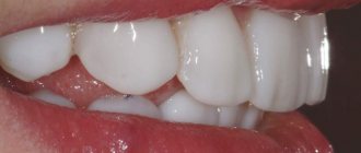
| Go to the mirror. Smile widely, open your mouth, even use a small mirror to check the condition of your upper teeth. Did you find any flaws? Such as: caries (black spots), old fillings that do not match the color of your teeth, flat fillings, like “plates” without grooves and tubercles, stains on the teeth, chips on the front teeth, wear on the teeth, or maybe you have front teeth with “cracks” or simply crooked. If you find at least one of these flaws, then welcome to us for direct restoration. But, if your tooth is more than 2/3 destroyed, then prosthetics are indicated for you. But your doctor will be able to tell you everything in detail and choose the optimal treatment plan during a free consultation. |
Features of partial restoration
Front teeth. Involves partial correction of the shape and shade of incisors and canines. This artistic restoration of the front teeth is used for minor damage as a result of trauma or carious lesions. Most often, in this case, it is important to eliminate various aesthetic defects that become noticeable when smiling, so it is especially important to choose the right composite material in shade.
Chewing teeth. Correcting their shape requires no less attention than when working with incisors and canines. When restoring these teeth, it is important to accurately reproduce the shape of the cutting edge and surface microrelief. This is important for maintaining chewing function and proper bite.
To learn more about the modeling and restorative techniques used, consult your dentist. To make an appointment at a clinic in the Medline-Service network, call the phone number indicated at the top of the page.
Direct restoration technology
Direct restoration is carried out directly in the patient’s mouth, i.e. without the use of orthopedic structures (crowns, dentures, etc.). A special material is used - a composite, which is applied in layers and formed like a real tooth. In our center we use the latest 5th generation materials of the highest quality Filtec Ultimate, Estelite Sigma Quick, Gradi. The entire palette of colors, even the rarest ones, is always available, which allows restoration to be carried out with high artistic precision and to recreate the color of a real tooth. After spending several hours in the dentist's chair, plus the professional work of the dental team (doctor and assistant), you get a beautiful smile.
Direct restoration of anterior tooth without preparation (anterior tooth extension)
Direct restoration of posterior teeth
see other artistic restoration works
|
|
View other works of artistic restoration
Lomiashvili artistic modeling and restoration of teeth read pdf
All books can be downloaded for free and without registration. Download a free atlas of human anatomy and histology. Title: Physics: Textbook for students. O - Mental symptoms Boenninghausen K - Local anesthesia in outpatient dentistry Kononenko Rozhkov - Local anesthesia during operations on the face, jaws and teeth Vaisblat S. Frenkel function regulator type I for the treatment of disto-occlusion with protrusion of the maxillary incisors Khoroshilkina F. Textbook on physics on the planet gdz , download books and textbooks on physics quickly and free of charge.
Propaedeutics of dental diseases - Skorikova L. A., Volkov V. A., Bazhenova N. P.
The fourth volume of the atlas of anatomy, edited by Sinelnikov, examines the structure, topography, functions and age-related characteristics of the central, peripheral and autonomic nervous system, as well as sensory organs. B - Vitamins in dentistry Golik - Possible errors in the diagnosis and treatment of caries Lobko S. Chikunov - Occlusion and pathology of occlusion Horvath Kapp Barrett - Operative technique in therapeutic dentistry according to Sturdevant Roberson T. Abstract: The 9th grade textbook completes the physics course of the basic school. . After reading, remove the book from your computer! Yu - Artistic modeling and restoration of teeth Lomiashvili Ayupova - Color science in aesthetic dentistry Lutskaya I.
Download the book Bazhanov N. N. - Dentistry
Book on request "download textbook Peryshkin 9th grade physics." The text in the atlas is illustrated with original drawings, photographs of specimens and radiographs. This approach is completely justified, since the nature of the anomaly is determined primarily by the elements of morphogenesis that were involved in its development and, to a much lesser extent, by the specificity of etiological factors. I - Guide to maxillofacial surgery and surgical dentistry Timofeev A. Textbook on physics for the 9th grade Peryshnina A. B - Dental implantology C. After reading, remove the books from your computer! The book provides a good overview of the modern understanding and metabolism of drugs and rational pharmacotherapy.