Content:
- How do they manifest themselves?
- Where do they come from?
- Consequence of dental diseases
- Aphthae of traumatic nature
- Result of common diseases
- How to cure
Defects of the mucous membranes of the oral cavity are not uncommon.
Many people encounter them. They can occur due to infectious and viral diseases, mechanical damage to tissues. Most often, dentists encounter white sores on the gums. These are painful areas that prevent the patient from enjoying life. They make eating problematic and do not allow for high-quality hygiene measures. Any touch to the affected area provokes acute pain.
It is important not to ignore the presence of white patches on the gums. If they appear, you should contact your dentist. The doctor will determine the cause of the disease and tell you how to eliminate it.
Complications
In the absence of timely and high-quality treatment, the disease can cause serious complications. A frequent consequence of suppuration during periodontitis is periodontal disease, a disease characterized by the formation of very deep gum pockets and the spread of foci of infection and suppuration throughout the gums. Over a long period of time, periodontal disease usually leads to loosening of teeth and their loss. In addition, inflammation can lead to the formation of gumboil, a large abscess that causes the gums and cheek to swell and lose their natural shape. Treatment for flux usually requires surgery.
The presence of a constant source of infection in the body negatively affects the functioning of all organs and systems, in particular the functioning of the immune system. Abscesses and other chronic infectious diseases lead to a steady decrease in immune status, which creates favorable conditions for the development of a whole range of diseases.
We should not forget that the muscles and nerves of the face are located in close proximity to the gums. For example, the formation of a large abscess on the gingival part of the upper jaw can lead to damage to the trigeminal nerve, which innervates the nasolabial triangle. Sharp pain of a neurogenic nature, paresis and paralysis of the facial muscles are rare, but still probable consequences of gingival abscesses.
Where do they come from?
Conventionally, all provoking factors can be divided into the following groups:
- mechanical or thermal damage to tissues;
- dental diseases;
- systemic pathologies.
In other words, the problem may be much more serious than it seems at first glance.
Consequence of dental diseases
Among the diseases of the oral cavity that cause the appearance of painful white spots are:
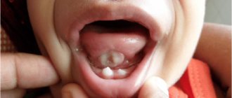
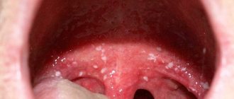
- Aphthous stomatitis. Has a relapsing course. A person develops painful aphthae. They can be located on the cheeks, tongue, palate, lips. They always hurt a lot. Heal within 7-10 days. If such an aphtha is damaged by carelessness, the regeneration processes will proceed very slowly and after the end of regeneration, a non-absorbable scar will remain. Until now, scientists have not established why aphthous structures appear. Most researchers believe that reduced immunity is to blame.
- Herpetic stomatitis. Caused by the herpes virus. A huge number of small white erosions appear in the patient’s mouth. Women over the age of thirty are prone to the disease. Aphthae occur on the tongue and on the surface of the floor of the mouth. They do not have clear boundaries. “They drag on” for one to a week and a half. In addition to ulcers, this form of stomatitis causes increased body temperature, enlarged subcervical lymph nodes, and general weakness.
- Necrotizing periadenitis. Another name for this pathology is Setton's aphthae. The disease is difficult to correct. It often takes several years to treat it. First, compactions appear under the mucous membrane. Soon, instead of them, ulcers with protruding edges form. They can be strewn with the surface of the cheeks, tongue, lips. Setton's aphthae are always very painful. They interfere with normal conversation and eating.
- Vincent's ulcerative necrotic stomatitis. Infectious lesion. It is caused by associations of fusobacteria and spirochetes. Painful areas form in the patient's mouth. The condition is aggravated by muscle pain and headaches. To eliminate tumors in the mouth, doctors carry out local etiotropic therapy, use antibiotics and antiprotozoal drugs. It has been noticed that Vincent's disease is more often encountered by men over the age of thirty with reduced immunity and vitamin deficiency.
Associated symptoms
If you find red spots in your mouth, you should consult a specialist. As a rule, most of the listed diseases are also accompanied by the following manifestations:
- local pain,
- itching and burning,
- bad breath,
- swelling and inflammation of the gums,
- local temperature rise,
- discomfort when chewing, swallowing, communicating.
If these symptoms appear, you should consult a doctor.
Aphthae of traumatic nature
Ulcers resulting from trauma include:
- Afty Bednar. Happens only to small children. They are traumatic ulcerations. They arise due to the habit of constantly gnawing hard objects, poor hygiene, and gross mechanical irritation of the upper palate. Usually such white areas are covered with a yellowish coating, which can be easily removed.
- Traumatic aphthae. These include ulcers formed due to sudden movements with a toothbrush, carelessly performed dental treatment, rubbing of dentures, exposure to acids, alkalis, and medications. Treatment of such neoplasms does not take much time and is not complicated. The main thing here is to remove the influence of the provoking factor, then the tissues will recover fairly quickly.
Result of common diseases
Some diseases have a systemic effect on the body. This means:
- Tuberculosis. When the lungs are affected, negative symptoms can spread to the mucous membranes of the mouth. This occurs due to the penetration of the pathogen into the oral cavity through damaged epithelium. Then the patient develops ulcers on the cheeks and under the tongue. First, these are always specific tuberculous tubercles, and then aphthae. They grow quite quickly. At the same time, an increase in the amount of plaque on the tongue, weakness, and loss of body weight are recorded.
- Syphilis. Pathology caused by Treponema pallidum. During the incubation period, which lasts about three weeks, aphthae occur. They are round or oval. Their edges are usually raised, swollen, and the color is red. Such damage takes a long time to heal - up to three months. Syphilis erosions are not as painful as tuberculosis ones. After their healing, retracted scars form.
- HIV infection. It has been noticed that every third HIV-infected person has inflamed oral tissues. In this case, therapy must be specific. It is not the dentist who is responsible for its implementation, but the infectious disease specialist. As soon as the patient’s health improves and the immune system is strengthened, the aphthae will disappear on its own.
Abscess on the gum with periodontitis -
Gum suppuration can look completely different, but in any case, the formation of an abscess on the gum always occurs in the projection of the causative tooth. If the cause of suppuration is an infection in the root canals, then you will always see an old filling or crown on the causative tooth, or the tooth will be partially destroyed. In this case, infection in the root canals gradually leads to the development of a focus of chronic inflammation at the apex of the tooth root.
In dentistry, such inflammation of the tooth is called chronic periodontitis. From time to time, an exacerbation of chronic inflammation may occur, and in this case, pus begins to form at the apex of the tooth root, which comes out through the bone tissue and penetrates under the gum, forming an abscess there. Therefore, please note that with periodontitis, an abscess on the gum most often forms not at the gingival edge, but closer to the projection of the apex of the root of the causative tooth.
Abscess on the gum with periodontitis: photo
Symptoms - an abscess with periodontitis may look like a slight swelling of the gums in the area of 1 tooth or in the area of several teeth, if a significant purulent abscess has formed - flux (Fig. 4). Typically, the appearance of gum swelling is preceded by pain when biting one of the teeth. The pain can be acute, but sometimes purulent inflammation can occur without pain. Sometimes, in the projection of gum suppuration, swelling of the soft tissues of the face appears.
The abscess formed under the mucous membrane of the gums can burst with the formation of a fistula opening (Fig. 7-8). The fistula opening is connected through a fistulous tract to the source of inflammation at the apex of the tooth root. Therefore, gradual discharge of pus may be observed from the fistula openings. As soon as the acute inflammation at the root apex subsides and the process of pus formation stops, the fistula openings can close, but only until the process worsens again.
X-ray diagnostics - if you have a fistula or a purulent sac on your gum, treatment is only possible at the dentist. Before starting treatment, you need to take an x-ray to confirm the presence of inflammation at the apex of the tooth root (24stoma.ru). The image will also show the quality of root canal filling, if it was performed previously. It’s sad, but according to official statistics, dentists fill root canals poorly in as many as 60-70% of cases.
The most common mistake dentists make is not filling root canals up to the apex of the tooth root, as a result of which infection begins to multiply in the part of the root canal that is not filled with filling material. As a result, a focus of chronic inflammation develops at the apex of the tooth root, for example, in the form of a cyst or granuloma. Below you can see what cysts and granulomas look like on a diagram, an x-ray, and also on the apex of the root of an extracted tooth.
Treatment of gum suppuration during periodontitis -
If, against the background of an exacerbation of chronic periodontitis, an abscess appears on the gum, then what to do will depend on the results of examining the tooth and analyzing the x-ray. In some cases, it may turn out that the tooth can no longer be treated, and then you will be referred to a surgeon for removal. However, in most cases, it is possible to cure such a tooth, and then the doctor’s algorithm for further actions will depend on whether root canal filling was previously performed in this tooth.
1) If the root canals are not filled –
This greatly facilitates the doctor’s work, because... in this case, the dentist will not have to go through the trouble of unsealing poorly filled root canals. In this case, a standard method of treating periodontitis is used, which includes mechanical treatment of the root canals + treatment of the inflammation at the root apex. On your first visit, the dentist will remove your old filling or crown, drill out tooth tissues destroyed by caries, and perform a mechanical expansion of the root canals to allow pus to drain out through them.
After this, the dentist will prescribe you antibiotics, antiseptic rinses, and will most likely send you to a surgeon to make an incision in the gums, which is necessary to create a good outflow of pus. After 3-4 days, the dentist will make an appointment for you again to complete the mechanical treatment of the root canals and seal them. If the lesion is small at the apex of the tooth root, the doctor can immediately perform a permanent filling of the root canals with gutta-percha. But usually, the canals first have to be filled with temporary medicinal paste for a period of 1-2 months, and only after that permanent filling with gutta-percha is carried out.
How the gum incision is made - the incision is usually completely painless and is performed under local anesthesia. If the abscess is small, the size of the incision usually does not exceed 5-7 mm, but if a large purulent abscess develops (as in the video below), the incision is made up to 1.5 cm. After opening, pus comes out of the gum, then the wound is washed with antiseptics and into it a small rubber drain is inserted. The latter prevents the edges of the wound from sticking together, which is necessary so that the inflammatory purulent exudate does not accumulate, but continues to separate from the wound. The drainage is usually left for several days.
2) If the canals in the tooth are sealed poorly -
If the dentist sees on an x-ray that the cause of suppuration lies in poor-quality root canal filling performed earlier, then there are 2 possible treatment options. This is either a standard treatment of periodontitis with preliminary filling of the root canals, or a minor surgical operation (resection of the root apex)…
- Standard therapeutic treatment - on the first visit, the dentist drills out the old filling, removes the crown and tries to unfill poorly filled root canals.
Next, the canals are washed with antiseptics, the tooth is left open for several days + antibiotics are prescribed. If necessary, the patient is also sent to a surgeon for a gum incision. Thus, unlike the previous treatment option, only 1 point has been added here - unsealing the root canals. When the inflammation subsides after a few days, first, either temporary filling of the canals with medicinal paste, or immediate permanent filling with gutta-percha can be carried out. The medicinal paste is left in the canals for a period of 1 to 3 months. During this period, an x-ray will be taken to record a decrease in the size of the focus of chronic inflammation at the apex of the tooth root. When the lesion disappears or becomes small, the canals will be sealed with gutta-percha and a permanent filling or crown will be placed. - Resection of the root apex (Fig. 13) –
This method allows you not to re-treat root canals or remove the crown from the tooth.
It consists of making a small incision in the projection of the apex of the tooth root on the gum, through which the doctor uses a drill to cut off the apex of the root with the unfilled part of the root canal from the tooth. Also, a cyst or granuloma is scraped out of the wound. However, the operation can only be performed on those patients whose root canal is poorly filled only at the very apex of the root. Root resection is a very simple operation and usually takes only 25-35 minutes. It is easiest to carry out on the front teeth, much more difficult on the side teeth. Low cost of the operation + no need to spend money on replacing the crown and re-treating the tooth. The operation is performed after the acute inflammation has been relieved, which may require an incision and antibiotic therapy.
→ Root apex resection operation
How to cure
Treatment for damage to the mucous membranes is always selected individually. There is no single regimen that is suitable for every clinical situation. First, the doctor determines the cause of the complication, and only then tells how it can be eliminated. The emphasis is on the area of the affected surface and the features of its localization, the presence of concomitant diseases, and the severity of symptoms.
To stimulate local regenerative processes the following can be used:
- topical agents - gels, solutions, creams, ointments;
- medications administered orally, intravenously or intramuscularly (broad-spectrum antibiotics, painkillers, immunostimulants, anti-inflammatory drugs, vitamins);
- physiotherapy.
In some cases, it is necessary to carry out professional hygiene, treat identified dental diseases, and replace old prosthetic systems with new ones. If necrotic tissue needs to be removed, the doctor uses anesthetics. They make the intervention painless.
It is very important that a person does not ignore painful areas on the gums. If they appear and do not go away within three to five days, you should definitely consult a dentist. After all, it is possible that the disorder is not associated with stomatitis, but with some serious pathology of the internal organs. In any case, you won’t be able to find out at home.
Of course, it is easier to prevent the development of a dental disorder than to treat it. Preventive measures to prevent the occurrence of erosion on the gums come down to following the rules of hygiene, undergoing specialized dental examinations, timely treatment of all diseases, and proper nutrition. People who care about their health encounter gum ulcers much less frequently.