From this article you will learn:
- how much does it cost to do a dental x-ray,
- what you need to know about radiation doses,
- Is it possible to take a dental photograph during pregnancy?
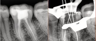
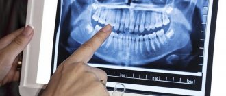
Dental X-ray is a traditional way to determine the quality of root canal filling, as well as to diagnose inflammation at the apex of the tooth root (with apical periodontitis). X-rays of human teeth in dentistry are most often carried out using targeted X-rays. A targeted photograph of a tooth received this name because it is small in size and shows the condition of only a few teeth and the bone tissue around them (Fig. 1-2).
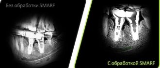
Modern targeted X-rays produce a very low dose of X-ray radiation (compared to what it was 15-20 years ago). This is due both to the advent of ultra-sensitive photographic films, which require a significantly lower dose of radiation, and secondly, to the advent of special intraoral sensors that capture the image digitally and display it on a computer screen. Such digital images require several times less X-ray radiation - even compared to modern films.
Sighting and panoramic radiography in dentistry –
However, targeted images are not suitable for planning bite correction, assessing the condition of bone tissue before implantation, or planning the installation of future implants. They are not very convenient for planning treatment and prosthetics for a large number of teeth, and often do not allow detection of perforations and cracks in the tooth root. Focused photographs of the teeth may not be enough in cases where it is necessary to find problems with root canal filling in order to be able to properly treat the teeth.
Therefore, dentists very often refer patients to more complex options for x-ray examination of teeth and jaw bone tissue -
- dental computed tomography (CT),
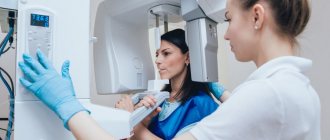
- orthopantomogram (Fig. 3),
- teleroentgenogram (for bite correction).
In this article, we will go into detail about the pros and cons of targeted dental x-rays, and what you need to pay attention to if you need to have a dental x-ray (for your own safety). For detailed reviews on the remaining specified methods of radiography in dentistry, read the links above.
Reviews about the clinic on independent portals:
Features of the appointment of dental x-rays
If you are a new patient, you will likely undergo dental x-rays so that the new dentist can get a clear picture of your dental health. This is especially important if you do not have x-rays from your previous dentist. Children may need dental x-rays more often than adults because their dentists need to monitor the growth and development of their teeth. This is important because it can help the dentist determine whether baby teeth need to be removed to prevent complications, such as adult teeth growing behind baby teeth.
X-ray of teeth
Modern computer digital technologies used in the Novodent clinic have made it possible to significantly reduce the radiation dose to patients, protect staff, and improve the quality of the resulting image.
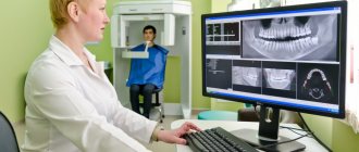
An initial consultation with a dentist does not always provide complete information about the condition of the dentition, so to obtain a complete clinical picture, the patient may be referred for dental x-rays. The procedure is a highly informative diagnostic method that allows us to identify pathological changes in the internal tissues of teeth, gums, and jawbone.
X-ray examination of teeth at the Novodent clinic is carried out using modern and high-quality equipment, which allows you to obtain clear images of one or several teeth or the entire jaw. The results obtained are the basis for making a correct diagnosis and drawing up a treatment regimen.
Summary: important points
As a practicing dentist who knows the system from the inside, I want to draw your attention to the following points that are important for your safety. If the clinic has an X-ray machine, then a license must be obtained for it, the issuance of which presupposes the mandatory presence of a certified radiologist on the staff of the dental clinic. However, in reality, even in large clinics and public clinics, it is not always the case that x-rays will be taken by a trained specialist.
Even if he is, he may go on vacation or get sick, and a regular nurse (dental assistant) will take the pictures instead. This is a gross violation that leads to both the production of low-quality images and an increase in the radiation dose. In small clinics, the risks of receiving a poor-quality x-ray examination are much higher, and the first thing that makes you suspect a forgery is if the picture is taken not by a special employee, but by a nurse from the dentist you came to see.
The second very important point: if you see that the x-ray is not done in a special room, but the x-ray machine is located right next to the dentist’s chair, then you should change the clinic and the doctor. The fact is that all objects in this office (including the dental chair and doctor’s instruments) will have an increased background radiation, and this is no longer safe for health. We hope that our article on the topic: X-ray of the jaw and teeth was useful to you!
Sources:
1. Higher prof. the author’s education in therapeutic and surgical dentistry, 2. Based on personal experience in therapeutic and surgical dentistry, 3. National Library of Medicine (USA), 4. “Digital and film radiography in outpatient dentistry” (Chibisova), 5. “X-ray diagnostics in dentistry" (Lutskaya I.K.).
When is a dental x-ray necessary?
Dental X-ray is the most practical and affordable way to study pathologies of the dental system. Thanks to the technique, it is possible to determine the condition of the internal structures of the tooth and obtain information about the features of the anatomical structure of the dentition.
Based on the results of the x-ray, the doctor is able to determine the following disorders:
- the degree of damage to dental tissues due to periodontitis, pulpitis, caries;
- inflammatory processes of the gums and pulp;
- purulent masses in root canals;
- destruction of dental structures under the crown;
- cysts or tumors;
- draw up a plan for surgical intervention;
- determine the size, volume, structure of bone tissue before implantation;
- assess the correctness of occlusion;
- detect anomalies of the dental system;
- determine the position of erupting wisdom teeth;
- assessing the effectiveness of previous treatment.
X-rays are carried out before the start of orthodontic treatment, implantation, prosthetics, before and after filling the tooth canals.
Are there any contraindications to X-ray examination?
Dental X-rays are not performed on patients with severe bleeding in the mouth, or those who are unconscious or in critical condition.
Pregnancy is considered a relative contraindication. In this condition, women should not undergo dental procedures involving radiation until 4-5 months. But in each individual case, the expectant mother needs to personally consult a doctor, since situations are different.
A breastfeeding woman can undergo an X-ray examination. But it is advisable to resort to digital methods and skip one or two feedings after the session.
What is included in the service
If there is a need to take an X-ray of a tooth, the clinic provides a high-quality X-ray machine that allows you to get a clear image of the area being examined.
Depending on the purpose of diagnosis, images come in several types:
- Targeted – allows you to assess the condition of one or more teeth and periodontal tissues: dentin, root canals, blood vessels, periodontium.
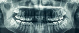
- Panoramic (OPTG, orthopantomogram) is a detailed examination method that allows you to obtain a flat image of the jaws, teeth, joints, and sinuses. Panoramic X-rays of teeth can easily detect carious cavities, cysts, and defects in fillings, and will also allow the doctor to draw up a plan for implantation and prosthetics.
- Occlusal - more often prescribed to children or in the absence of complete information after targeted radiography.
In addition to the main types of images, photographs can be film and digital. Transmitted using a special sensor or computer.
At the Novodent clinic, in addition to standard types of x-rays, teleradiography is performed, which is recommended when planning prosthetics; it studies the structure of the facial skull and soft tissues. If there is a need to obtain accurate results, a computed tomography scan is offered, which will assess the area under study with millimeter accuracy.
How is dental x-ray examination performed?
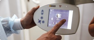
The photo is taken in a special room with lead-protected walls and floors. The doctor asks the patient to remove metal jewelry and accessories, as they may distort the results of the study. The vital organs of the body are protected by an apron with lead inserts. Features of the study depend on the method used.
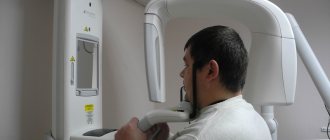
When receiving a targeted image, the doctor directs an x-ray beam to the area of the jaw being examined from the side of the face or to an area of the oral cavity. An orthopantomogram is obtained in a different way. The patient fixes his head in a special device, grabs the plate in a disposable cover with his teeth and remains motionless while the mobile part of the device makes a full revolution around the head.
Is there an alternative to CT
There are many other diagnostic imaging methods (x-ray, orthopantomogram, ultrasound, etc.), but only CT provides the possibility of highly accurate, separate images of all types of tissue at different angles and to different depths. Although a panoramic dental photograph remains an equally important diagnostic tool for a dentist today, it can only provide a general overview. In turn, a 3D tomogram allows you to obtain not a single flat image of the jaw, but a whole series of sequential multiplanar images in different projections and without the distortions inherent in a panoramic image.
Example:
due to the different density of bone structures exposed to X-ray radiation, it is impossible to see less dense bone in a 2-dimensional image; accurate information is provided by a 3- D image of the teeth.
Possible harm
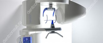
Cone beam dental computed tomography is the safest and fastest diagnostic method. Thanks to the use of a conical X-ray beam, the radiation dose received during the study is 10 times less than when using spiral CT. And the pulsating mode of the X-ray beam further reduces the radiation dose. The three-dimensional computed tomograph SOREDEX Scanora 3D is one of the safest devices in terms of X-ray radiation dose - only 0.035 m3v.
However, despite the safety of the study, CT also has contraindications. If we just talk about dental tomography, it is not performed during pregnancy (in the 1st trimester). 3D dental x-rays with contrast are prohibited for pregnant and lactating women, patients with endocrine disorders (diabetes mellitus, thyroid pathologies), renal failure and intolerance to iodine-containing drugs.
Why are dental x-rays prescribed?
A 3D photograph of teeth is performed if the following indications exist:
- Injuries of the maxillofacial area.
- Preparation for endodontic treatment (structure of root canals, pathological processes in the periodontium, degree of pulp damage, etc.).
- Diagnosis of neoplasms (cysts, abscesses, granulomas, tumors).
- Anomalies of development and deformation of the maxillofacial apparatus.
- Quality control of filling and implant installation.
- Planning of orthodontic treatment (identification of impacted and dystopic teeth, analysis of the condition of the tissues around each tooth, etc.).
- Detection of hidden periodontal cavities and pockets.
- Implantation planning (assessment of jaw bone parameters, indications for sinus lift or osteoplasty, modeling the result of implantation).
- Endogenous pathologies of the maxillary sinuses.
Three-dimensional x-ray examination is the gold standard when planning any complex dental procedure or surgery. CT allows you to quickly make an accurate diagnosis, competently plan treatment or dental prosthetics, and monitor the results.
Advantages of the method
- The ability to rotate, enlarge, and examine images in any projection and section, which is impossible with conventional 2-dimensional scanning.
- The examination lasts only a few seconds (8-20 seconds).
- Complete diagnostic information.
- Maximum security.
- Digital information format.
- Detection of any pathological processes at an early stage.
- No prior preparation required.
- 3D reconstruction without distortion or artifacts.
- A wide range of purposes - from endodontic dental treatment and implantation to maxillofacial operations.
How does the procedure and decoding work?
To take a 3D photograph of teeth, a standing or sitting patient needs to bite a special plate and fix his position in the device using a fixing stand. During the entire scanning time, you must remain absolutely still.
The tomograph sensor makes a series of revolutions around the patient’s head for 8-20 seconds, producing about 200 images in different projections. Processing digital data takes 5-15 minutes, after which the information is written to a disk or flash drive. No preparation is required, you just need to remove all metal jewelry from your neck, ears, and hair before the procedure.