Symptoms and clinical picture of lymphadenitis in children
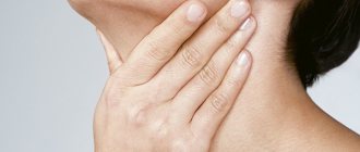
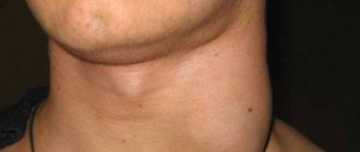
Signs of lymphadenitis in children are expressed in a significant increase in lymph nodes (mainly cervical and submandibular), sometimes so much that it is visible to the naked eye. On palpation, the child feels acute pain, the outer skin is hyperemic.
Symptoms of lymphadenitis in children vary depending on its type.
General - there are signs of intoxication, fever, chills, deterioration in general health, drowsiness, weakness, dizziness, sweating, etc.
Purulent - the symptoms described above are even more pronounced, the skin in places of swelling is noticeably reddened, there is increased swelling and local pain.
Manifestations of lymphadenitis can acquire varying degrees of severity, depending on the patient’s age, complexity and duration of the disease (the acute period can last from 2 to 4 weeks).
Causes of enlarged lymph nodes in a child
If your doctor suspects that your lymphadenopathy is not the result of an infection, the first step is to perform a physical examination and take a detailed medical history. Hard, dense nodes that do not move under the fingers will be the basis for referring the child for laboratory tests, possibly also for imaging. Only if the test results are concerning, the doctor will order a histopathological examination.
Basically, there can be two reasons for enlarged lymph nodes in a child. The first is the proliferation of nodal cells, mainly lymphocytes. The second reason is infiltration caused by foreign cells - inflammatory and malignant. Neoplastic diseases that present with lymphadenopathy include lymphocytic lymphoma and Hodgkin lymphoma. However, cancer is not the main cause of enlarged lymph nodes. More often, lymphadenopathy is caused by: - viral infections;
- allergies; — vaccination; - autoimmune diseases.
Basic treatment methods and contraindications
Treatment is carried out using an integrated approach and conservative methods of therapy.
The following medications are prescribed:
- antibiotics of narrow or broad spectrum of action;
- probiotics - to preserve intestinal microflora;
- antihistamines - to prevent allergic reactions and reduce tissue swelling.
Physiotherapeutic methods are represented by UHF and EHF therapy, which has an anti-inflammatory and antiseptic effect.
Severe infections may require surgery.
Contraindications:
- suspicion of the presence of a tumor formation;
- Do not apply warm compresses or heat areas of swelling.
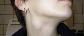
Enlarged lymph nodes in a child's neck
Enlarged lymph nodes in a child's neck are of greatest concern to parents and doctors because they may be a sign of Hodgkin's lymphoma. The disease most often occurs in adolescent children, less often in young children. In most cases, the cause of enlarged nodes in the neck is viral infections, incl. mononucleosis, that is, a disease caused by the EBV virus, and bacterial - inflammation of the tonsils and throat.
In the case of lymphadenopathy located in the neck, you can be sure that the pediatrician will prescribe basic diagnostic tests, incl. blood analysis.
However, it is worth considering that an increased level of lymphocytes in a child under 6 years of age is perceived as normal. Doctors call this physiological lymphocytosis. Additionally, you should be aware that the standards set by the laboratory apply to adults, so any test results should be discussed with your pediatrician. Comparing the data to the standards provided by the laboratory is not sufficient to correctly interpret a child's test results.
General information
Lymphadenitis is called inflammation of the lymph nodes (hereinafter referred to as LN) due to the presence of infectious or non-infectious inflammation. Thus, it is a secondary disease caused by the underlying disease.
In pediatrics, this phenomenon is not uncommon. The main reason that the submandibular lymph nodes (and in other parts of the body) often become inflamed lies in the incomplete formation of the immune system of the young body.
Note. The disease is most often registered in children aged from one to six and, as a rule, has a fairly pronounced course.
Causes of enlarged lymph nodes in children
Lymph nodes belong to the organs of the immune system.
Role of lymph nodes:
Barrier. Lymph nodes (LN) are the “first line of defense” against the penetration of various foreign agents into the child’s body. A natural reaction to this “acquaintance” is an enlargement of the lymph nodes.
Filtration. Various substances, microbial cells, and particles of the body’s own tissues settle in the lymph nodes.
Parents often call enlarged lymph nodes “glands.”
The lymph node itself is a small oval formation (several millimeters in diameter). On the outside it is covered with a special capsule of connective tissue, and on the inside it is divided into separate sections by partitions. These elements of the immune system are located in groups throughout the body at the confluence of several lymphatic vessels. In newborn babies, the capsule of the lymph nodes is still very tender and thin, so it is difficult to feel them under the skin. By the age of one year, lymph nodes can already be felt in almost all healthy children.
The maximum number of lymph nodes appears by 10 years of age. In an adult, their total number is 420-460.
The size and condition of the lymph nodes are assessed by palpation. In this case, it is necessary to palpate the lymph nodes in all groups (there are 15 of them) - from the occipital to the popliteal. If the lymph nodes are significantly enlarged, parents or the child themselves can pay attention to this symptom. When a lymph node becomes inflamed (lymphadenitis) due to the appearance of pain, children clearly indicate the localization of the process.
Normally, in healthy children, no more than three groups of lymph nodes are usually palpable.
Should not be palpable:
- chin;
- supraclavicular;
- subclavian;
- chest;
- elbows;
- popliteal lymph nodes.
Signs of normal lymph nodes:
- their size does not exceed the diameter of a small pea,
- they are isolated
- soft elastic consistency.
- mobile,
- not connected to the skin and to each other (doctors say “not fused”),
- painless.
An increase in the size of lymph nodes with a change in their consistency is called lymphadenopathy, a quantitative increase in lymph nodes is called polyadenia (from aden- gland, since previously lymph nodes were mistakenly considered glands).
Causes of enlarged lymph nodes in children:
- infections;
- tumor processes;
- a special metabolic disorder called “storage diseases”.
Local (regional) isolated enlargement of lymph nodes is always caused by changes in the corresponding zone (region) of the body from which lymph comes.
In all cases of unusual local enlargement of lymph nodes, a thorough examination of the child is necessary to determine the extent to which the process has generalized (spread).
Stages of diagnosis for isolated enlarged lymph nodes:
Clinical diagnosis. It consists in the fact that the doctor first makes a diagnosis based on the complaints of the child or parents, clinical data and the results of blood, urine, x-ray, etc. tests.
Tuberculin diagnostics. Necessary to exclude the possibility of tuberculosis.
Special (serological) blood tests (performed if another infection is suspected).
Usually, with an isolated increase in any group of lymph nodes, antibacterial agents are prescribed for 8-10 days. An improvement in the child's condition and a decrease in swelling of the lymph nodes is regarded as confirmation of the bacterial nature of the disease.
Biopsy of a lymph node or puncture with examination of the lymph node tissue under a microscope. It is carried out in the most difficult diagnostic cases.
Features of damage to lymph nodes in certain areas:
Enlarged occipital lymph nodes: found in inflammatory processes of the scalp (purulent rash, boils, osteomyelitis of the calvarial bones, fungal infection). With rubella, in addition to the occipital ones, other groups of lymph nodes are enlarged to a lesser extent.
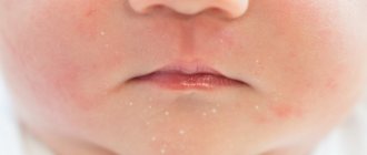
Enlarged parotid lymph nodes: characteristic of inflammation of the middle and outer ear, purulent inflammation of the scalp (pyoderma), lice, furunculosis. Often these groups of lymph nodes react to infection of an allergic rash in atopic dermatitis and eczema, especially when it is localized behind the ears.
Enlarged lymph nodes behind the angle of the lower jaw and along the posterior muscles of the neck. Develops during inflammatory processes in the nasopharynx or after them: tonsillitis, infectious mononucleosis (as a manifestation of a generalized process), with chronic tonsillitis and adenoid growths, with tuberculosis of the tonsils.
Enlarged lymph nodes behind the angle of the lower jaw and in the middle triangle of the neck: severe forms of tonsillitis, scarlet fever. With diphtheria of the tonsils, there is a symmetrical increase in lymph nodes to the size of an apple. At the same time, they are elastic and painful. Swelling of the surrounding tissues develops with an increase in the size of the neck. In the case of severe angina, acute inflammation of the lymph nodes can develop - lymphadenitis and even purulent lesions. In case of cat scratch disease (caused by a special microorganism), this group of lymph nodes may increase. The combination of sore throat, lymphadenitis and peeling of the palms and feet is characteristic of streptococcal infection (streptococcal tonsillitis, scarlet fever). Several more reasons for the reaction of this group of lymph nodes: Kawasaki syndrome (combined with damage to the eyes, skin, coronary arteries, fever, rash), toxoplasmosis, tumors of the blood and lymphatic system (Hodgkin's disease - lymphogranulomatosis, non-Hodgkin's lymphoma - lymphosarcoma).
Enlarged lymph nodes in the lateral triangle of the neck may be a sign of infection in the nasopharynx, tuberculosis of the lymph nodes, or tumors.
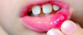
Enlargement of the lymph nodes in the mental area develops with an abscess in areas of the jaw, with damage to the front teeth - incisors, stomatitis, inflammation of the lower lip.
Enlarged submandibular lymph nodes are characteristic of inflammation of the jaw due to dental damage, stomatitis and gingivitis (inflammation of the gums). After treatment with antibiotics, the lymph nodes shrink and restore their mobility when palpated.
Enlargement of the axillary lymph nodes occurs with infectious diseases of various causes in the arm and shoulder area (purulent skin lesions, chicken pox). Unilateral lymph node enlargement can develop after vaccination or injury to the arm, such as cat scratch disease.
Enlarged ulnar lymph nodes are a sign of infection in the hand or forearm.
Enlarged inguinal lymph nodes indicate an infection in the lower extremities, localized in the bones, muscles or skin. This symptom appears with inflammation of the joints, with severe diaper dermatitis, with furunculosis in the gluteal region, inflammation of the genitals, after BCG vaccination when the vaccine is administered to the thigh area. Moreover, an isolated increase in lymph nodes for 3 months after BCG is normal, and a longer persistence of the symptom is regarded as an indirect sign of a decrease in the activity of the immune system or high virulence of the vaccination material, or as an individual feature of the child’s reactivity. A biopsy of such lymph nodes may reveal large numbers of blood cells called macrophages. After BCG, LNs can be soaked in lime and palpated through thin skin for many years. In case of infection penetration through the skin of the leg in cat scratch disease, the group of inguinal lymph nodes also reacts.
Frequent wounds to the skin of the legs and feet, infection of these wounds in children of preschool and school age lead to the fact that in most of them significantly enlarged lymph nodes are clearly palpable in the groin area. Such children are not considered sick in the absence of other signs of pathology.
Many diseases begin with an increase in lymph nodes in any one zone, and then the lymph nodes increase in almost all groups. This picture is observed with influenza, measles, rubella, infectious mononucleosis, viral pneumonia, viral hepatitis, widespread eczema, congenital syphilis, toxoplasmosis, etc.
In addition to the lymph nodes, other formations can also raise the skin and be palpated under the skin. One of the reasons for this is lymphangioma (parents are more familiar with a nearby formation of blood vessels - hemangioma), a soft swelling under the skin without clear boundaries. Lymphangioma is localized mainly in the neck and can cause difficulties during childbirth, swallowing and breathing. Often requires surgical treatment.
It must be remembered that LNs are located both in the chest and in the abdominal cavity.
Signs of enlarged lymph nodes in the chest:
- breathing problems;
- difficulty swallowing;
- prolonged hiccups.
The reaction of lymph nodes in the abdominal cavity to a viral or bacterial infection can manifest itself as very severe abdominal pain, which sometimes requires a differential diagnosis with appendicitis.
Parents should know that if one or more enlarged lymph nodes are detected in one group, they need to carefully examine the child and palpate lymph nodes in other areas. Before consulting a doctor and having a blood test, you should not heat the lymph nodes. Children who have enlarged glands in most groups must be examined using ultrasound of internal organs to assess the condition of the liver, spleen and abdominal lymph nodes.
May our children be healthy!
Doctor T.A. Konon
Classification
Often, along with the submandibular lymph nodes, the cervical lymph nodes also become inflamed.
Depending on the nature of the course, submandibular lymphadenitis occurs:
- acute – up to 14 days;
- subacute – from two weeks to a month;
- chronic – more than one month.
According to its genesis and origin, the disease can be specific or nonspecific.
Based on the type of transformation, pathogenesis is classified into:
- serous or infiltrating;
- purulent - the formation of pyogenic processes in the lymph nodes;
- necrotic – destruction of lymph nodes;
- adenophlegmons.
Based on their etiology, they distinguish between odontogenic (associated with dental pathology) and non-odontogenic.
Clinical picture and diagnosis
In children, lymph nodes most often become inflamed in the neck and under the lower jaw, usually symmetrically and on both sides.
Signs depend on the stage:
- On the serous (in the first days) lymph nodes are enlarged, painful, dense to the touch, mobile, without hyperemia of the skin above them. The temperature may be slightly normal or slightly elevated.
- In the acute stage (3-7 days), the young patient’s condition noticeably worsens: fever, chills, febrility, weakness, poor sleep and appetite, as well as other signs characteristic of intoxication. The soreness of the lymph nodes intensifies, the sensation is sharp, shooting, hyperemia and swelling of the skin over the area of inflammation appears. There is a threat of the formation of adenophlegmon in the maxillofacial area with the spread of the pyogenic process beyond the focus, and secondary complications in the form of sinus thrombosis and sepsis are not uncommon.
Chronic submandibular lymphadenitis in children is often part of general lymphadenitis, which can be either primary (with a weakly virulent pathogenic microflora) or as a continuation of the acute form. In this case, the nodes become poorly mobile and compacted.
As a rule, there is no pain, and the child’s well-being at the remission stage remains satisfactory. Diagnostic procedures are listed in the table.
Table. Diagnosis of submandibular lymphadenitis in children:
| Method | A comment |
| The doctor examines the child, palpates the neck and jaws. The parents are asked for details of their medical history and symptoms. |
| The presence of peripheral blood leukocytes is determined. |
| The structure of the inflamed lymph nodes is examined. |
| Cytohistological analysis of the selected material is carried out to exclude a malignant process. |
Note. If there is a weak infectious focus in the body of a young patient that supports lymphadenitis, then over time the lymph nodes slowly succumb to destruction, and granulation tissue forms instead.
In addition to the indicated diagnostic examinations, the following may be prescribed by the doctor’s decision:
- tuberculin tests;
- X-ray;
- puncture;
- tank-seeding of the obtained material.
Treatment
Therapeutic tactics are determined by the stage of the pathology, the nature of the pathogenesis, the condition of the child and the type of primary inflammation. It is important not only to eliminate the symptoms, but also to overcome the infectious elements that led to this condition.
In case of purulent lymphadenitis, surgical intervention is indicated to cleanse and antiseptic the lesion
Conservative treatment is possible for serous and chronic nonspecific lymphadenitis, for this purpose the following is prescribed:
- antibacterial drugs: cephalosporins, macrolides and others;
- desensitizing agents;
- physiotherapeutic procedures: UHF, heating, compresses, etc.;
- mineral and vitamin complexes;
- immunomodulators;
- restoratives.
If a purulent process is diagnosed or conservative therapy does not help, then treatment is continued in a hospital setting. Through surgery, the diseased lymph node is opened and cleaned; if necessary, the lymph node is completely removed. During the rehabilitation period, detoxification and anti-inflammatory drugs are prescribed.
Note. In the presence of submandibular lymphadenitis in children of tuberculous etiology, treatment is carried out in special hospitals.