until January 31
Free* consultation with dentists (generalist, surgeon, orthodontist) More details All promotions
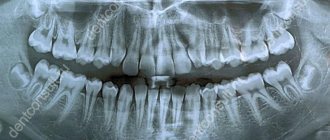
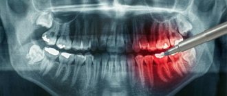
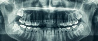
Orthopantomogram
– a high-precision panoramic image of the teeth, allowing you to get a complete picture of the condition of the oral cavity. An orthopantomogram is done using a special device - an orthopantomograph, a type of X-ray equipment that has a circular rotating element. Compared to a traditional X-ray machine, the orthopantomograph provides a reduced radiation dose, so it can be safely used when the need for a panoramic image arises.
An orthopantomogram shows teeth, maxillofacial joints, facial bones of the skull, and nasal sinuses. The main advantage of an orthopantomogram is the ability to simultaneously obtain a panoramic image of the upper and lower jaws. It is the need to see the whole picture that immediately determines the need to use a panoramic photograph.
Orthopantomogram
When is an orthopantomogram needed?
Orthopantomogram is used:
- in therapeutic dentistry. A panoramic photograph of the teeth allows you to identify periodontal problems, as well as carious cavities, and helps to create a comprehensive treatment plan;
- in surgical dentistry - before tooth extraction operations;
- in orthodontics. An orthopantomogram allows you to get a general idea of the state of your bite and select the right technique and hardware to correct it;
- in implantology – for planning implantation decisions.
Are there any restrictions
This examination option is considered safe and harmless, however, there are some contraindications. The procedure is not performed if the patient has malignant neoplasms in the head and neck area, as well as blood cancers - leukemia, lymphoma and others. In the first case, under the influence of radiation, the tumor can begin to grow and mutate, which will lead to even more severe complications. In the second case, even a slight effect of radiation on the bone marrow, a hematopoietic organ, can provoke and accelerate the development of the disease, including fatal consequences.
Also, performing OPTG is not always possible in a small child or in a person with serious mental disorders - when the patient is unable to remain still for even 1-2 minutes.
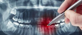
What does an orthopantomogram show?
A panoramic photograph of the teeth helps to detect:
- dental anomalies;
- caries, including at the initial stage;
- periodontal pockets;
- tooth cyst;
- foci of inflammation.
Using an orthopantomogram it is possible to establish:
- the need to begin correcting malocclusion;
- condition of periodontal tissues;
- degree of formation of tooth roots;
- when installing implants (for example, the condition of the bone at the site of intended implantation);
- and much more.
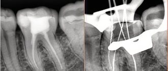
What types of photographs are there and what is better to choose?
As for the classification of OPTG, there are 2 types of panoramic X-rays of the jaw - film and digital orthopantomogram. Sometimes you can also hear about three-dimensional, but in fact this type of OPTG does not exist - all X-ray results here are only two-dimensional, “flat”.
- 1. film – this type of OPTG appeared first. Initially, models of devices were used that involved the use of a special film. It was placed on one side of the patient's head, while the X-ray tube was on the other side. After the procedure, the image was transferred to film, after which it was processed and developed,
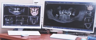
- 2. digital – a modern version of the examination involves recording images on a medium in digital format. The obtained data is loaded into a special computer program, and the doctor can carefully study the already processed image from the computer screen.
What is better to choose – digital or film orthopantomogram? Of course, it is better to give preference to the digital option. It is more modern, accurate, and the image can be enlarged many times on a computer to examine the pathological area in detail.
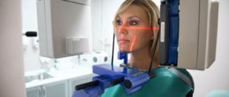
How is an orthopantomogram done?
No special preparation is required for the study. You will need to remove glasses, earrings, hair clips, other metal objects, and dentures. An orthopantomogram is usually done in a standing position. A special X-ray protective apron-cape is put on the chest. The position of the head is fixed with special clamps. As directed by the doctor, you will need to bite a special plastic mark (which is located in a disposable cover) - this will allow the image to be correctly centered. The tongue should be kept pressed to the palate. During an orthopantomogram, the module will move around the head, this will take 15-20 seconds, and during this time you must stand strictly still in order for the image to be of the proper quality.
You can get an orthopantomogram in Moscow at Family Doctor JSC.
Sign up for diagnostics Do not self-medicate. Contact our specialists who will correctly diagnose and prescribe treatment.
Why do you need a panoramic photo?
A panoramic photograph of the dentition is taken to diagnose the general condition of the oral cavity. The procedure is not painful. The only thing the patient faces in this case is the need to open his mouth wide while standing.
The procedure has its own indications, which are determined by the attending physician. Usually they are provided for in the future of any serious procedures and are as follows:
- dental implantation;
- its alignment;
- treatment of periodontal disease;
- inflammation of the soft tissues of the oral cavity;
- assessment of tooth growth rate;
- surgical intervention.
It is worth considering that before an orthopantogram you should remove all objects that interfere with the passage of x-rays (jewelry, etc.).
Patient protection is mandatory during the procedure. To carry it out, he wears an apron made with lead and a special collar on the neck area. This helps avoid radiation exposure and subsequent problems from the harmful effects of X-rays.
It is worth understanding that the safety of x-rays is achieved only when all necessary measures are followed. In addition, the better the device, the faster and better quality the picture will be. The X-ray of a digital orthopantomograph is better than that of a film one.
How is X-ray performed?
As already mentioned, the patient must stand. His chest should be pressed against the platform, which determines the correct position in space. The conditions of the procedure stipulate that he will need to bite a special plastic stick, after first pressing his tongue against the soft palate. Lips must be closed.
The doctor may ask you to change the position of the head so that the x-ray can cover the required area and the diagnosis is correct.
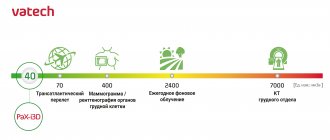
Currently, the procedure is performed using a safer PaX-i3d “Green” tomograph. Its main differences from devices of previous generations are that it is safer and scans an order of magnitude faster, protecting not only the patient, but also the clinic staff from exposure to X-rays.
What are the advantages and disadvantages of OPTG
An orthopantogram can significantly improve the quality of diagnosis, and today dental treatment is practically not carried out without radiography, except for minor problems. Among the undeniable advantages of the technique, experts highlight the following points:
- speed of the procedure - literally in a couple of minutes the image is ready for study;
- modern equipment makes it possible to adjust the height and trajectory of the emitter, which made diagnostics possible for children and wheelchair users;
- minimal radiation exposure and absolute safety for the body;
- high quality and accuracy of the obtained images;
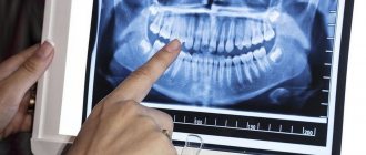
- the ability to zoom in on the area under study and study it in detail on a computer monitor.
With an orthopantomogram, minimal radiation exposure is used. If we talk about the disadvantages, the main disadvantage of OPTG is obtaining a flat two-dimensional image. As a result, the image may slightly distort the dimensions of teeth, root canals and bone tissue. Such deviations can vary from 15 to 30%. To obtain a more accurate image, it is better to resort to computed tomography.
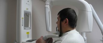
How is OPTG performed?
The patient is put on a special protective apron and asked to sit inside the orthopantomograph. In this case, the hands are placed on the handles of the orthopantomograph, the chin is placed on a special holder, and a special mouthpiece is placed in the teeth.
After this, the orthopantomograph is turned on, after 20-30 seconds the procedure is completed, and the study data is transferred to the PC.
Acceptability during pregnancy
Panoramic tomography is extremely undesirable for patients during pregnancy - in such cases it is performed only if there is an urgent need. The procedure poses an increased risk during the first trimester. In later stages of pregnancy, X-ray radiation will not have such a severe effect, but can still cause certain disturbances in the functioning of internal systems. For example, the thyroid gland is especially sensitive to the effects of radiation.
Features of conducting OPTG for children
We figured out what is visible in panoramic photographs and why they are taken, and found out that this is a completely safe diagnostic method. Now it should be noted that an orthopantomogram for children is carried out taking into account some nuances. A radiation exposure of 55 μSv (1 shot) is completely safe for a child, so a similar procedure is allowed from 5-6 years of age. The examination assumes that the patient must stand still for some time, but for young children this task can be quite difficult . When it comes to small patients, the procedure is carried out in exposure mode - the area of exposure is reduced, and the sensor is adjusted to a trajectory of movement corresponding to the small size of the jaws.
“My son also recently had a panoramic photo taken. He's 10, everything went great. Now there are new technologies everywhere, everything is safe. The procedure took less than a minute, and the doctors assured me that there was nothing wrong with it. How else can you start correcting your bite? The orthodontist prescribed OPTG for us. So far I’ve made the plate, but in a couple of years we’ll get braces. There’s no place here without pictures!”
In pediatric dentistry, OPTG is used to detect the rudiments of permanent teeth, assess the correctness of their formation, to diagnose adentia and identify malocclusions. Most often, this procedure is prescribed by an orthodontist before starting to correct defects in bite and teeth position. The procedure is completely safe for the child
Cost of dental examination
The cost of an orthopantomogram in clinics in Moscow and the regions starts from 700 rubles. But sometimes patients undergo it for free, for example, during turnkey implantation, when the total price of dental restoration already includes all the necessary diagnostic methods, consultations, implants, dentures and the work of specialists.
Chibisova M.A. Digital and film radiography in outpatient dentistry, 2004.
Author: Bespalov R. D. (Thank you for your help in writing the article and the information provided)
Potential dangers and possible complications
Many patients in dental clinics are interested in quite natural questions regarding whether an orthopantomogram is harmful and how often it can be done so as not to cause harm to health. Let's start with the fact that if you follow the technology of the procedure, you should not be afraid of any problems. One of the potential complications may be excessive radiation exposure, however, with modern equipment this situation is excluded, even though OPTG will have to be performed at least 2-3 times during implantation, as well as several more times during the year after it to monitor the results of treatment .
Numerous studies on the safety of using an orthopantomograph have proven that with a single procedure, the patient receives a radiation dose corresponding to values in the range from 10 to 40 μSv . This is an acceptable and completely harmless dosage that does not pose any threat to the human body. For comparison: with a single fluorography procedure, the radiation dose is up to 500 μSv.
Beam prosthesis from RUB 170,000!
Preparation of the oral cavity, individual bar with installation, taking impressions, manufacturing, installation and fitting of dentures. The price is for 1 jaw. Lifetime guarantee! Consultation with a doctor is free! Call now: +7 (495) 215-52-31