Doctors Cost
Price list Doctors clinic
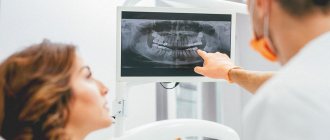
The Family Doctor clinic on Usacheva has installed an advanced X-ray Cone-beam computed tomograph - ORTOPANTOMOGRAPH OP300 Maxio (from Kavo), specially designed for dentistry and otorhinolaryngology.
Computed tomography is one of the most accurate diagnostic methods, allowing to obtain comprehensive information about the condition of bone and other tissues. It is widely used in the field of dentistry and maxillofacial surgery. Cone beam computed tomography of teeth allows you to quickly obtain a three-dimensional model of the patient’s dental system.
What is 3D dental computed tomography
Description
Three-dimensional x-ray examination of teeth is one of the types of x-rays, as a result of which the doctor receives an image of the highest possible quality. In this case, both hard and soft tissues of the oral cavity are visible in the image. Thanks to the advent of this research method, the quality of diagnosis has significantly improved. As a result, the risk of making any errors during the treatment process is minimized.
During the examination, the dentist can view the image in different projections for a more accurate analysis. CT is best suited for assessing the condition of dental hard tissues. The quality of the resulting image largely depends on the power of the equipment used.
Difference from X-ray
The main difference is that the operating principle of CT and X-ray is based on very different physical phenomena. During a three-dimensional examination, the familiar X-rays are used, and during magnetic resonance imaging, a magnetic field is used.
Today, computed tomography is performed using a variety of equipment; there are several types of devices. Some allow you to obtain images of the highest quality, while others provide a fairly strong radiation load on the patient’s body. That is why it is recommended to contact only trusted clinics where the most modern equipment is installed.
Scope of technology application
Intraoral digital scanners have been used in dental practice for a long time. Device manufacturers are constantly improving models, increasing scanning speed and improving the quality of the resulting three-dimensional images. 3D technology is a worthy alternative to the standard procedure for taking jaw impressions.
The device is used in several areas of dentistry.
- Orthodontics - by examining the oral cavity in several planes, the doctor will be able to make a more accurate diagnosis, examine the clinical picture in detail, and predict the result of therapy.
- Orthopedics – makes it possible to produce prosthetics (veneers, crowns, bridges and others) with an accuracy of hundredths of a mm.
- Implantology – based on the data obtained, surgical templates are created, and the condition of soft and bone tissues is diagnosed before installing implants.
- Gnathology – recommended for the manufacture of individual splints and splints.
The technology reflects advances in computer developments for the medical field. The President dentistry in Moscow has long been using equipment with innovative software, which has a positive effect on the quality of services provided to patients.
How to do a 3D computed tomography of teeth
Preparation
It is worth noting that no preliminary preparation is required for a dental CT scan. Before the procedure, it is enough to remove all metal items, jewelry, watches, and take out your mobile phone from your pockets. These items may interfere with the examination. This also applies to metal structures in the oral cavity; they will also have to be removed for a while.
Next, the specialist must put a special protective apron on the patient; it will cover the upper part of the body and prevent the negative impact of the device on the body. It is very important that the clinic employs real professionals trained in this matter, and not ordinary nurses.
Conversation with a doctor
Even at the stage of consultation with your dentist, it is very important to warn him about all previous interventions in the oral cavity, orthopedic treatment, etc. It is important for the doctor to know whether there are any structures, crowns in the mouth, whether operations have been performed previously, etc.
This is required only so that the doctor can change the tomograph settings in time. As a result, you can get the most accurate result and a clear picture of the jaw. If any structures can be removed before the procedure, it is best to do this in advance.
Carrying out tomography
During the procedure itself, the patient can be in different positions: he can sit, stand, or even lie on a special couch. It all depends on the device used by the clinic. The mouth can also be closed or open. The equipment will rotate on its own around the desired area, so it is very important to take a comfortable position and not move.
The entire study lasts no more than 1 minute (provided the entire jaw is examined); one tooth requires no more than 10 seconds of time. In the case of cone beam tomography, the device will rotate around the head. As a result, the doctor can receive up to 200 regular images, which can later be converted into 3D.
Panoramic or 3D photo
The only difference between a CT scan and a standard orthopantomogram is that the image is three-dimensional and more detailed. This method is more advanced and allows you to obtain more reliable data. The use of CT is especially important if there is a suspicion of serious pathologies of bone tissue.
When working with a CT scanner, images can be combined with each other and saved on a variety of media for further use by other specialists. In addition, the procedure is much faster compared to other research options.
Methods for diagnosing dental condition
Most methods for diagnosing the condition of the teeth and oral cavity fall into the category of visualization, that is, they can clearly show everything that happens in this part of the body. At a minimum, it is now completely impossible to imagine dentistry without radiography. Now this type of examination is included in the standard set of procedures that a patient undergoes when visiting the clinic with any complaint.
On a note! An examination is necessary not only when diagnosing the condition of a specific tooth, but also if the patient is faced with problems with the functioning of the entire maxillofacial apparatus.
Table. Main types of diagnostics in dentistry.
| View | Description |
| This is the most common diagnostic option, which can be carried out in any, even the most modest clinic. It provides the opportunity to correctly and accurately assess the condition of the canals in the roots of the tooth and identify developing pathologies in the area of hard tissues. Often used for endotonia. An X-ray image is a small black and white image on a special film or on a CD. | |
| This is otherwise called a panoramic image of the dental system. Using it, the doctor will be able to identify pathologies in the development of the jaw in general and teeth in particular, detect all carious cavities, understand how much hard tissue is destroyed during periodontal disease, clarify whether endotonic treatment helped the patient, etc. The image will also allow you to see whether there are any or deviations or changes in the condition of the lower sections of the maxillary sinuses. An orthopantomogram is also done in relation to the temporomandibular joints. It is often prescribed before prosthetics. | |
| This is a picture of not just the jaw, but the entire skull in one projection or another. It is necessary for measuring parts of the facial part (cephalometry). Based on this image, you can plan treatment with an orthodontist. | |
| It is this picture that is called 3D, which is discussed in the article. We'll talk about it separately below. |
Types of 3D computed tomography
Step-by-step CT
This method was the first to appear many years ago, so the results of such a study have minimal accuracy. The device produces a low-quality image, while the radiation exposure to the human body in just one procedure is almost 2 times higher than the recommended annual radiation dose.
Step-by-step tomography involves scanning each desired layer one by one. Gradually the table moves, all other layers are examined. There can be more than 10 of them in total, it all depends on how much jaw volume needs to be checked. Time spent in the device: from 10 to 25 minutes.
Multi-slice CT (MSCT)
This device involves less load, and the examination time is also reduced. However, the use of such a device is currently considered quite hazardous to health. In this case, the clarity of the resulting image will not be high enough. Spiral devices are recommended for studying soft tissues, such as gums, muscles, salivary glands, etc.
Multislice or multislice CT is one of the most modern diagnostic methods. The main difference is that the X-ray tube moves at a much higher speed, and the machine itself has more than two detectors that receive the X-ray beams.
Cone beam CT (CBCT)
This type of research is considered one of the most modern and convenient. It is designed to study hard and soft tissues. At the same time, the equipment provides ideal picture quality, and the radiation exposure to the body remains minimal. This study is usually classified as volumetric digital. Most often it is used to study the jaw.
It is very important that the patient keeps his head still during the procedure. A device will rotate around the head, on one side which has an X-ray source, and on the other - a small receiver. As a result, the doctor has a three-dimensional model at his disposal.
Peculiarities
In the most modern devices, the radiation dose depends on several factors, for example, on the body weight of a particular person. For example, an adult will receive about 55 μ3 V, and a child during the same examination will receive no more than 15 μ3 V.
To view ready-made volumetric images of the jaw, the dentist needs special programs on his work computer. There, the resulting images can be edited, processed and combined with each other. It is important to note that the image quality will also depend on the software installed on the PC.
Advantages
Indeed, doctors used to do without computed tomography. However, the possibilities of prosthetics or orthodontics were then limited: it’s easy to recall the terrifying false jaws, in which it was impossible to eat or speak, or dental plates, in which only a few decided to walk due to the colossal inconvenience and terrible external kind.
Modern advances in dentistry also require cutting-edge diagnostic methods. Dental CT is just such a method. Many experts call modern tomographic examination the term “3D computed tomography.”
The main advantages of this diagnostic method are:
- identification of infection localizations;
- diagnosis of maxillodental anomalies or defects (for example, identifying bone pockets, joint diseases, determining the degree of gum inflammation);
- diagnostics for complex injuries or congenital pathologies;
- the need to calculate down to the millimeter the movement of teeth when using braces;
- control over the development of jaw pathologies;
- clarification of the location of the lesion before surgery.
Dental CT is often prescribed for the following disorders:
- previously undiagnosed jaw diseases (developmental defects, difficult or spontaneous movements of the jaws, the appearance of clicks when moving, pain during chewing, etc.);
- injuries;
- prosthetics of bone organs;
- complex tooth extraction;
- spasms of the jaw muscles when moving the mouth;
- complications after treatment or tooth extraction;
- identification of hidden diseases of the pulp, teeth, gums;
- detection of cysts or tumors;
- hidden cavities in the mouth;
- before implantation in combination with reconstructive operations;
- malocclusion;
- unerupted teeth;
- examination of any soft tissues of the mouth.
3D computed tomography of teeth without pain
Safety
Many patients put off visiting a dental clinic simply because they fear that all procedures cause pain and discomfort. This issue is especially relevant if it is necessary to send young children for dental treatment. However, no need to worry! Most modern procedures within the walls of a dental clinic are absolutely painless.
This is due to the use of the most advanced equipment and technologies. In addition, if necessary, the specialist may suggest the administration of a mild anesthetic. All drugs used in the work are harmless, the risk of an allergic reaction to them is minimal.
Benefits of use
Until recently, classical X-ray machines were used to examine the oral cavity and take pictures of the jaw; an orthopantogram, that is, a panoramic photograph, was taken. Today, doctors have more modern equipment at their disposal that allows them to obtain better quality images. As a result, it is possible to more closely monitor the treatment process.
CT provides a three-dimensional image without any distortion in shape and size. This guarantees the study of the clinical picture with 100% accuracy, so the dentist can identify the presence of individual structural features of the dentition of each patient.
Advantages of an intraoral 3D scanner
The modern technique has many advantages:
- highest accuracy - the error of data from intraoral 3D scanners is only 12 microns versus 100 microns in the case of making an impression using silicone materials;
- maximum convenience for the doctor and the patient - the manipulations are simple and do not cause discomfort;
- the device scans each print in just 24 seconds;
- excellent information content - the three-dimensional model visualizes all defects and shortcomings of the maxillofacial system.
Digital impressions are sent to the laboratory or other dental clinic without delay. They show as accurately as possible the condition of the teeth and the jaw as a whole. The dentist can study the impression in detail and suggest the most suitable treatment option, taking into account all the features of the case.
Contraindications
Absolute
Absolute contraindications to CT scanning include the presence of serious diseases such as complex mental disorders, problems with the heart or cerebral vessels.
It is also not recommended to undergo examination if there are malignant tumors in the body, tuberculosis, diabetes mellitus, etc. Also, this device is not suitable for those who suffer from a fear of closed spaces (treatment in this case can cause serious psychological trauma).
Relative
An obstacle to diagnosis may be the teenager’s too young age or increased activity (as a result of which it is impossible to hold him motionless in one place even for several minutes), pregnancy in any trimester, breastfeeding, etc.
Patients may also experience allergic reactions to the substances used if they need to be introduced into the blood. Kidney failure, colds and fever are also temporary contraindications.
How to prepare for research
Dental tomography usually does not require any special preparation. It can be performed on both an inpatient and outpatient basis. If dental tomography is performed without the use of contrast, then the patient does not need to behave in any special way before the procedure. The only restriction is to abstain from food for 4-5 hours before the study.
On the appointed day, the patient usually comes to the tomography room for a certain time. At the same time, he is asked to remove any metal objects from himself: jewelry, accessories, belts, braces. This is necessary to ensure that the research results do not have errors.
If contrast is used, a little preparation before the study is necessary. Often, a few days before the tomography, the patient undergoes a blood test to identify contrast allergens, as well as to determine normal or reduced urinary function of the kidneys.
Rehabilitation after 3D computed tomography of teeth
Harm from tomography
Modern devices pose virtually no threat to human health, even with frequent examinations. Before the procedure, you must remove all metal products and put on a special vest. This will help reduce the negative impact from the emitter to zero.
During the examination, you must sit still, otherwise the picture may turn out unclear, and the picture will have to be taken again. There is no pain; the procedure does not require prior administration of an anesthetic.
Duration of the procedure
It is worth paying attention to the fact that computer tomography machines come in a variety of different types. During dental treatment, the patient does not need to lie down inside a large device; only a small device will be sufficient.
To study the jaw, dentists use miniature devices, so the duration of the procedure will be minimal (no more than a couple of minutes). In this case, the device will have time to take more than 300 photographs of the jaw from different sides.
Dental scanning – digital dental impression
Recently, devices have appeared - intraoral scanners! These are special computers that allow you to take an impression of a person’s jaw without silicone mass. The doctor moves a camera connected to a computer around the teeth in the mouth, and special software builds a three-dimensional virtual model of the patient's jaw and bite.
Comparison of traditional silicone impressions and jaw scanning (digital impressions)
| Traditional silicone dental impressions | Digital impressions – 3D scanning of teeth |
| Takes about 10 minutes. | Takes about 10 minutes. |
| The mass is introduced into the mouth and remains there. May provoke a gag reflex. | The mass is not administered into the mouth. People with a gag reflex find it easier to carry a camera in their mouth than a mass. |
| Need a place to store models. | They are stored on the server and do not require physical space. |
| There are difficulties with forwarding, for example, for consultation with a doctor living in another country (city). | Sending digital impressions occurs in a minute via the Internet. |
| From one cast, only one model can be made from plaster with high quality and accuracy. | You can make as many models as you like from a digital impression without losing quality. |
Why is 3D computed tomography necessary?
Indications
There are situations where you can do without a 3D CT scan, but not always.
It will be indispensable for studying the anatomy and structure of the hard tissues of the patient’s jaw, if necessary, diagnosing serious pathologies, detecting impacted units (for example, wisdom teeth), identifying hidden carious cavities and much more.
Also, this procedure should be carried out before any orthodontic treatment, before implantation and prosthetics, as well as if the presence of tumors in the tissue is suspected.
Advantages
CT demonstrates the most accurate and reliable picture of the condition of the patient’s jaw in all projections. This allows you to select the optimal treatment and carry it out as quickly as possible.
This study allows us to identify malignant tumors at the earliest stages and remove them in time. If suspicions are confirmed, then further study of the formations is recommended using MRI.
Indications for this type of examination
3D tomography has a lot of possibilities, for which it is valued by doctors. Thus, it will allow you to assess the condition of not only the jaw, but also the gums, see the quality of previously installed fillings, and reflect all pathologies and anomalies. Indications for this may include:
- abnormalities in the structure of the jaw and the shape of the teeth;
- suspicions of pathological changes in dental tissues;
- identification of the rudiments of permanent teeth in children, assessment of the condition of milk teeth;
- the presence of cysts, inflammation, cracks in the roots of the tooth;
- suspicion of traumatic injuries to the jaw;
- preparation for implant installation;
- the presence of tumors in the jaw area;
- preparation for plastic surgery;
- preparation for operations in the jaw area.
Thus, a detailed three-dimensional image is necessary in all complex cases that require detail and a thorough assessment of the patient's current condition. It is especially important in assessing abnormally growing teeth, as well as in case of complex fractures and dental pathologies.
On a note! A CT scan is also often performed during preparation for implant placement. Moreover, its efficiency and effectiveness are much higher than that of orthopantomography. It is often necessary when performing a so-called sinus lift.
Table. Areas of application of CT.
| Sphere | Explanation |
| Orthodontics | The ability to assess how distorted the bite is. |
| Surgery | Assessment of the possibility of certain manipulations by the surgeon, as well as the possibility of installing teeth. |
| Maxillofacial Surgery | Assessment of the condition of the jaw and surrounding areas of the skull. |
| Therapy | The ability to detect deep-lying caries and choose the right treatment option. |
Cost of 3D computed tomography in Moscow
You can get a 3D computed tomography scan of your teeth at the Kingdom of Smiles clinic. The final cost for this service consists of several factors: the number of units studied, the age of the patient, and the need for related procedures.
Payment for services provided can be either a one-time payment or in installments without interest. New promotions and lucrative discount offers appear regularly on the company's website. If you are worried about toothache, do not delay, sign up for a free consultation!
| Service | Cost, rub. |
| X-ray with printout | 500 |
| CT area 6*6cm (3 teeth) | 1700 |
| CT area 6*10 (one jaw) | 2900 |
| CT area 7.5*14.5 (both jaws) | 4200 |
previous post
Installation of crowns in Moscow
next entry
Where can I take a photo of a tooth?
Computed tomography is becoming an increasingly common procedure offered by dentistry in Moscow. However, unlike a digital orthopantomogram, it can so far only be found in large clinics offering a wide range of services. You can always find out where to take a 3D photograph of a tooth in Moscow from the specialist who referred you for this study. The price for a computed tomography scan of the jaw, or 3D dental image, in Moscow varies from clinic to clinic and starts from approximately 2,500 rubles.
Publisher: Expert magazine about dentistry Startsmile.ru
Author of the material: Ekaterina Gasparova